Review
. 2020 Nov 10;6(5):1388-1401.
doi: 10.1016/j.bioactmat.2020.10.021. eCollection 2021 May.

Advances in 3D bioprinting technology for cardiac tissue engineering and regeneration

Affiliations
Review

Advances in 3D bioprinting technology for cardiac tissue engineering and regeneration

Nanbo Liu et al. Bioact Mater. .

Abstract

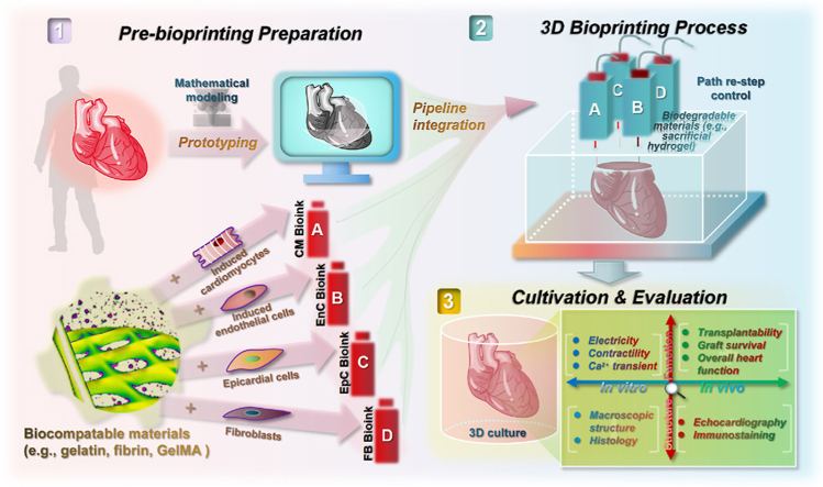
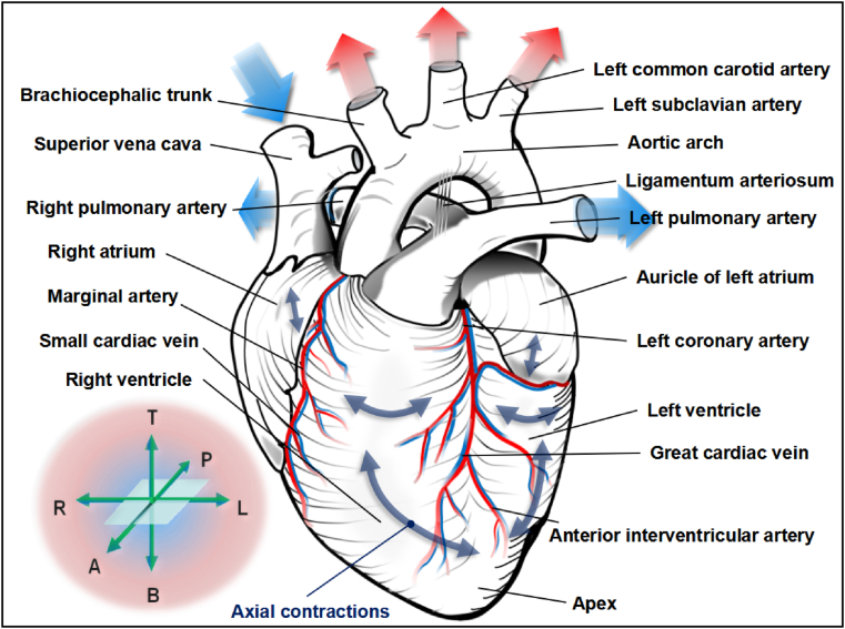
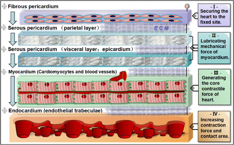
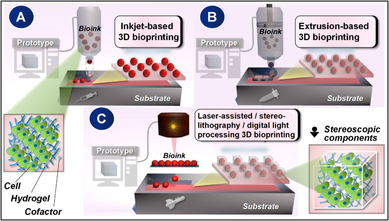
Cardiovascular disease is still one of the leading causes of death in the world, and heart transplantation is the current major treatment for end-stage cardiovascular diseases. However, because of the shortage of heart donors, new sources of cardiac regenerative medicine are greatly needed. The prominent development of tissue engineering using bioactive materials has creatively laid a direct promising foundation. Whereas, how to precisely pattern a cardiac structure with complete biological function still requires technological breakthroughs. Recently, the emerging three-dimensional (3D) bioprinting technology for tissue engineering has shown great advantages in generating micro-scale cardiac tissues, which has established its impressive potential as a novel foundation for cardiovascular regeneration. Whether 3D bioprinted hearts can replace traditional heart transplantation as a novel strategy for treating cardiovascular diseases in the future is a frontier issue. In this review article, we emphasize the current knowledge and future perspectives regarding available bioinks, bioprinting strategies and the latest outcome progress in cardiac 3D bioprinting to move this promising medical approach towards potential clinical implementation.

Keywords: 3D bioprinting; Bioink; Heart repair and regeneration; Stem cell therapy.


Conflict of interest statement

All the authors agree that no conflict of interest is declared.

Figures

Image 1
Graphical abstract
Fig. 1
Fig. 1
Anatomy and function of human heart. The unique structure of the heart's strong muscle and large/small blood vessel that closely merge together as one is the mechanical force and energy basis for the human body in performing various physiological activities. In particular, the striated muscle fibrous tissue that undergoes autonomic contraction at the same electro-physiological rhythm is a critical feature of coordination between the left heart and the right heart, in order to efficiently handle the repeated remodeling of blood biochemistry. Coordinate icons in the lower left corner are “T” for top, “P” for posterior, “A” for anterior, “R” for right, “L” for left, “B” for bottom.
Fig. 2
Fig. 2
Five cross-sectional layers of myocardial tissue from exterior to interior. The first layer is fibrous pericardium consisting of fibroblasts that can secure the heart's anatomical position. The second and third layer is both serous pericardium, which can lubricate mechanical force of myocardium. The forth layer is the thick myocrdium consisting of active cardiomyocytes and supportive blood vessels. The fifth layer is the robust endocardium that has endothelial trabeculae in order to increase pumping force and area inside. Each layer of myocardial tissue is physically and orderly bond by the components of ECM.
Fig. 3
Fig. 3
Major classification of tissue engineering-targeted 3D bioprinting technology. Briefly, a standardized micro-construction as designed by the digital cubic prototype can be achieved either in use of gravity hanging drops controlled by an inkjet-based 3D bioprinter (A), by facilitating mechanical squeezing forces through the regulation of an extrusion-based 3D bioprinter (B), or through the direct-writing function of a laser-guided, stereolithography, or digital light processing 3D bioprinter (C). The above three printing strategies can be used either individually or in combination according to the specific needs of tissue engineering, the purpose of which is to achieve a closer simulation of native tissues and organs.
Fig. 4
Fig. 4
Full-fledged and high-resolution cardiac structures engineered by advanced 3D bioprinting. Through accurate printing and cross-linking of cell-loading cardiac bioink, the original structure of heart can be realistically reproduced in construction of: linear columnar (A) [85], grid-like pattern (B) [67], spherical cluster (C) [148], rectangular parallelepiped (D) [86], valentine heart-shaped material (E) [72], cube-shaped block (F) [71], perforated column (G) [84], conical container (H) [102], four-chamber sagittal plane (I) [102], heart valve-like structure (J & K) [68,102], left heart with large vessel-like construction (L) [68], right ventricle-like construction (M & N) [90], and micro vascularized HEHT (O) [117].
Fig. 5
Fig. 5
Evaluations of the representative advanced 3D bioprinted cardiac tissues. A. Immunostaining of cardiac biomarkers may include views of whole (a1) [117] and part (a2) [69]. B. Spontaneous muscle-like contraction after stabilization can be documented by video (b1 and b2) [72,90] and quantification (b3) [86]. C. The micro-engineered cardiac tissues can also excite stable cardiomyocyte-like depolarization-repolarization action potentials (c1 and c2) [102,148]. D. Dynamic calcium transient processes of the advanced 3D bioprinted functional cardiomyocytes may be real-time detected (d1 and d2) [72,90]. E. The 3D bioprinted patch-like tissues composed of iPSC-CMs and blood vessel endothelial cells could be well integrated into mice physiological subcutaneous tissue (e1) [71] and rat myocardial infarcted heart (e2) [73] for in vivo measurements, respectively.

Similar articles

Cited by

References

    1. Virani S.S., Alonso A., Benjamin E.J., Bittencourt M.S., Callaway C.W., Carson A.P. Heart disease and stroke statistics-2020 update: a report from the American heart association. Circulation. 2020;141(9):e139–e596. - PubMed
    1. Riggs K.W., Zafar F., Radzi Y., Yu P., Bryant R., Morales D.L.S. Adult congenital heart disease: current early expectations after cardiac transplantation. Ann. Thorac. Surg. 2020;109(2):480–486. - PubMed
    1. Hernandez G.A., Lemor A., Clark D., Blumer V., Burstein D., Byrne R. Heart transplantation and in-hospital outcomes in adult congenital heart disease patients with Fontan: a decade nationwide analysis from 2004 to 2014. J. Card. Surg. 2020;35(3):603–608. - PubMed
    1. Tomov M.L., Gil C.J., Cetnar A., Theus A.S., Lima B.J., Nish J.E. Engineering functional cardiac tissues for regenerative medicine applications. Curr. Cardiol. Rep. 2019;21(9):105. - PMC - PubMed
    1. Takahashi K., Tanabe K., Ohnuki M., Narita M., Ichisaka T., Tomoda K. Induction of pluripotent stem cells from adult human fibroblasts by defined factors. Cell. 2007;131(5):861–872. - PubMed

LinkOut - more resources