

Classifying Anal Intraepithelial Neoplasia 2 Based on LAST Recommendations
- PMID: 33210115
- PMCID: PMC8130877
- DOI: 10.1093/ajcp/aqaa188
Classifying Anal Intraepithelial Neoplasia 2 Based on LAST Recommendations
Abstract
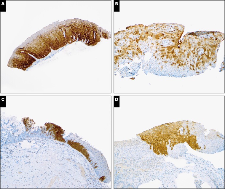
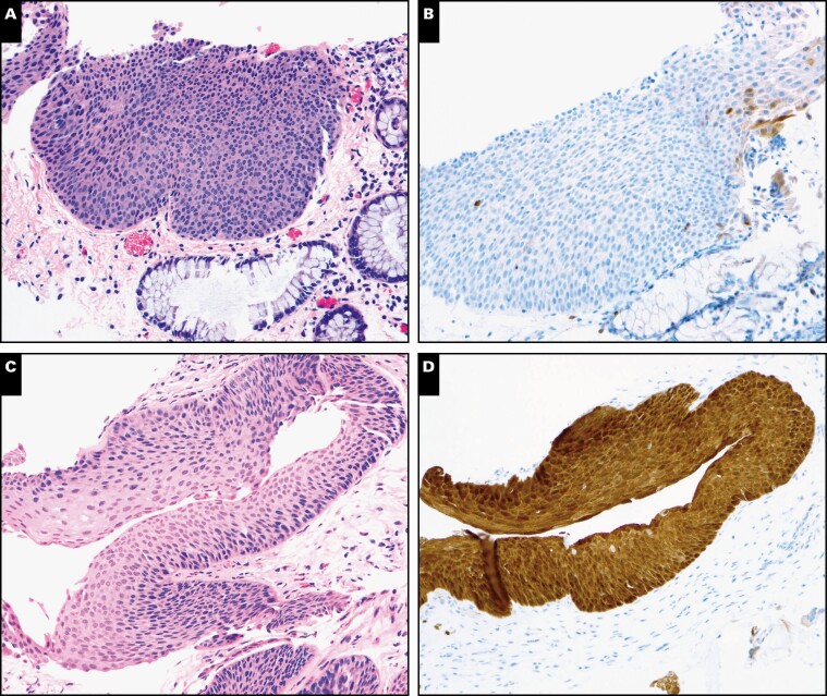
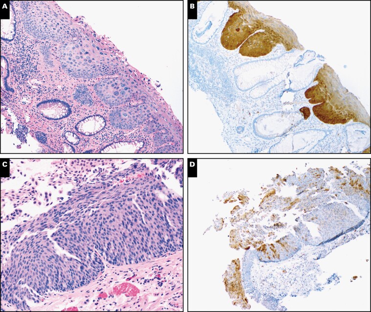
Objectives: The Lower Anogenital Squamous Terminology (LAST) recommendations classify human papillomavirus-associated squamous lesions into low- and high-grade squamous intraepithelial lesions (LSILs/HSILs). Our study aimed to assess interobserver agreement among 6 experienced pathologists in assigning 40 anal lesions previously diagnosed as anal intraepithelial neoplasia 2 (AIN 2) to either HSIL or non-HSIL categories.
Methods: Agreement based on photomicrographs of H&E alone or H&E plus p16 immunohistochemistry was calculated using κ coefficients.
Results: Agreement was fair based on H&E alone (κ = 0.42; 95% confidence interval [CI], 0.34-0.52). Adding p16 improved agreement to moderate (κ = 0.55; 95% CI, 0.54-0.62). On final diagnosis, 21 cases (53%) had unanimous diagnoses, and 19 (47%) were divided. When designating p16 results as positive or negative, agreement was excellent (κ = 0.92; 95% CI, 0.83-0.95). Among variables (staining location, extent, and intensity), staining of the basal/parabasal layers was a consistent feature in cases with consensus for positive results (20/20). Of the 67 H&E diagnoses with conflicting p16 results, participants modified 32 (48%), downgrading 23 HSILs and upgrading 9 non-HSILs.
Conclusions: Although p16 increased interobserver agreement, disagreement remained considerable regarding intermediate lesions. p16 expression, particularly if negative, can reduce unwarranted HSIL diagnoses and unnecessary treatment.
Keywords: Anal intraepithelial neoplasia 2; Human papillomavirus; Interobserver agreement; p16 Immunohistochemistry.
© American Society for Clinical Pathology, 2020. All rights reserved. For permissions, please e-mail: journals.permissions@oup.com.
Figures
References
-
- Darragh TM, Colgan TJ, Cox JT, et al. . Members of LAST Project Work Groups . The Lower Anogenital Squamous Terminology standardization project for HPV-associated lesions: background and consensus recommendations from the College of American Pathologists and the American Society for Colposcopy and Cervical Pathology. Arch Pathol Lab Med. 2012;136:1266-1297. - PubMed
-
- Stoler MH. Human papillomaviruses and cervical neoplasia: a model for carcinogenesis. Int J Gynecol Pathol. 2000;19:16-28. - PubMed
-
- WHO Classification of Tumours. Lyon, France: International Agency for Research on Cancer; 2019.
-
- Castle PE, Stoler MH, Solomon D, et al. . The relationship of community biopsy-diagnosed cervical intraepithelial neoplasia grade 2 to the quality control pathology-reviewed diagnoses: an ALTS report. Am J Clin Pathol. 2007;127:805-815. - PubMed
-
- Castle PE. A LASTing impression: incorporating p16 immunohistochemistry into routine diagnosis of cervical neoplasia. Pathol Case Rev. 2013;18:154-157.
Publication types
MeSH terms
Substances
Grants and funding
LinkOut - more resources
Full Text Sources
Medical
