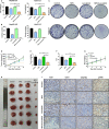
Sophoridine suppresses lenvatinib-resistant hepatocellular carcinoma growth by inhibiting RAS/MEK/ERK axis via decreasing VEGFR2 expression
- PMID: 33210432
- PMCID: PMC7810959
- DOI: 10.1111/jcmm.16108
Sophoridine suppresses lenvatinib-resistant hepatocellular carcinoma growth by inhibiting RAS/MEK/ERK axis via decreasing VEGFR2 expression
Abstract
Hepatocellular carcinoma (HCC) is one of the most lethal cancer types with insufficient approved therapies, among which lenvatinib is a newly approved multi-targeted tyrosine kinase inhibitor for frontline advanced HCC treatment. However, resistance to lenvatinib has been reported in HCC treatment recently, which limits the clinical benefits of lenvatinib. This study aims to investigate the underlying mechanism of lenvatinib resistance and explore the potential drug to improve the treatment for lenvatinib-resistant (LR) HCC. Here, we developed two human LR HCC cell lines by culturing with long-term exposure to lenvatinib. Results showed that the vascular endothelial growth factor receptors (VEGFR)2 expression and its downstream RAS/MEK/ERK signalling were obviously up-regulated in LR HCC cells, whereas the expression of VEGFR1, VEGFR3, FGFR1-4 and PDGFRα/β showed no difference. Furthermore, ETS-1 was identified to be responsible for VEGFR2 mediated lenvatinib resistance. The cell models were further used to explore the potential strategies for restoration of sensitivity of lenvatinib. Sophoridine, an alkaloid extraction, inhibited the proliferation, colony formation, cell migration and increased apoptosis of LR HCC cells. In vivo and in vitro results showed Sophoridine could further sensitize the therapeutic of lenvatinib against LR HCC. Mechanism studies revealed that Sophoridine decreased ETS-1 expression to down-regulate VEGFR2 expression along with downstream RAS/MEK/ERK axis in LR HCC cells. Hence, our study revealed that up-regulated VEGFR2 expression could be a predicator of the resistance of lenvatinib treatment against HCC and provided a potential candidate to restore the sensitivity of lenvatinib for HCC treatment.
Keywords: VEGFR2; hepatocellular carcinoma; lenvatinib resistance; sophoridine.
© 2020 The Authors. Journal of Cellular and Molecular Medicine published by Foundation for Cellular and Molecular Medicine and John Wiley & Sons Ltd.
Conflict of interest statement
The authors confirm that there are no conflicts of interest.
Figures


Similar articles
-
Oxysophocarpine suppresses FGFR1-overexpressed hepatocellular carcinoma growth and sensitizes the therapeutic effect of lenvatinib.Life Sci. 2021 Jan 1;264:118642. doi: 10.1016/j.lfs.2020.118642. Epub 2020 Oct 24. Life Sci. 2021. PMID: 33148422
-
Dual effects of targeting neuropilin-1 in lenvatinib-resistant hepatocellular carcinoma: inhibition of tumor growth and angiogenesis.Am J Physiol Cell Physiol. 2024 Oct 1;327(4):C1150-C1161. doi: 10.1152/ajpcell.00511.2024. Epub 2024 Sep 9. Am J Physiol Cell Physiol. 2024. PMID: 39250819
-
Lenvatinib inhibits angiogenesis and tumor fibroblast growth factor signaling pathways in human hepatocellular carcinoma models.Cancer Med. 2018 Jun;7(6):2641-2653. doi: 10.1002/cam4.1517. Epub 2018 May 7. Cancer Med. 2018. PMID: 29733511 Free PMC article.
-
New insights into the mechanism of resistance to lenvatinib and strategies for lenvatinib sensitization in hepatocellular carcinoma.Drug Discov Today. 2024 Aug;29(8):104069. doi: 10.1016/j.drudis.2024.104069. Epub 2024 Jun 25. Drug Discov Today. 2024. PMID: 38936692 Review.
-
The action and resistance mechanisms of Lenvatinib in liver cancer.Mol Carcinog. 2023 Dec;62(12):1918-1934. doi: 10.1002/mc.23625. Epub 2023 Sep 6. Mol Carcinog. 2023. PMID: 37671815 Free PMC article. Review.
Cited by
-
Advances in hepatocellular carcinoma drug resistance models.Front Med (Lausanne). 2024 Jul 31;11:1437226. doi: 10.3389/fmed.2024.1437226. eCollection 2024. Front Med (Lausanne). 2024. PMID: 39144662 Free PMC article. Review.
-
Targeted inhibition of PDGFRA with avapritinib, markedly enhances lenvatinib efficacy in hepatocellular carcinoma in vitro and in vivo: clinical implications.J Exp Clin Cancer Res. 2025 May 7;44(1):139. doi: 10.1186/s13046-025-03386-8. J Exp Clin Cancer Res. 2025. PMID: 40336047 Free PMC article.
-
Sophoridine Counteracts Obesity via Src-Mediated Inhibition of VEGFR Expression and PI3K/AKT Phosphorylation.Int J Mol Sci. 2024 Jan 19;25(2):1206. doi: 10.3390/ijms25021206. Int J Mol Sci. 2024. PMID: 38279206 Free PMC article.
-
Roles of clinical application of lenvatinib and its resistance mechanism in advanced hepatocellular carcinoma (Review).Am J Cancer Res. 2024 Sep 15;14(9):4113-4171. doi: 10.62347/UJVP4361. eCollection 2024. Am J Cancer Res. 2024. PMID: 39417171 Free PMC article. Review.
-
RNF8 depletion attenuates hepatocellular carcinoma progression by inhibiting epithelial-mesenchymal transition and enhancing drug sensitivity.Acta Biochim Biophys Sin (Shanghai). 2023 May 6;55(4):661-671. doi: 10.3724/abbs.2023076. Acta Biochim Biophys Sin (Shanghai). 2023. PMID: 37154586 Free PMC article.
References
-
- Forner A, Reig M, Bruix J. Hepatocellular carcinoma. Lancet. 2018;391:1301‐1314. - PubMed
-
- Villanueva A. Hepatocellular carcinoma. N Engl J Med. 2019;380:1450‐1462. - PubMed
-
- Llovet JM, Zucman‐Rossi J, Pikarsky E, et al. Hepatocellular carcinoma. Nat Rev Dis Primers. 2016;2:16018. - PubMed
-
- Chen W, Zheng R, Baade PD, et al. Cancer statistics in China, 2015. CA Cancer J Clin. 2016;66:115‐132. - PubMed
-
- Bray F, Ferlay J, Soerjomataram I, Siegel RL, Torre LA, Jemal A. Global cancer statistics 2018: GLOBOCAN estimates of incidence and mortality worldwide for 36 cancers in 185 countries. CA Cancer J Clin. 2018;68:394‐424. - PubMed
Publication types
MeSH terms
Substances
Grants and funding
- 2019ZDYF09/the Key R&D Program of Lishui City
- 2019ZDYF17/the Key R&D Program of Lishui City
- WKJ-ZJ-1932/the Jointly Built Key projects of provincial and ministry of the National Health Commission
- 81803778/National Natural Science Foundation of China
- LGD19H160002/the Public Welfare Technology Research Program of Zhejiang Province
LinkOut - more resources
Full Text Sources
Medical
Miscellaneous

