. 2021 Jan;11(1):e01953.
doi: 10.1002/brb3.1953. Epub 2020 Nov 18.

Altered functional activity in bipolar disorder: A comprehensive review from a large-scale network perspective

Affiliations

Altered functional activity in bipolar disorder: A comprehensive review from a large-scale network perspective

Sujung Yoon et al. Brain Behav. 2021 Jan.

Abstract

Background: Growing literature continues to identify brain regions that are functionally altered in bipolar disorder. However, precise functional network correlates of bipolar disorder have yet to be determined due to inconsistent results. The overview of neurological alterations from a large-scale network perspective may provide more comprehensive results and elucidate the neuropathology of bipolar disorder. Here, we critically review recent neuroimaging research on bipolar disorder using a network-based approach.

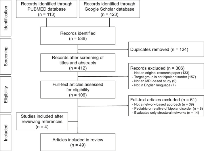
Methods: A systematic search was conducted on studies published from 2009 through 2019 in PubMed and Google Scholar. Articles that utilized functional magnetic resonance imaging technique to examine altered functional activity of major regions belonging to a large-scale brain network in bipolar disorder were selected.

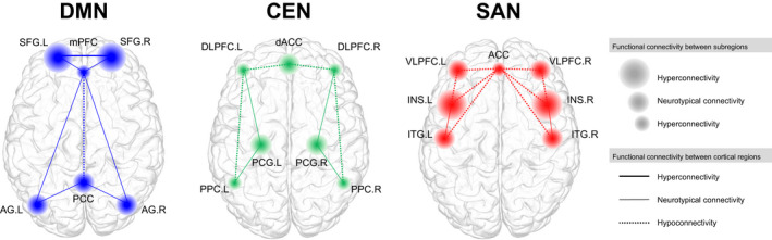
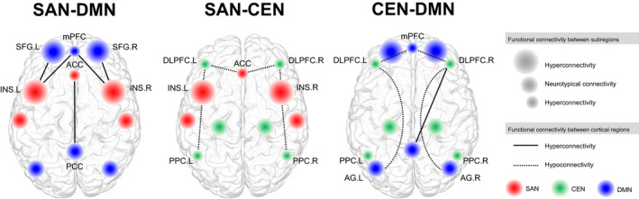
Results: A total of 49 studies were reviewed. Within-network hypoconnectivity was reported in bipolar disorder at rest among the default mode, salience, and central executive networks. In contrast, when performing a cognitive task, hyperconnectivity among the central executive network was found. Internetwork functional connectivity in the brain of bipolar disorder was greater between the salience and default mode networks, while reduced between the salience and central executive networks at rest, compared to control.

Conclusion: This systematic review suggests disruption in the functional activity of large-scale brain networks at rest as well as during a task stimuli in bipolar disorder. Disrupted intra- and internetwork functional connectivity that are also associated with clinical symptoms suggest altered functional connectivity of and between large-scale networks plays an important role in the pathophysiology of bipolar disorder.

Keywords: emotion; executive control; functional magnetic resonance imaging (fMRI); psychiatric disorders.


Conflict of interest statement

None declared.

Figures

FIGURE 1
FIGURE 1
Flowchart of literature search and identification of eligible articles. The initial database search resulted in 113 records from PubMed and 423 records from Google Scholar, together which resulted in 412 records after the exclusion of duplicates. The first screening of titles and abstracts excluded 306 records for the following reasons: 133 records were not an original research paper, 157 records did not target bipolar disorder, 9 records did not include MRI data, and 7 records were not in English. A second screening of the 106 full‐text articles led to the exclusion of 61 records as a result of the following: 39 records did not use a network‐based approach, 8 records were based on pediatric or relatives of bipolar disorder, and 14 records evaluated structural brain networks. An additional four records were also included from cross‐referencing, resulting in a total of 49 records for this review. MRI, magnetic resonance imaging.
FIGURE 2
FIGURE 2
Intranetwork alteration in functional activity at rest based on current articles reviewed. Within‐network alteration in functional connectivity in bipolar disorder based on the triple network model. For each network, the key regions that belong to the network are shown in colored nodes. The size of the nodes represents hyperconnectivity, hypoconnectivity, or statistically similar (neurotypical) functional connectivity between the respective region and its nearby subregional structures, such as the functional connectivity between the dorsal and ventral part of the region, as compared to the healthy control group. The functional connectivity between regions are shown in edges, where the type of edge represents either hyperconnectivity, hypoconnectivity or statistically similar (neurotypical) functional connectivity as compared to the healthy control group. ACC, anterior cingulate cortex; AG, angular gyrus; CEN, central executive network; dACC, dorsal anterior cingulate cortex; DLPFC, dorsolateral prefrontal cortex; DMN, default mode network; INS, insula; ITG, inferior temporal gyrus; L, left; mPFC, medial prefrontal cortex; PCC, posterior cingulate cortex; PCG, precentral gyrus; PPC, posterior parietal cortex; SAN, salience network; SFG, superior frontal gyrus; R, right; VLPFC, ventrolateral prefrontal cortex
FIGURE 3
FIGURE 3
Internetwork alteration in functional connectivity at rest based on current articles reviewed. Internetwork alteration in functional connectivity in bipolar disorder based on the triple network model. Red represents major regions of the salience network, green represents major regions of the central executive network, and blue represents major regions of the default mode network. The size of the nodes represents hyperconnectivity, hypoconnectivity, or statistically similar (neurotypical) functional connectivity between the respective region and its nearby subregional structures, such as the functional connectivity between the dorsal and ventral part of the region, as compared to the healthy control group. In summary, the results from the 49 studies reviewed indicates a significant increased connectivity between the salience network and default mode network. Decreased functional connectivity was reported between the salience network and central executive network, as well as between the central executive network and the default mode network. It is noteworthy that increased functional connectivity was also found between the right dorsolateral prefrontal cortex of the central executive network and the posterior cingulate cortex of the default mode network, although this relationship was not reported bilaterally. ACC, anterior cingulate cortex; Angular gyrus; CEN, central executive network; DLPFC, dorsolateral prefrontal cortex; DMN, default mode network; INS, insula; L, left; mPFC, medial prefrontal cortex; PCC, posterior cingulate cortex; PPC, posterior parietal cortex; R, right; SAN, salience network; SFG, superior frontal gyrus

References

    1. Adler, C. M. , Levine, A. D. , DelBello, M. P. , & Strakowski, S. M. (2005). Changes in gray matter volume in patients with bipolar disorder. Biological Psychiatry, 58(2), 151–157. 10.1016/j.biopsych.2005.03.022 - DOI - PubMed
    1. Agnew‐Blais, J. , & Danese, A. (2016). Childhood maltreatment and unfavourable clinical outcomes in bipolar disorder: A systematic review and meta‐analysis. The Lancet Psychiatry, 3(4), 342–349. 10.1016/S2215-0366(15)00544-1 - DOI - PubMed
    1. American Psychiatric Association (2013). Diagnostic and statistical manual of mental disorders (DSM‐5®). American Psychiatric Pub.
    1. Anand, A. , Li, Y. , Wang, Y. , Lowe, M. J. , & Dzemidzic, M. (2009). Resting state corticolimbic connectivity abnormalities in unmedicated bipolar disorder and unipolar depression. Psychiatry Research: Neuroimaging, 171(3), 189–198. 10.1016/j.pscychresns.2008.03.012 - DOI - PMC - PubMed
    1. Angst, J. , & Cassano, G. (2005). The mood spectrum: Improving the diagnosis of bipolar disorder. Bipolar Disorders, 7, 4–12. 10.1111/j.1399-5618.2005.00210.x - DOI - PubMed

Publication types