Review
. 2021 Sep;28(9):2422-2435.
doi: 10.1007/s43032-020-00393-2. Epub 2020 Nov 19.

Fetal Growth Restriction: Does an Integrated Maternal Hemodynamic-Placental Model Fit Better?

Affiliations
Review

Fetal Growth Restriction: Does an Integrated Maternal Hemodynamic-Placental Model Fit Better?

F Mecacci et al. Reprod Sci. 2021 Sep.

Abstract

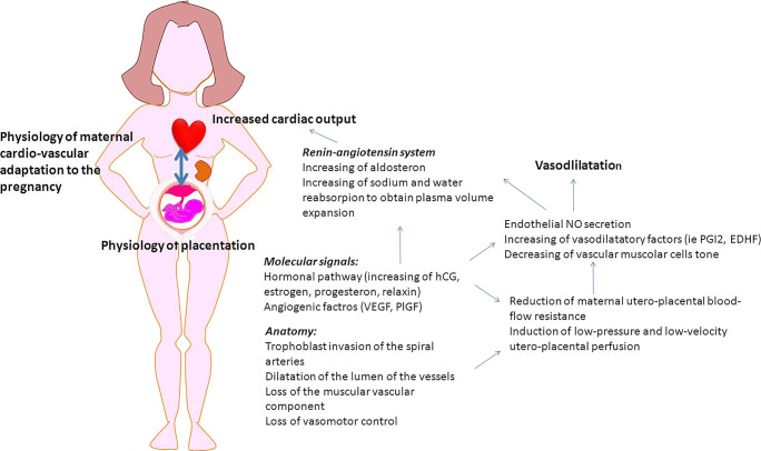
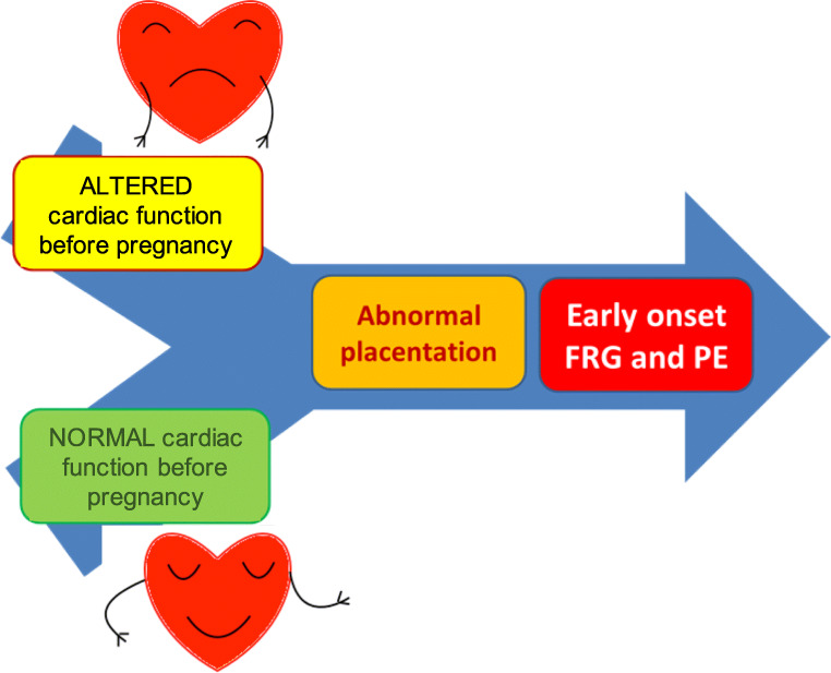
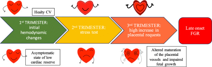
In recent years, a growing interest has arisen regarding the possible relationship between adverse pregnancy outcomes (APOs) and inadequate maternal hemodynamic adaptations to the pregnancy. A possible association between "placental syndromes," such as preeclampsia (PE) and fetal growth restriction (FGR), and subsequent maternal cardiovascular diseases (CVD) later in life has been reported. The two subtypes of FGR show different pathogenetic and clinical features. Defective placentation, due to a poor trophoblastic invasion of the maternal spiral arteries, is believed to play a central role in the pathogenesis of early-onset PE and FGR. Since placental functioning is dependent on the maternal cardiovascular system, a pre-existent or subsequent cardiovascular impairment may play a key role in the pathogenesis of early-onset FGR. Late FGR does not seem to be determined by a primary abnormal placentation in the first trimester. The pathological pathway of late-onset FGR may be due to a primary maternal cardiovascular maladaptation: CV system shows a flat profile and remains similar to those of non-pregnant women. Since the second trimester, when the placenta is already developed and increases its functional request, a hypovolemic state could lead to placental hypoperfusion and to an altered maturation of the placental villous tree and therefore to an altered fetal growth. Thus, this review focalizes on the possible relationship between maternal cardiac function and placentation in the development of both early and late-onset FGR. A better understanding of maternal hemodynamics in pregnancies complicated by FGR could bring various benefits in clinical practice, improving screening and therapeutic tools.

Keywords: Abnormal placentation; Cardiac output; Cardiovascular diseases; Fetal growth restriction; Maternal hemodynamics; Systemic vascular resistance.


Conflict of interest statement

The authors declare that they have no conflict of interest.

Figures

Fig. 1
Fig. 1
How placenta and maternal systemic cardiovascular systems interact for the women adaptation to the pregnancy. Spiral artery modification (obtained thanks to the trophoblast invasion process) reduces the utero-placental flow resistance. This artery modifications are associated with the placental secretion of angiogenic factors (i.e., vascular endothelial growth factor VEGF, placental growth factor PlGF) increasing nitric oxide (NO) and other vasodilatory factors such as prostacyclin (PGI2) and endothelium-derived hyperpolarizing factor (EDHF). Pregnancy hormones (human chorion gonadotropin HCG, estrogen, progesterone, relaxin) and placental secreted factors show many functions during pregnancy. They act as angiogenic factors and are able to activate the renin-angiotensin system, inducing the increase of aldosterone level and water and sodium reabsorption from the kidney, leading in turn to plasma volume expansion and hemodilution. Moreover, estrogens are indirectly able to intervene in the regulation of the pathway of NO and relaxin acts in the maternal hemodynamic adaptation, playing a role in the reduction of renal artery resistance. The increased NO in renal arteries also contributed to the reduction of renal vascular resistance. The result is an increased maternal blood volume with increased cardiac output and reduction in peripheral resistance. A detailed review of placental signaling of the maternal organism has been recently performed by Osol et al. [15]
Fig. 2
Fig. 2
Pathogenetic hypotheses of early-onset FRG. In the pathogenesis of early FGR, pre-pregnancy cardiovascular dysfunction and abnormal placentation could coexist
Fig. 3
Fig. 3
Pathogenetic hypotheses of late-onset FRG. How placenta responds to the pump deficit? At about 25 weeks of gestation, there are maximal divergences between the performance of a healthy CV system and one with a low cardiac reserve
Fig. 4
Fig. 4
Oriented physiological explanation of the types of FGR according to the maternal cardiac function and placental development. The hypothesis of origin of early-onset FGR (black arrows) and late-onset FGR (gray dotted arrows) is shown. Details are described in the text

References

    1. Resnik R. Intrauterine growth restriction. Obstet Gynecol. 2002;99(3):490–496. - PubMed
    1. Burton GJ, Jauniaux E. Pathophysiology of placental-derived fetal growth restriction. Am J Obstet Gynecol. 2018;218(2):S745–S761. doi: 10.1016/j.ajog.2017.11.577. - DOI - PubMed
    1. Gordijn SJ, Beune IM, Wynia K. Consensus definition of fetal growth restriction : a Delphi procedure. Ultrasound Obstet Gynecol. 2016;48:333–339. doi: 10.1002/uog.15884. - DOI - PubMed
    1. Figueras F, Caradeux J, Crispi F, Eixarch E. Diagnosis and surveillance of late-onset fetal growth restriction. Am J Obstet Gynecol. 2018;218(2S):S790–S802.e1. doi: 10.1016/j.ajog.2017.12.003. - DOI - PubMed
    1. Gruenwald P. Abnormalities of placental vascularity in relation to intrauterine deprivation and retardation of fetal growth. Significance of avascular chorionic villi. N Y State J Med. 1961;61:1508–1513. - PubMed

Publication types

LinkOut - more resources