

Resting-State Functional Connectivity Predicts STN DBS Clinical Response
- PMID: 33211330
- PMCID: PMC7987812
- DOI: 10.1002/mds.28376
Resting-State Functional Connectivity Predicts STN DBS Clinical Response
Abstract
Background: Deep brain stimulation of the subthalamic nucleus is a widely used adjunctive therapy for motor symptoms of Parkinson's disease, but with variable motor response. Predicting motor response remains difficult, and novel approaches may improve surgical outcomes as well as the understanding of pathophysiological mechanisms. The objective of this study was to determine whether preoperative resting-state functional connectivity MRI predicts motor response from deep brain stimulation of the subthalamic nucleus.
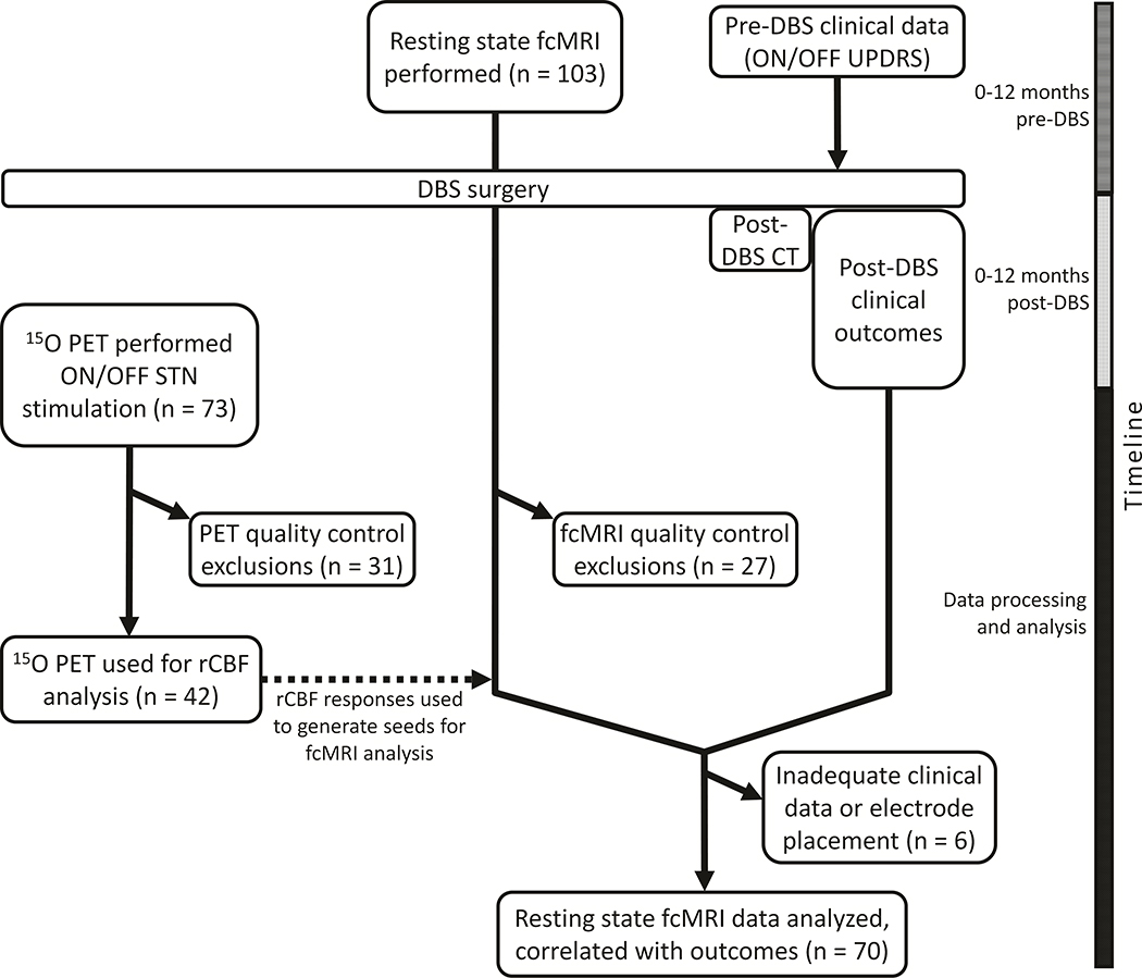
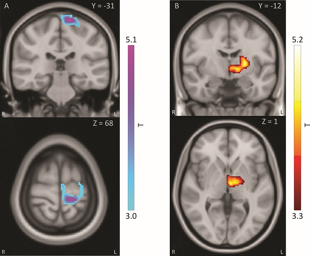
Methods: We collected preoperative resting-state functional MRI from 70 participants undergoing subthalamic nucleus deep brain stimulation. For this cohort, we analyzed the strength of STN functional connectivity with seeds determined by stimulation-induced (ON/OFF) 15 O H2 O PET regional cerebral blood flow differences in a partially overlapping group (n = 42). We correlated STN-seed functional connectivity strength with postoperative motor outcomes and applied linear regression to predict motor outcomes.
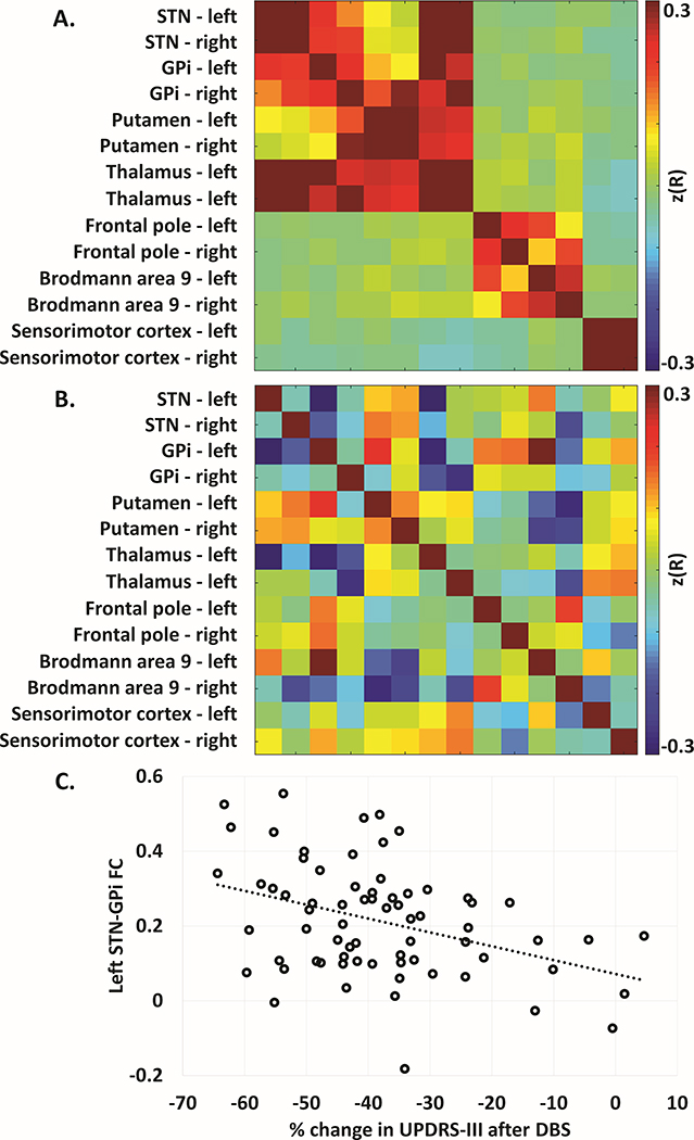
Results: Preoperative functional connectivity between the left subthalamic nucleus and the ipsilateral internal globus pallidus correlated with postsurgical motor outcomes (r = -0.39, P = 0.0007), with stronger preoperative functional connectivity relating to greater improvement. Left pallidal-subthalamic nucleus connectivity also predicted motor response to DBS after controlling for covariates.
Discussion: Preoperative pallidal-subthalamic nucleus resting-state functional connectivity predicts motor benefit from deep brain stimulation, although this should be validated prospectively before clinical application. These observations suggest that integrity of pallidal-subthalamic nucleus circuits may be critical to motor benefits from deep brain stimulation. © 2020 International Parkinson and Movement Disorder Society.
Keywords: DBS; Parkinson's disease; functional connectivity.
© 2020 International Parkinson and Movement Disorder Society.
Figures
References
-
- Deuschl G, Schade-Brittinger C, Krack P, et al. A randomized trial of deep-brain stimulation for Parkinson’s disease. N. Engl. J. Med. 2006;355(9):896–908. - PubMed
-
- Follett KA, Weaver FM, Stern M, et al. Pallidal versus subthalamic deep-brain stimulation for Parkinson’s disease. N. Engl. J. Med. 2010;362(22):2077–2091. - PubMed
-
- Stefani A, Cerroni R, Mazzone P, et al. Mechanisms of action underlying the efficacy of deep brain stimulation of the subthalamic nucleus in Parkinson’s disease: central role of disease severity. Eur. J. Neurosci. 2019;49(6):805–816. - PubMed
-
- Lozano AM, Lipsman N. Probing and regulating dysfunctional circuits using deep brain stimulation. Neuron 2013;77(3):406–24. - PubMed
-
- Welter ML, Houeto JL, Tezenas du Montcel S, et al. Clinical predictive factors of subthalamic stimulation in Parkinson’s disease. Brain 2002;125(Pt 3):575–83. - PubMed
Publication types
MeSH terms
Grants and funding
- U19 NS110456/NS/NINDS NIH HHS/United States
- R01 DK064832/DK/NIDDK NIH HHS/United States
- U54 NS116025/NS/NINDS NIH HHS/United States
- RF1 AG064937/AG/NIA NIH HHS/United States
- R01 NS075321/NS/NINDS NIH HHS/United States
- R01 NS075527/NS/NINDS NIH HHS/United States
- K23 NS041248/NS/NINDS NIH HHS/United States
- R01 NS097799/NS/NINDS NIH HHS/United States
- P01 NS080675/NS/NINDS NIH HHS/United States
- R01 HD085930/HD/NICHD NIH HHS/United States
- RF1 NS075321/NS/NINDS NIH HHS/United States
- R01 NS109487/NS/NINDS NIH HHS/United States
- R01 NS107281/NS/NINDS NIH HHS/United States
- R01 ES025991/ES/NIEHS NIH HHS/United States
- R01 AG050263/AG/NIA NIH HHS/United States
- F31 NS071639/NS/NINDS NIH HHS/United States
- R01 NS058797/NS/NINDS NIH HHS/United States
- C06 RR020092/RR/NCRR NIH HHS/United States
- R01 EB009352/EB/NIBIB NIH HHS/United States
- R01 NS103957/NS/NINDS NIH HHS/United States
- P30 NS098577/NS/NINDS NIH HHS/United States
- R01 NS041509/NS/NINDS NIH HHS/United States
- U54 TR001456/TR/NCATS NIH HHS/United States
- R01 HD070855/HD/NICHD NIH HHS/United States
- P30 NS048056/NS/NINDS NIH HHS/United States
- R01 NS097437/NS/NINDS NIH HHS/United States
- R01 ES029524/ES/NIEHS NIH HHS/United States
- U54 NS065701/NS/NINDS NIH HHS/United States
- UL1 TR000448/TR/NCATS NIH HHS/United States
- R21 AG063974/AG/NIA NIH HHS/United States
- U24 CA204854/CA/NCI NIH HHS/United States
- R61 AT010753/AT/NCCIH NIH HHS/United States
- U10 NS077384/NS/NINDS NIH HHS/United States
- UL1 TR002345/TR/NCATS NIH HHS/United States
- UH2 TR002065/TR/NCATS NIH HHS/United States
- R01 NS092865/NS/NINDS NIH HHS/United States
- R01 OH011661/OH/NIOSH CDC HHS/United States
- T32 EB021955/EB/NIBIB NIH HHS/United States
LinkOut - more resources
Full Text Sources
Medical
Miscellaneous
