

Artificial intelligence and algorithmic computational pathology: an introduction with renal allograft examples
- PMID: 33211332
- PMCID: PMC8715391
- DOI: 10.1111/his.14304
Artificial intelligence and algorithmic computational pathology: an introduction with renal allograft examples
Abstract
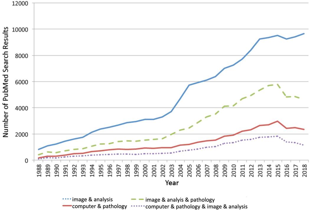
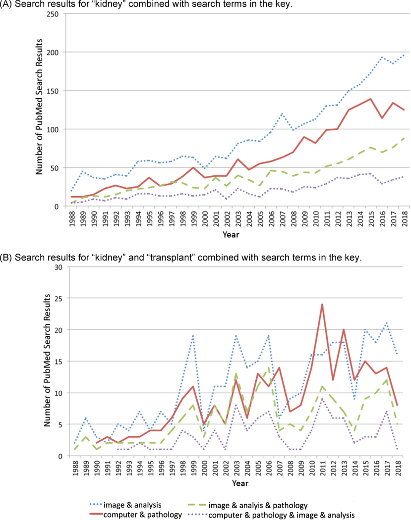
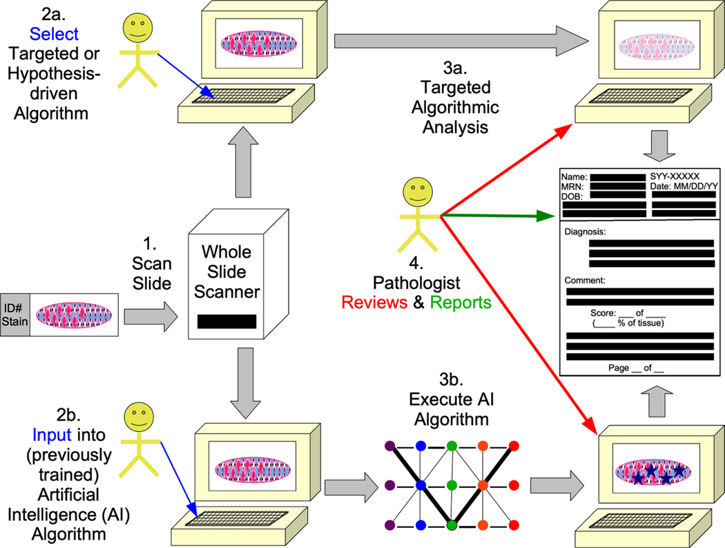
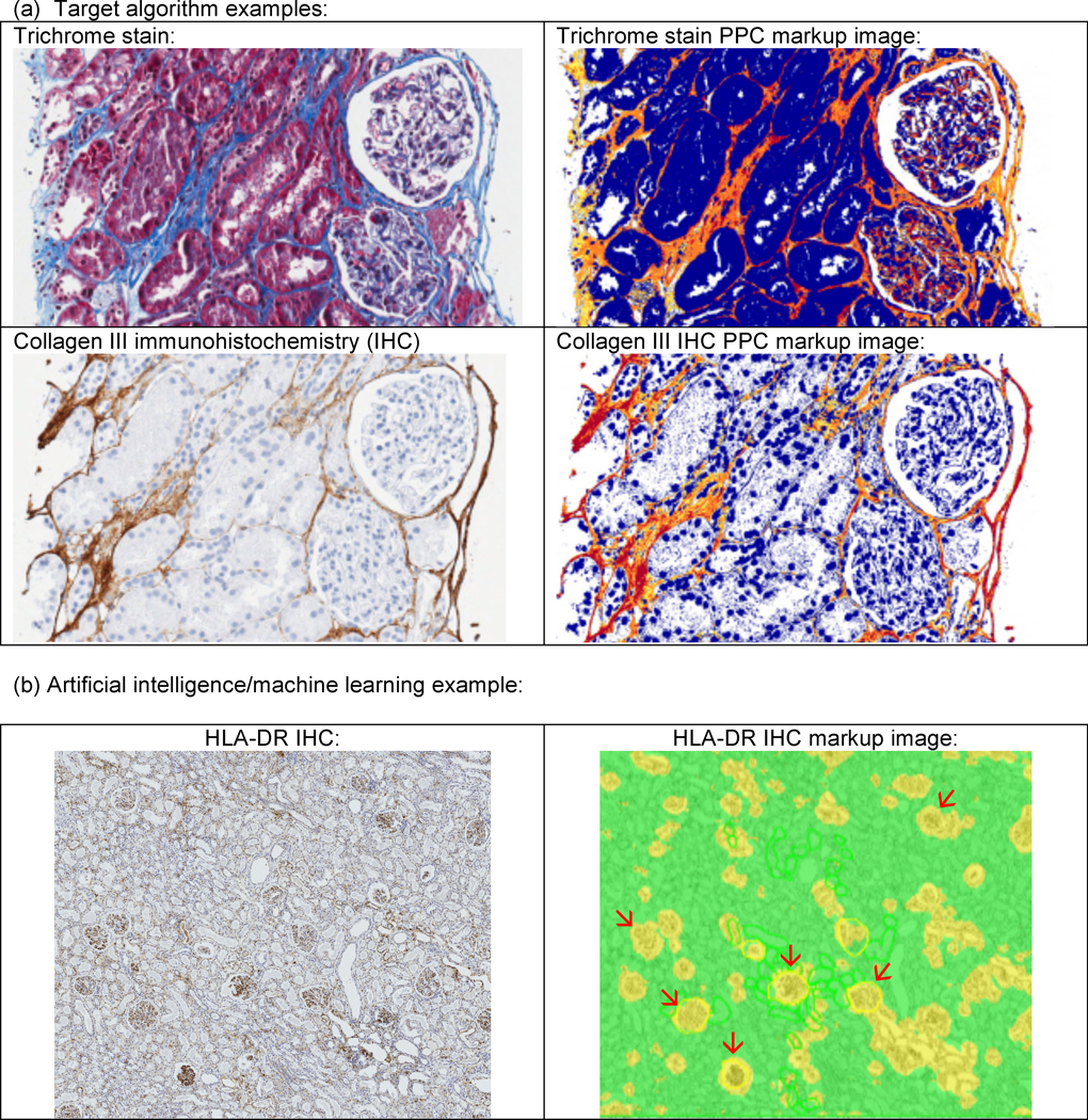
Whole slide imaging, which is an important technique in the field of digital pathology, has recently been the subject of increased interest and avenues for utilisation, and with more widespread whole slide image (WSI) utilisation, there will also be increased interest in and implementation of image analysis (IA) techniques. IA includes artificial intelligence (AI) and targeted or hypothesis-driven algorithms. In the overall pathology field, the number of citations related to these topics has increased in recent years. Renal pathology is one anatomical pathology subspecialty that has utilised WSIs and IA algorithms; it can be argued that renal transplant pathology could be particularly suited for whole slide imaging and IA, as renal transplant pathology is frequently classified by use of the semiquantitative Banff classification of renal allograft pathology. Hypothesis-driven/targeted algorithms have been used in the past for the assessment of a variety of features in the kidney (e.g. interstitial fibrosis, tubular atrophy, inflammation); in recent years, the amount of research has particularly increased in the area of AI/machine learning for the identification of glomeruli, for histological segmentation, and for other applications. Deep learning is the form of machine learning that is most often used for such AI approaches to the 'big data' of pathology WSIs, and deep learning methods such as artificial neural networks (ANNs)/convolutional neural networks (CNNs) are utilised. Unsupervised and supervised AI algorithms can be employed to accomplish image or semantic classification. In this review, AI and other IA algorithms applied to WSIs are discussed, and examples from renal pathology are covered, with an emphasis on renal transplant pathology.
Keywords: artificial intelligence; digital pathology; image analysis; machine learning; renal transplant pathology.
© 2020 John Wiley & Sons Ltd.
Conflict of interest statement
Disclosure/Statement of Competing Financial Interests
The authors of this manuscript have no conflicts of interest to disclose as described by
Conflict of interest: The authors declare no conflicts of interest related to this manuscript.
Figures
Similar articles
-
Whole Slide Imaging, Artificial Intelligence, and Machine Learning in Pediatric and Perinatal Pathology: Current Status and Future Directions.Pediatr Dev Pathol. 2025 Mar-Apr;28(2):91-98. doi: 10.1177/10935266241299073. Epub 2024 Nov 18. Pediatr Dev Pathol. 2025. PMID: 39552500 Review.
-
Deep learning-enabled classification of kidney allograft rejection on whole slide histopathologic images.Front Immunol. 2024 Jul 5;15:1438247. doi: 10.3389/fimmu.2024.1438247. eCollection 2024. Front Immunol. 2024. PMID: 39034991 Free PMC article.
-
Artificial intelligence applied to breast pathology.Virchows Arch. 2022 Jan;480(1):191-209. doi: 10.1007/s00428-021-03213-3. Epub 2021 Nov 18. Virchows Arch. 2022. PMID: 34791536 Review.
-
Revolutionizing Digital Pathology With the Power of Generative Artificial Intelligence and Foundation Models.Lab Invest. 2023 Nov;103(11):100255. doi: 10.1016/j.labinv.2023.100255. Epub 2023 Sep 26. Lab Invest. 2023. PMID: 37757969 Review.
-
Artificial intelligence driven next-generation renal histomorphometry.Curr Opin Nephrol Hypertens. 2020 May;29(3):265-272. doi: 10.1097/MNH.0000000000000598. Curr Opin Nephrol Hypertens. 2020. PMID: 32205581 Free PMC article. Review.
Cited by
-
The Puzzle of Preimplantation Kidney Biopsy Decision-Making Process: The Pathologist Perspective.Life (Basel). 2024 Feb 15;14(2):254. doi: 10.3390/life14020254. Life (Basel). 2024. PMID: 38398762 Free PMC article. Review.
-
Artificial intelligence in renal pathology: Current status and future.Biomol Biomed. 2023 Mar 16;23(2):225-234. doi: 10.17305/bjbms.2022.8318. Biomol Biomed. 2023. PMID: 36378066 Free PMC article. Review.
-
Naturally occurring autoimmune disease in (NZB X NZW) F1 mice is correlated with suppression of MZ B cell development due to aberrant B Cell Receptor (BCR) signaling, which is exacerbated by exposure to inorganic mercury.Toxicol Sci. 2023 Nov 11;197(2):211-21. doi: 10.1093/toxsci/kfad120. Online ahead of print. Toxicol Sci. 2023. PMID: 37952249 Free PMC article.
-
An Exploratory Review on the Potential of Artificial Intelligence for Early Detection of Acute Kidney Injury in Preterm Neonates.Diagnostics (Basel). 2023 Sep 5;13(18):2865. doi: 10.3390/diagnostics13182865. Diagnostics (Basel). 2023. PMID: 37761232 Free PMC article.
-
Artificial Intelligence Assists in the Detection of Blood Vessels in Whole Slide Images: Practical Benefits for Oncological Pathology.Biomolecules. 2023 Aug 29;13(9):1327. doi: 10.3390/biom13091327. Biomolecules. 2023. PMID: 37759727 Free PMC article. Review.
References
Publication types
MeSH terms
Grants and funding
LinkOut - more resources
Full Text Sources
Other Literature Sources
Medical
Miscellaneous
