

A loss-of-function mutation p.T256M in NDRG4 is implicated in the pathogenesis of pulmonary atresia with ventricular septal defect (PA/VSD) and tetralogy of Fallot (TOF)
- PMID: 33211401
- PMCID: PMC7876499
- DOI: 10.1002/2211-5463.13044
A loss-of-function mutation p.T256M in NDRG4 is implicated in the pathogenesis of pulmonary atresia with ventricular septal defect (PA/VSD) and tetralogy of Fallot (TOF)
Abstract
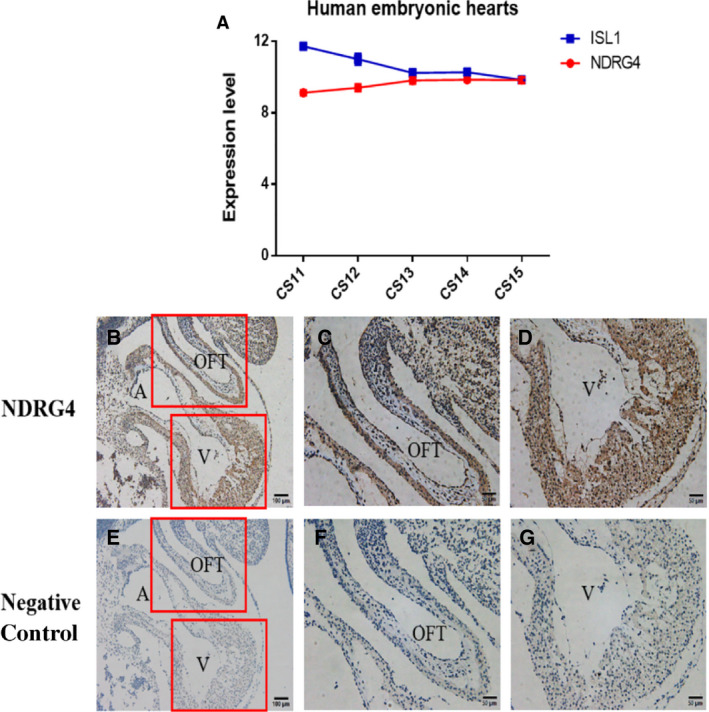
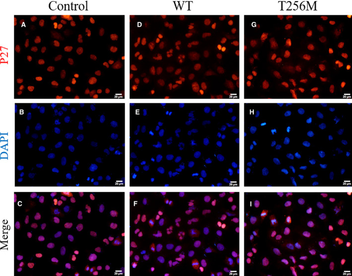
Pulmonary atresia with ventricular septal defect (PA/VSD) is a rare congenital heart disease (CHD) characterized by a lack of luminal continuity and blood flow from either the right ventricle or the pulmonary artery, together with VSDs. The prevalence of PA/VSD is about 0.2% of live births and approximately 2% of CHDs. PA/VSD is similar to tetralogy of Fallot (TOF) in terms of structural and pathological characteristics. The pathogenesis of these two CHDs remains incompletely understood. It was previously reported that N-myc downstream-regulated gene (NDRG)4 is required for myocyte proliferation during early cardiac development. In the present study, we enrolled 80 unrelated patients with PA/VSD or TOF and identified a probably damaging variant p.T256M of NDRG4. The p.T256M variant impaired the proliferation ability of human cardiac myocytes (hCM). Furthermore, the p.T256M variant resulted in G1 and G2 arrest of hCM, followed by an increase in p27 and caspase-9 expression. Our results provide evidence that the p.T256M variant in NDRG4 is a pathogenic variant associated with impaired hCM proliferation and cell-cycle arrest and likely contributes towards the pathogenesis of PA/VSD and TOF.
Keywords: NDRG4; PA/VSD; TOF; cardiac myocytes; p27; proliferation.
© 2020 The Authors. FEBS Open Bio published by John Wiley & Sons Ltd on behalf of Federation of European Biochemical Societies.
Conflict of interest statement
The authors declare that they have no conflicts of interest.
Figures


Similar articles
-
Relation of genotype 22q11 deletion to phenotype of pulmonary vessels in tetralogy of Fallot and pulmonary atresia-ventricular septal defect.Heart. 1998 Feb;79(2):186-90. doi: 10.1136/hrt.79.2.186. Heart. 1998. PMID: 9538314 Free PMC article.
-
22q11.2 deletion syndrome is associated with increased mortality in adults with tetralogy of Fallot and pulmonary atresia with ventricular septal defect.Int J Cardiol. 2020 May 1;306:56-60. doi: 10.1016/j.ijcard.2020.02.064. Epub 2020 Feb 27. Int J Cardiol. 2020. PMID: 32145937
-
Anterograde blood flow associated with modified Blalock-Taussig shunt does not modify pulmonary artery growth compared with modified Blalock-Taussig shunt alone.Arch Cardiovasc Dis. 2021 Apr;114(4):268-276. doi: 10.1016/j.acvd.2020.11.007. Epub 2021 Jan 25. Arch Cardiovasc Dis. 2021. PMID: 33509744
-
Perioperative and Anesthetic Considerations in Tetralogy of Fallot With Pulmonary Atresia.Semin Cardiothorac Vasc Anesth. 2021 Sep;25(3):218-228. doi: 10.1177/10892532211027395. Semin Cardiothorac Vasc Anesth. 2021. PMID: 34380349 Review.
-
Stenting the complex patent ductus arteriosus in tetralogy of Fallot with pulmonary atresia: challenges and outcomes.Future Cardiol. 2018 Jan;14(1):55-73. doi: 10.2217/fca-2017-0053. Epub 2017 Dec 4. Future Cardiol. 2018. PMID: 29199861 Review.
Cited by
-
Echocardiography phenotyping in murine genetic reference population of BXD strains reveals significant QTLs associated with cardiac function and morphology.Physiol Genomics. 2023 Feb 1;55(2):51-66. doi: 10.1152/physiolgenomics.00120.2022. Epub 2022 Dec 19. Physiol Genomics. 2023. PMID: 36534598 Free PMC article.
-
Cardiomyocyte proliferation and regeneration in congenital heart disease.Pediatr Discov. 2024 Sep;2(3):e2501. doi: 10.1002/pdi3.2501. Epub 2024 Aug 12. Pediatr Discov. 2024. PMID: 39308981 Free PMC article.
-
Genetic insights into non-syndromic Tetralogy of Fallot.Front Physiol. 2022 Oct 6;13:1012665. doi: 10.3389/fphys.2022.1012665. eCollection 2022. Front Physiol. 2022. PMID: 36277185 Free PMC article. Review.
-
NOTCH2, ATIC, MRI1, SLC6A13, ATP13A2 Genetic Variations are Associated with Ventricular Septal Defect in the Chinese Tibetan Population Through Whole-Exome Sequencing.Pharmgenomics Pers Med. 2023 Apr 27;16:389-400. doi: 10.2147/PGPM.S404438. eCollection 2023. Pharmgenomics Pers Med. 2023. PMID: 37138656 Free PMC article.
References
-
- Digilio MC, Marino B, Grazioli S, Agostino D, Giannotti A and Dallapiccola B (1996) Comparison of occurrence of genetic syndromes in ventricular septal defect with pulmonic stenosis (classic tetralogy of Fallot) versus ventricular septal defect with pulmonic atresia. Am J Cardiol 77, 1375–1376. - PubMed
-
- Tchervenkov CI and Roy N (2000) Congenital heart surgery nomenclature and database project: pulmonary atresia–ventricular septal defect. Ann Thorac Surg 69, S97–S105. - PubMed
-
- Abid D, Elloumi A, Abid L, Mallek S, Aloulou H, Chabchoub I, Bouraoui A, Thabet A, Gargouri L, Zribi M et al (2014) Congenital heart disease in 37,294 births in Tunisia: birth prevalence and mortality rate. Cardiol Young 24, 866–871. - PubMed
Publication types
MeSH terms
Substances
Supplementary concepts
Associated data
- Actions
- Actions
- Actions
LinkOut - more resources
Full Text Sources
Other Literature Sources
