. 2021 May 1;130(5):1305-1316.
doi: 10.1152/japplphysiol.00421.2020. Epub 2020 Nov 19.

Intravenous sulforhodamine B reduces alveolar surface tension, improves oxygenation, and reduces ventilation injury in a respiratory distress model

Affiliations

Intravenous sulforhodamine B reduces alveolar surface tension, improves oxygenation, and reduces ventilation injury in a respiratory distress model

You Wu et al. J Appl Physiol (1985). .

Abstract

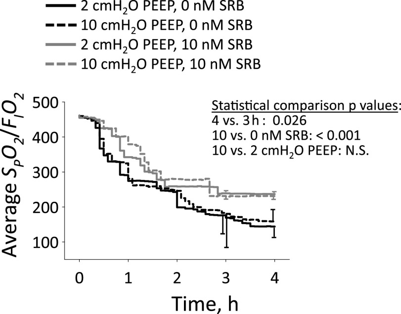
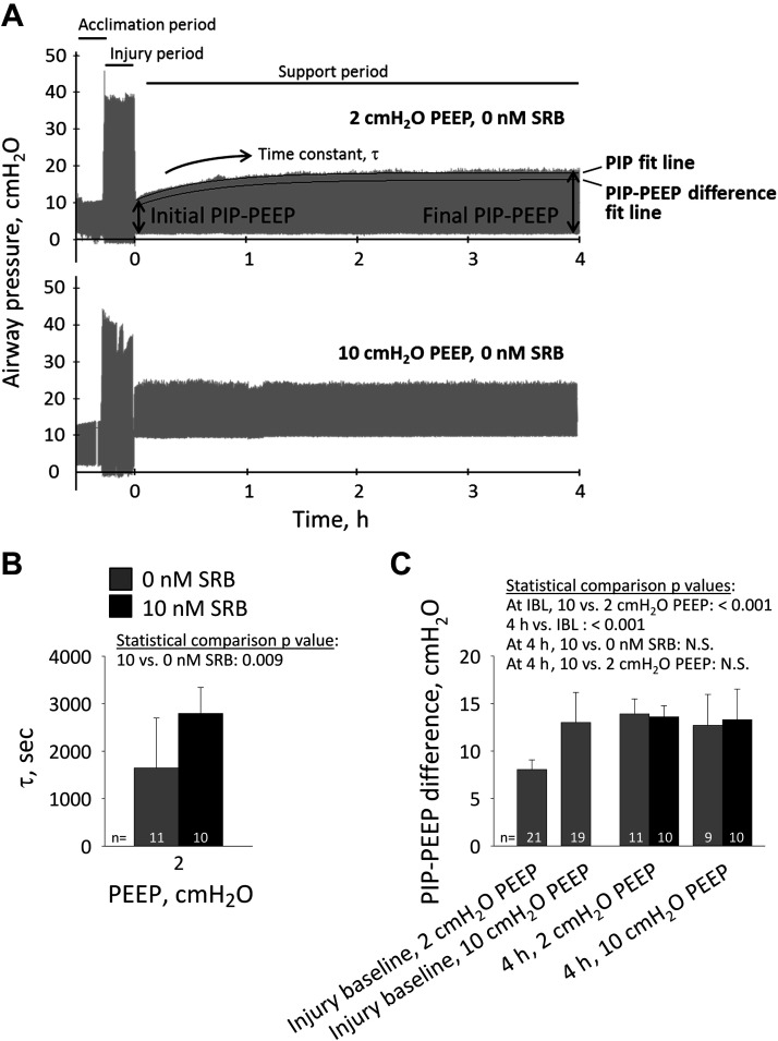
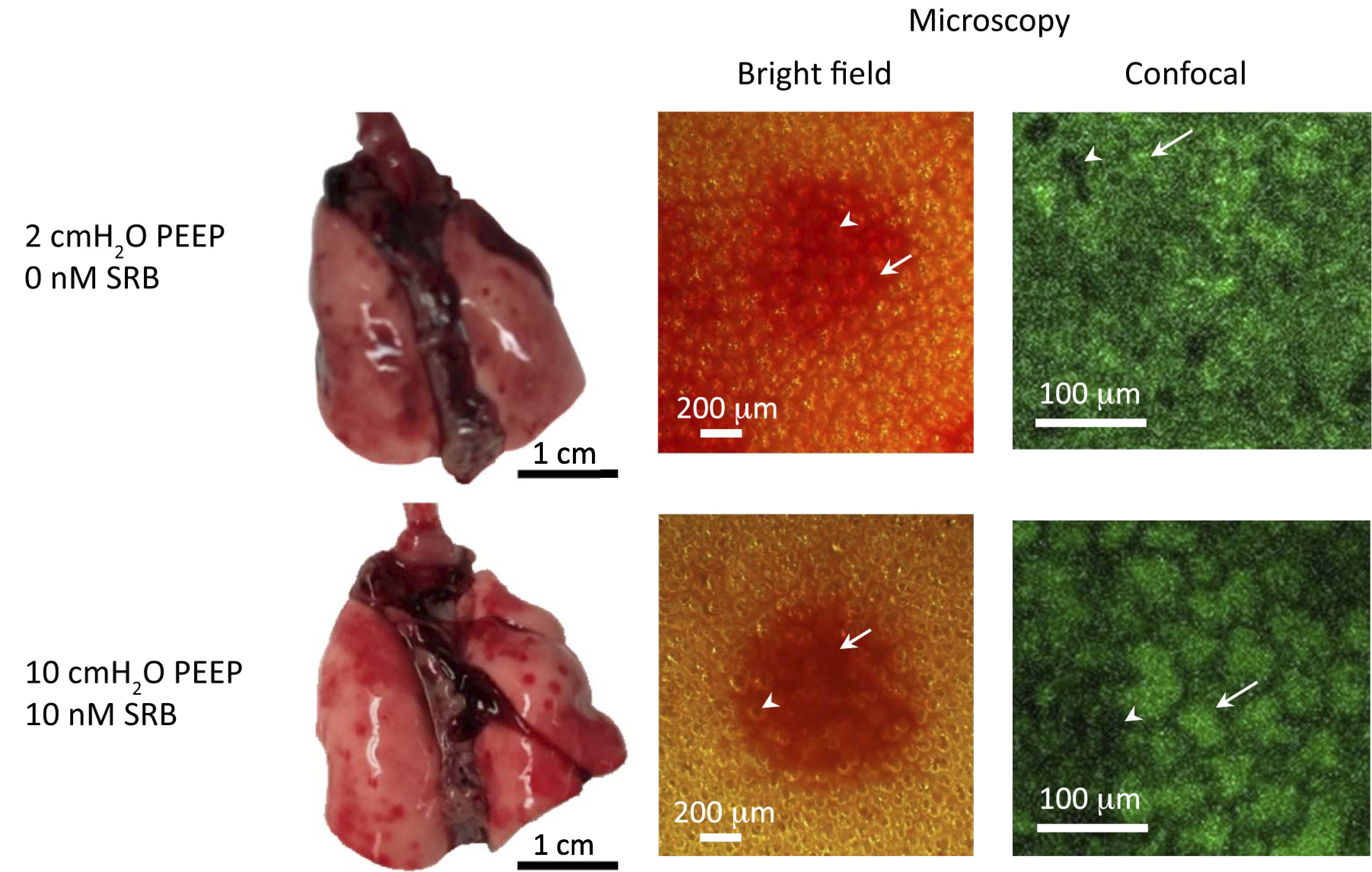
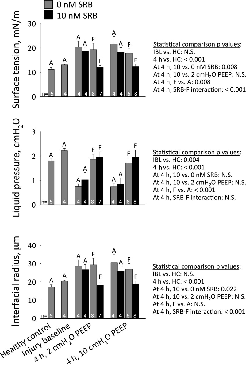
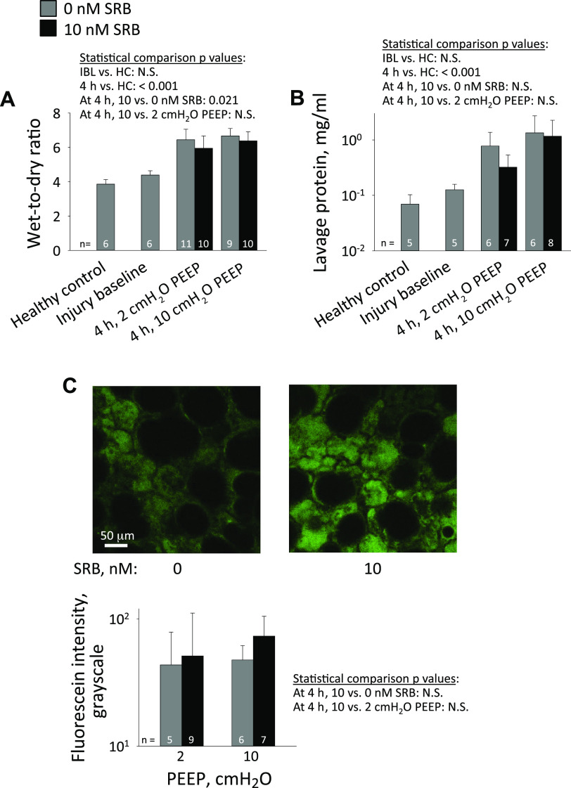
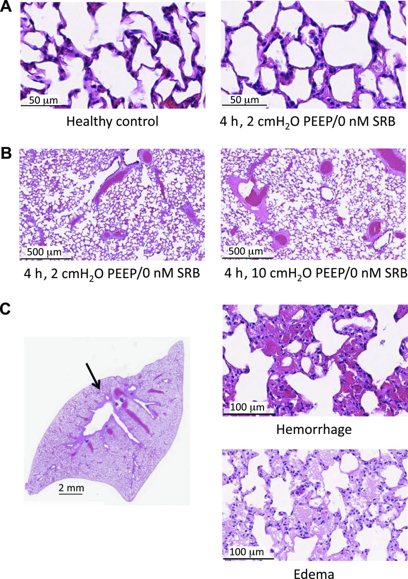
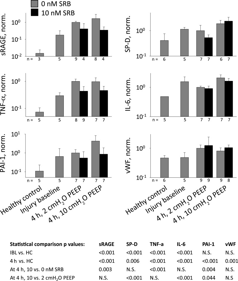
In the neonatal respiratory distress syndrome (NRDS) and acute respiratory distress syndrome (ARDS), mechanical ventilation supports gas exchange but can cause ventilation-induced lung injury (VILI) that contributes to high mortality. Further, surface tension, T, should be elevated and VILI is proportional to T. Surfactant therapy is effective in NRDS but not ARDS. Sulforhodamine B (SRB) is a potential alternative T-lowering therapeutic. In anesthetized male rats, we injure the lungs with 15 min of 42 mL/kg tidal volume, VT, and zero end-expiratory pressure ventilation. Then, over 4 h, we support the rats with protective ventilation-VT of 6 mL/kg with positive end-expiratory pressure. At the start of the support period, we administer intravenous non-T-altering fluorescein (targeting 27 µM in plasma) without or with therapeutic SRB (10 nM). Throughout the support period, we increase inspired oxygen fraction, as necessary, to maintain >90% arterial oxygen saturation. At the end of the support period, we euthanize the rat; sample systemic venous blood for injury marker ELISAs; excise the lungs; combine confocal microscopy and servo-nulling pressure measurement to determine T in situ in the lungs; image fluorescein in alveolar liquid to assess local permeability; and determine lavage protein content and wet-to-dry ratio (W/D) to assess global permeability. Lungs exhibit focal injury. Surface tension is elevated 72% throughout control lungs and in uninjured regions of SRB-treated lungs, but normal in injured regions of treated lungs. SRB administration improves oxygenation, reduces W/D, and reduces plasma injury markers. Intravenous SRB holds promise as a therapy for respiratory distress.NEW & NOTEWORTHY Sulforhodmaine B lowers T in alveolar edema liquid. Given the problematic intratracheal delivery of surfactant therapy for ARDS, intravenous SRB might constitute an alternative therapeutic. In a lung injury model, we find that intravenously administered SRB crosses the injured alveolar-capillary barrier thus reduces T specifically in injured lung regions; improves oxygenation; and reduces the degree of further lung injury. Intravenous SRB administration might help respiratory distress patients, including those with the novel coronavirus, avoid mechanical ventilation or, once ventilated, survive.

Keywords: acute respiratory distress syndrome; neonatal respiratory distress syndrome; oxygenation; surface tension; ventilation-induced lung injury.


Conflict of interest statement

No conflicts of interest, financial or otherwise, are declared by the authors.

Figures

Figure 1.
Figure 1.
Protocol. Schematic of experimental protocol. FIO2, fraction of inspired oxygen; PEEP, positive end-expiratory pressure; RR, respiratory rate; SPO2, peripheral arterial oxygen saturation; VT, tidal volume. Euthanasia requires 20–40, 15–25, and 10–20 min in healthy-control, injury-baseline, and 4-h time-point groups, respectively.
Figure 2.
Figure 2.
Oxygenation. Average SPO2/FIO2 versus time, from injury baseline onward. n = 9–11/group. Statistical comparisons performed at 3- and 4-h time points on log-transformed data.
Figure 3.
Figure 3.
Lung elastance. A: airway pressure traces, with indicated PEEP during support period, in the absence of SRB administration. On 2-cmH2O PEEP trace, top black curve shows exponential fit to peak inspiratory pressure (PIP); bottom black curve shows exponential fit to difference between PIP and PEEP, which is shifted 2 cmH2O down from and has different time constant than top curve. With fixed PEEP and VT, PIP-PEEP difference indicates elastance. B: time constant, τ, for increase in difference between PIP and PEEP, starting at injury baseline, in 2-cmH2O PEEP groups. C: difference between PIP and PEEP at injury-baseline time point just prior to tail vein injection and at 4-h time point at end of support period. For statistical comparisons: IBL, injury-baseline time point; 4 h, 4-h time point; N.S., not significant. At IBL, as SRB is not yet administered, 0- and 10-nM SRB groups are combined.
Figure 4.
Figure 4.
Lung appearance. Photographs show lungs isolated at 4-h time point and at PEEP volumes, from indicated groups. Lungs exhibit focal hemorrhagic surface injury. Microscope images are of injured regions, with lungs still held at PEEP volume. Higher-magnification confocal images show fluorescein present in alveolar liquid and are located within central, injured regions of lower-magnification bright-field images. Arrows indicate shrunken, flooded alveoli (lack of air-liquid interface in bright-field image, green-labeled edema liquid in fluorescent image). Arrowheads indicate aerated alveoli. Color variation between bright-field images is due to varying lamp illumination.
Figure 5.
Figure 5.
Surface tension and underlying measurements. Alveolar interfacial surface tension, T, is calculated from the Laplace relation after setting alveolar air pressure, PALV, to 15 cmH2O; determining alveolar liquid phase pressure, PLIQ, in the liquid lining layer in the corner of an aerated (A) alveolus or below the meniscus of a flooded (F) alveolus by servo-nulling pressure measurement; and determining three-dimensional interfacial radius of curvature, R, from a z-stack of confocal microscopic images. For statistical comparisons: HC, healthy-control group.
Figure 6.
Figure 6.
Permeability. A: wet-to-dry ratio—a global permeability metric indicating combined injury severity and extent—of right caudal lobe. B: total protein content of lavage liquid—an alternative global permeability metric—from middle right lobe. Statistical analysis for this metric performed on log-transformed data. C: alveolar liquid fluorescein intensity—a local permeability metric specifically indicating injury severity. In 4-h time-point lungs after inflation to 30 cmH2O and deflation to 15 cmH2O, confocal images are obtained at 20-µm subpleural depth in flooded regions. Representative images from 10-cmH2O PEEP group exhibit average fluorescein intensities of 44 and 68 gray levels with 0- and 10-nM SRB, respectively. For display purposes, contrast of both images increased to same degree. Note typical pattern of diminished-sized flooded alveoli and overexpanded aerated alveoli. Graph presents group data. Statistical analysis for this metric performed on log-transformed data.
Figure 7.
Figure 7.
Histology. A: representative images of uninjured regions in control and injured lungs. B: perivascular edema is more frequent at high than low PEEP. C: focal alveolar injury is always present adjacent to main airway branch point [arrow in left image of 2-cmH2O PEEP/0-nM SRB group lung]. Injury is sometimes hemorrhagic (upper right image, from same lung as on left), sometimes edematous (lower right image, from 10-cmH2O PEEP/10-nM SRB group lung). Treatment with SRB does not alter histologic findings.
Figure 8.
Figure 8.
Injury markers. Injury markers quantified by enzyme-linked immunosorbent assays of blood plasma: soluble receptor for advanced glycation end products (sRAGE), surfactant protein D (SP-D), tumor necrosis factor α (TNF-α), interleukin 6 (IL-6), plasminogen activator inhibitor 1 (PAI-1), and von Willbrand factor (vWF). Data normalized by average value of 2-cmH2O PEEP/0-nM SRB group. Statistical analysis for all injury markers performed on log-transformed data.

Similar articles

Cited by

References

    1. Chen W, Chen Y-Y, Tsai C-F, Chen SC-C, Lin M-S, Ware LB, Chen C-M. Incidence and outcomes of acute respiratory distress syndrome: a nationwide registry-based study in Taiwan, 1997 to 2011. Medicine (Baltimore) 94: e1849, 2015. doi:10.1097/MD.0000000000001849. - DOI - PMC - PubMed
    1. Donda K, Vijayakanthi N, Dapaah-Siakwan F, Bhatt P, Rastogi D, Rastogi S. Trends in epidemiology and outcomes of respiratory distress syndrome in the United States. Pediatr Pulmonol 54: 405–414, 2019. doi:10.1002/ppul.24241. - DOI - PubMed
    1. Eworuke E, Major JM, Gilbert McClain LI. National incidence rates for acute respiratory distress syndrome (ARDS) and ARDS cause-specific factors in the United States (2006–2014). J Crit Care 47: 192–197, 2018. doi:10.1016/j.jcrc.2018.07.002. - DOI - PubMed
    1. Rubaltelli FF, Bonafe L, Tangucci M, Spagnolo A, Dani C. Epidemiology of neonatal acute respiratory disorders. A multicenter study on incidence and fatality rates of neonatal acute respiratory disorders according to gestational age, maternal age, pregnancy complications and type of delivery. Italian Group of Neonatal Pneumology. Biol Neonate 74: 7–15, 1998. doi:10.1159/000014005. - DOI - PubMed
    1. Rubenfeld GD, Caldwell E, Peabody E, Weaver J, Martin DP, Neff M, Stern EJ, Hudson LD. Incidence and outcomes of acute lung injury. N Engl J Med 353: 1685–1693, 2005. doi:10.1056/NEJMoa050333. - DOI - PubMed

Publication types