

In vitro mechanical vibration down-regulates pro-inflammatory and pro-fibrotic signaling in human vocal fold fibroblasts
- PMID: 33211714
- PMCID: PMC7676657
- DOI: 10.1371/journal.pone.0241901
In vitro mechanical vibration down-regulates pro-inflammatory and pro-fibrotic signaling in human vocal fold fibroblasts
Abstract
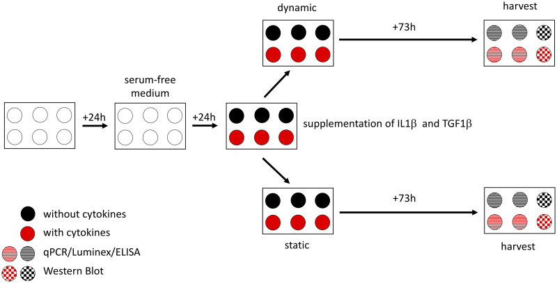
Introduction: Voice rest following phonotrauma or phonosurgery has a considerable clinical impact, but clinical recommendations are inconsistent due to inconclusive data. As biopsies of the vocal folds (VF) for molecular biology studies in humans are unethical, we established a new in vitro model to explore the effects of vibration on human vocal fold fibroblasts (hVFF) in an inflammatory and normal state, which is based on previously published models.
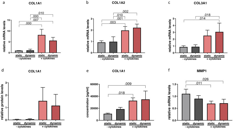
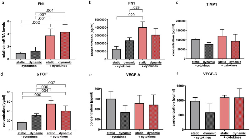
Methods: By using a phonomimetic bioreactor we were able to apply predefined vibrational stress patterns on hVFF cultured under inflammatory or normal conditions. Inflammatory and pro-fibrotic stimuli were induced by interleukin (IL)1β and transforming growth factor (TGF)β1, respectively. Mechanical stimulation was applied four hours daily, over a period of 72 hours. Outcome measurements comprised assessment of extracellular matrix (ECM)-related components, angiogenic factors, and inflammatory and fibrogenic markers on gene expression and protein levels.
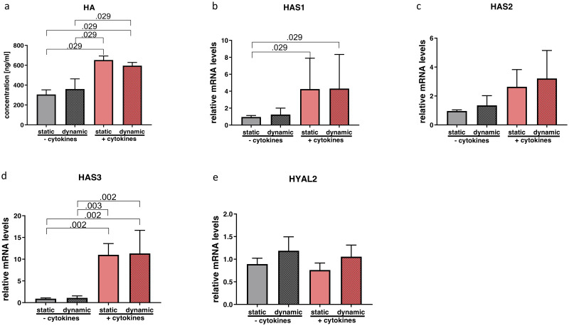
Results: Under inflammatory conditions, the inflammatory cytokine IL11, as well as the myofibroblast marker alpha smooth muscle actin (α-SMA) were significantly reduced when additional vibration was applied. The desirable anti-fibrotic ECM component hyaluronic acid was increased following cytokine treatment, but was not diminished following vibration.
Conclusion: Our experiments revealed the effect of vibrational stress on hVFF in an inflammatory state. Elevated levels of certain pro-inflammatory/pro-fibrotic factors could be mitigated by additional vibrational excitation in an in vitro setting. These findings corroborate clinical studies which recommend early voice activation following an acute event.
Conflict of interest statement
The authors have declared that no competing interests exist.
Figures


Similar articles
-
Biophysical aspects of mechanotransduction in cells and their physiological/biological implications in vocal fold vibration: a narrative review.Front Cell Dev Biol. 2025 Jan 27;13:1501341. doi: 10.3389/fcell.2025.1501341. eCollection 2025. Front Cell Dev Biol. 2025. PMID: 39931244 Free PMC article. Review.
-
Inhibition of the MRTF-A/SRF signaling axis alleviates vocal fold scarring.Matrix Biol. 2025 May;137:1-11. doi: 10.1016/j.matbio.2025.02.004. Epub 2025 Feb 14. Matrix Biol. 2025. PMID: 39956286
-
Exploring the Pathophysiology of Reinke's Edema: The Cellular Impact of Cigarette Smoke and Vibration.Laryngoscope. 2021 Feb;131(2):E547-E554. doi: 10.1002/lary.28855. Epub 2020 Jun 22. Laryngoscope. 2021. PMID: 32569447 Free PMC article.
-
Changes in cytokine signaling and extracellular matrix production induced by inflammatory factors in cultured vocal fold fibroblasts.Ann Otol Rhinol Laryngol. 2008 Mar;117(3):227-38. doi: 10.1177/000348940811700311. Ann Otol Rhinol Laryngol. 2008. PMID: 18444484
-
Response of fibroblasts to transforming growth factor-β1 on two-dimensional and in three-dimensional hyaluronan hydrogels.Tissue Eng Part A. 2012 Dec;18(23-24):2528-38. doi: 10.1089/ten.TEA.2012.0094. Epub 2012 Aug 21. Tissue Eng Part A. 2012. PMID: 22734649 Free PMC article.
Cited by
-
Influence of 40 Hz and 100 Hz Vibration on SH-SY5Y Cells Growth and Differentiation-A Preliminary Study.Molecules. 2022 May 23;27(10):3337. doi: 10.3390/molecules27103337. Molecules. 2022. PMID: 35630814 Free PMC article.
-
Periodic Mechanical Stress Inhibits the Development of Osteoarthritis via Regulating ATF3-Akt Axis.J Inflamm Res. 2023 Nov 28;16:5613-5628. doi: 10.2147/JIR.S419186. eCollection 2023. J Inflamm Res. 2023. PMID: 38046403 Free PMC article.
-
Knockdown of LncRNA NEAT1 inhibits myofibroblast activity in oral submucous fibrosis through miR-760/TPM1 axis.J Dent Sci. 2022 Apr;17(2):707-717. doi: 10.1016/j.jds.2021.11.003. Epub 2021 Dec 8. J Dent Sci. 2022. PMID: 35756787 Free PMC article.
-
Biophysical aspects of mechanotransduction in cells and their physiological/biological implications in vocal fold vibration: a narrative review.Front Cell Dev Biol. 2025 Jan 27;13:1501341. doi: 10.3389/fcell.2025.1501341. eCollection 2025. Front Cell Dev Biol. 2025. PMID: 39931244 Free PMC article. Review.
-
In Vitro Evaluation of Biomaterials for Vocal Fold Injection: A Systematic Review.Polymers (Basel). 2021 Aug 6;13(16):2619. doi: 10.3390/polym13162619. Polymers (Basel). 2021. PMID: 34451158 Free PMC article. Review.
References
-
- Hunt Berse, Morganelli Diegel, Brown Yeo, et al. Hypoxia augments cytokine (transforming growth factor-beta (TGF-beta) and IL-1)-induced vascular endothelial growth factor secretion by human synovial fibroblasts. Clin Exp Immunol. 1999. January;115(1):176–82. 10.1046/j.1365-2249.1999.00775.x - DOI - PMC - PubMed
MeSH terms
Substances
LinkOut - more resources
Full Text Sources
Research Materials
Miscellaneous
