. 2021 May;141(5):1207-1218.
doi: 10.1016/j.jid.2020.10.018. Epub 2020 Nov 16.

Epigenetic Alterations in Keratinocyte Carcinoma

Affiliations

Epigenetic Alterations in Keratinocyte Carcinoma

Qiuming Yao et al. J Invest Dermatol. 2021 May.

Abstract

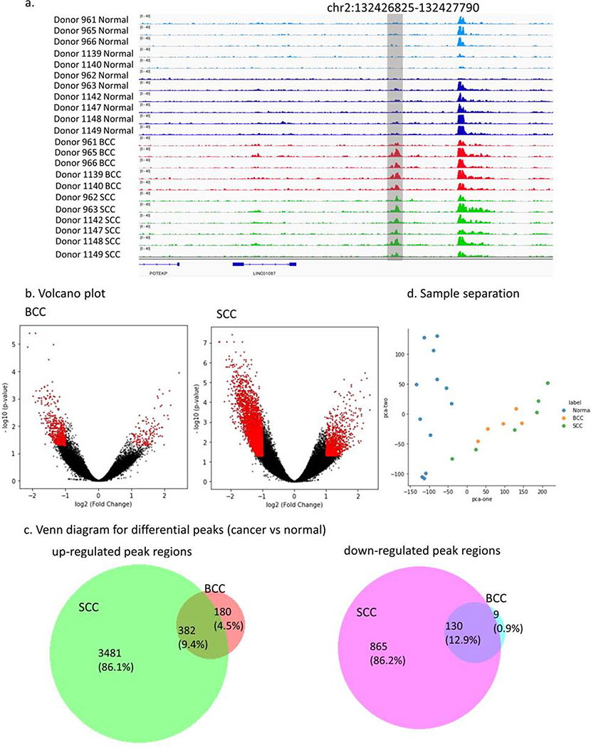
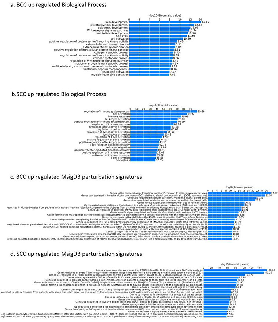
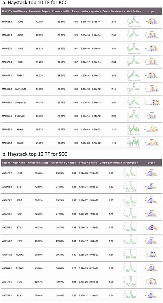
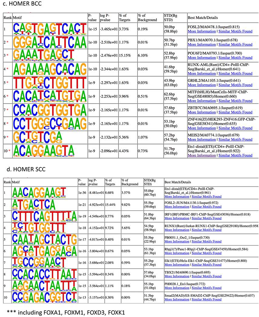
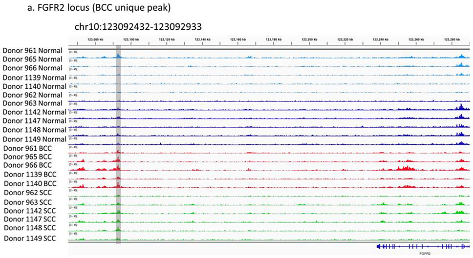
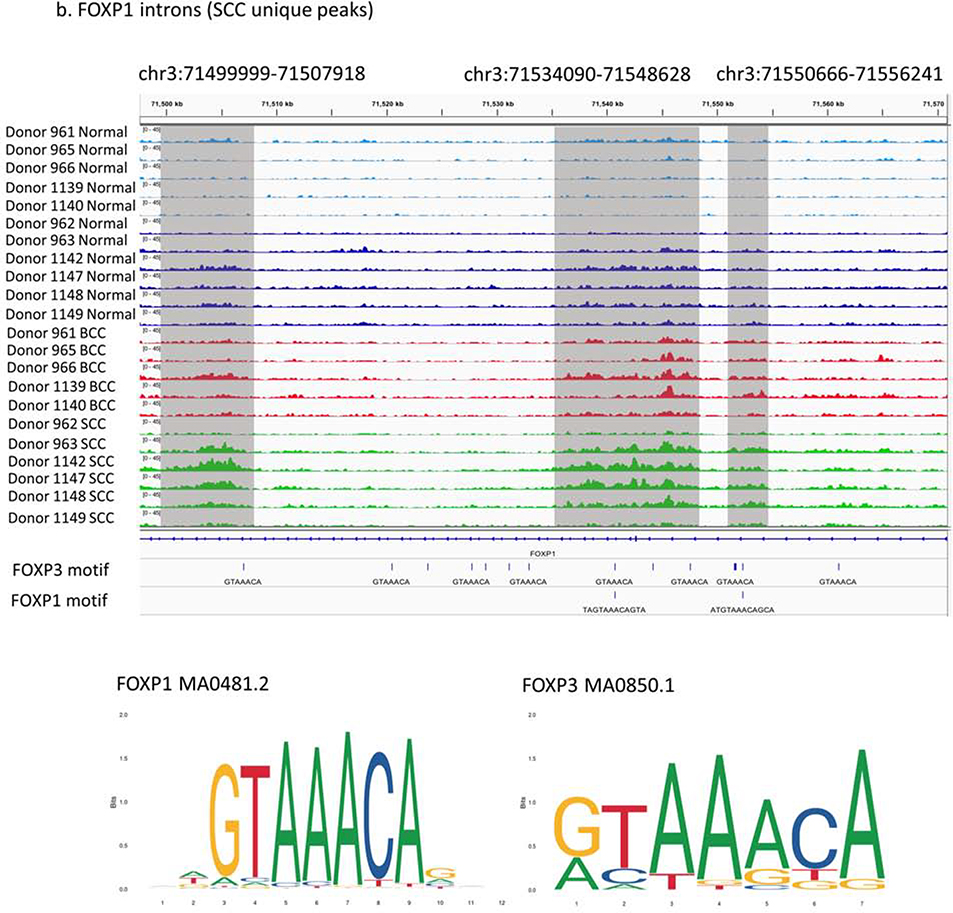
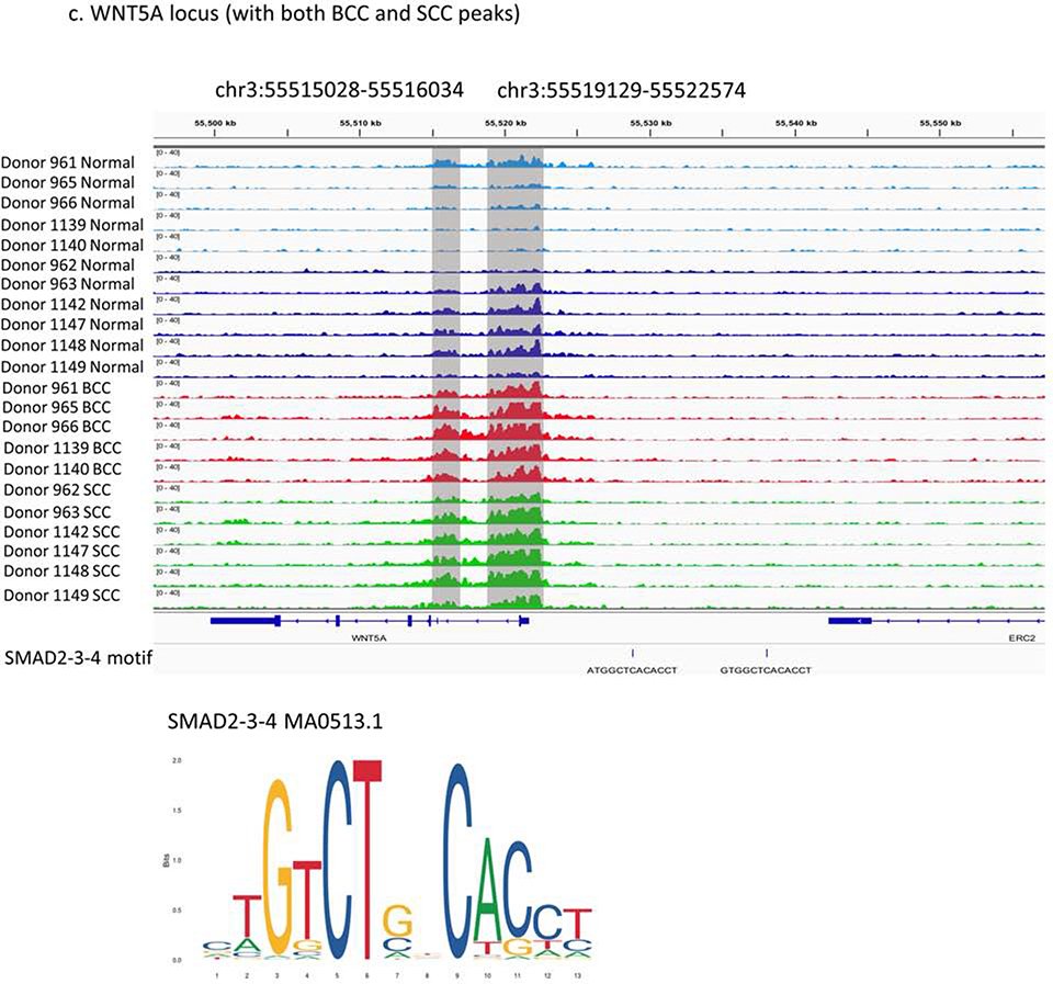
Basal cell carcinoma (BCC) and squamous cell carcinoma (SCC) are both derived from epidermal keratinocytes but are phenotypically diverse. To improve the understanding of keratinocyte carcinogenesis, it is critical to understand epigenetic alterations, especially those that govern gene expression. We examined changes to the enhancer-associated histone acetylation mark H3K27ac by mapping matched tumor-normal pairs from 11 patients (five with BCC and six with SCC) undergoing Mohs surgery. Our analysis uncovered cancer-specific enhancers on the basis of differential H3K27ac peaks between matched tumor-normal pairs. We also uncovered biological pathways potentially altered in keratinocyte carcinoma, including enriched epidermal development and Wnt signaling pathways enriched in BCCs and enriched immune response and cell activation pathways in SCCs. We also observed enrichment of transcription factors that implicated SMAD and JDP2 in BCC pathogenesis and FOXP1 in SCC pathogenesis. On the basis of these findings, we prioritized three loci with putative regulation events (FGFR2 enhancer in BCC, intragenic regulation of FOXP1 in SCC, and WNT5A promoter in both subtypes) and validated our findings with published gene expression data. Our findings highlight unique and shared epigenetic alterations in histone modifications and potential regulators for BCCs and SCCs that likely impact the divergent oncogenic pathways, paving the way for targeted drug discoveries.


Figures

Figure 1.
Figure 1.
H3K27ac specific regions for SCC and BCC. (a) H3K27ac signal tracks for tumor-normal samples at a single locus (chr2:132426825–132427790) show cancer-specific alteration. (b) Volcano plots show the log2 fold change and the p-value for BCC and SCC samples. Red dots are the regions with significant p-value (<5%) and absolute log2 fold change larger than 1. (c) Venn diagram for differential peaks unique and shared in BCC and SCC samples. (d) Significant H3K27ac peaks identified by DESeq2 separates tumor-normal samples using the first two principal components.
Figure 2.
Figure 2.
H3K27ac cancer-specific regions and GREAT analysis: (a) BCC up-regulated biological process, (b) SCC up-regulated biological process, (c) BCC up-regulated MsigDB perturbation signatures, and (d) SCC up-regulated MsigDB perturbation signatures
Figure 3.
Figure 3.
Putative transcription factors (TFs) located at regulatory elements defined by H3K27ac specific peaks in BCC and SCC samples from Haystack and Homer analyses: (a) Top 10 TFs for BCC from Haystack analyses, (b) Top 10 TFs for SCC from Haystack analyses, (c) Top 10 TFs in BCC from Homer analyses, and (d) Top 10 TFs in SCC from Homer analyses.
Figure 3.
Figure 3.
Putative transcription factors (TFs) located at regulatory elements defined by H3K27ac specific peaks in BCC and SCC samples from Haystack and Homer analyses: (a) Top 10 TFs for BCC from Haystack analyses, (b) Top 10 TFs for SCC from Haystack analyses, (c) Top 10 TFs in BCC from Homer analyses, and (d) Top 10 TFs in SCC from Homer analyses.
Figure 4.
Figure 4.
Three prioritized regions of differential regulation in KC samples: (a) BCC unique peak in FGFR2 locus (chr10:123092432–123092933), (b) SCC unique peaks in FOXP1 introns (chr3:71499999–71507918, chr3:71534090–71548628, chr3:71550666–71556241) and overlap with putative FOXP1/3 binding sites, and (c) both BCC and SCC specific peaks in WNT5A locus (chr3:55515028–55516034, chr3:55519129–55522574) and overlap with putative SMAD binding sites.
Figure 4.
Figure 4.
Three prioritized regions of differential regulation in KC samples: (a) BCC unique peak in FGFR2 locus (chr10:123092432–123092933), (b) SCC unique peaks in FOXP1 introns (chr3:71499999–71507918, chr3:71534090–71548628, chr3:71550666–71556241) and overlap with putative FOXP1/3 binding sites, and (c) both BCC and SCC specific peaks in WNT5A locus (chr3:55515028–55516034, chr3:55519129–55522574) and overlap with putative SMAD binding sites.

Comment in

References

    1. Asgari MM, Wang W, Ioannidis NM, Itnyre J, Hoffmann T, Jorgenson E, et al. Identification of Susceptibility Loci for Cutaneous Squamous Cell Carcinoma. J Invest Dermatol 2016;136(5):930–7. - PMC - PubMed
    1. Azzimonti B, Zavattaro E, Provasi M, Vidali M, Conca A, Catalano E, et al. Intense Foxp3+ CD25+ regulatory T-cell infiltration is associated with high-grade cutaneous squamous cell carcinoma and counterbalanced by CD8+/Foxp3+ CD25+ ratio. Br J Dermatol 2015;172(1):64–73. - PubMed
    1. Bottomley MJ, Thomson J, Harwood C, Leigh I. The Role of the Immune System in Cutaneous Squamous Cell Carcinoma. Int J Mol Sci 2019;20(8). - PMC - PubMed
    1. Brinkhuizen T, van den Hurk K, Winnepenninckx VJL, de Hoon JP, van Marion AM, Veeck J, et al. Epigenetic Changes in Basal Cell Carcinoma Affect SHH and WNT Signaling Components. PLoS ONE 2012;7(12). - PMC - PubMed
    1. Brown VL, Harwood CA, Crook T, Cronin JG, Kelsell DR, Proby CM. p16INK4a and p14ARF tumor suppressor genes are commonly inactivated in cutaneous squamous cell carcinoma. Journal of Investigative Dermatology 2004;122(5):1284–92. - PubMed

Publication types

Substances