

Laminar Differences in the Targeting of Dendritic Spines by Cortical Pyramidal Neurons and Interneurons in Human Dorsolateral Prefrontal Cortex
- PMID: 33212224
- PMCID: PMC7770119
- DOI: 10.1016/j.neuroscience.2020.10.022
Laminar Differences in the Targeting of Dendritic Spines by Cortical Pyramidal Neurons and Interneurons in Human Dorsolateral Prefrontal Cortex
Abstract
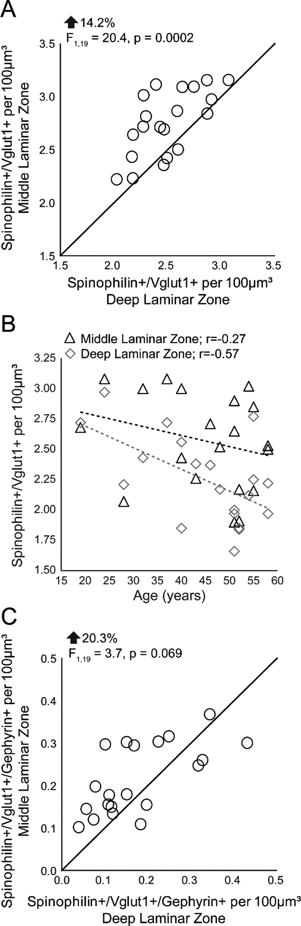
Activation of specific neural circuits in different layers of the primate dorsolateral prefrontal cortex (DLPFC) is essential for working memory, a core cognitive function. Recurrent excitation between pyramidal neurons in middle and deep layers of the DLPFC contributes to the laminar-specific activity associated with different working memory subprocesses. Excitation between cortical pyramidal neurons is mediated by glutamatergic synapses on dendritic spines, but whether the relative abundance of spines receiving cortical inputs differs between middle and deep cortical layers in human DLPFC is unknown. Additionally, GABAergic inputs to spines sculpt pyramidal neuron activity. Whether dendritic spines that receive a glutamatergic input from a cortical pyramidal neuron are targeted by GABAergic interneurons in the human DLPFC is unknown. Using triple-label fluorescence confocal microscopy, we found that 1) the density of spines receiving an input from a cortical pyramidal neuron is greater in the middle than in the deep laminar zone, 2) dendritic spines dually innervated by a cortical pyramidal neuron and an interneuron are present in the human DLPFC, and 3) the density of spines dually innervated by a cortical pyramidal neuron and an interneuron is also greater in the middle than in the deep laminar zone. Ultrastructural analyses support the presence of spines that receive a cortical pyramidal neuron synapse and an interneuron synapse in human and monkey DLPFC. These data support the notion that the DLPFC middle laminar zone is particularly endowed with a microcircuit structure that supports the gating, integrating and fine-tuning of synaptic information in recurrent excitatory microcircuits.
Keywords: GABA; axospinous; glutamate; layer 3; layer 5; ultrastructure.
Copyright © 2020 IBRO. Published by Elsevier Ltd. All rights reserved.
Conflict of interest statement
Declaration of Competing Interest The authors declare that they have no known competing financial interests or personal relationships that could have appeared to influence the work reported in this paper.
Figures


Similar articles
-
Ultrastructural analysis of parvalbumin synapses in human dorsolateral prefrontal cortex.J Comp Neurol. 2017 Jun 15;525(9):2075-2089. doi: 10.1002/cne.24171. Epub 2017 Mar 26. J Comp Neurol. 2017. PMID: 28074478 Free PMC article.
-
Strength of Excitatory Inputs to Layer 3 Pyramidal Neurons During Synaptic Pruning in the Monkey Prefrontal Cortex: Relevance for the Pathogenesis of Schizophrenia.Biol Psychiatry. 2023 Aug 15;94(4):288-296. doi: 10.1016/j.biopsych.2023.01.019. Epub 2023 Feb 2. Biol Psychiatry. 2023. PMID: 36736420 Free PMC article.
-
Close Homolog of L1 Regulates Dendritic Spine Density in the Mouse Cerebral Cortex Through Semaphorin 3B.J Neurosci. 2019 Aug 7;39(32):6233-6250. doi: 10.1523/JNEUROSCI.2984-18.2019. Epub 2019 Jun 10. J Neurosci. 2019. PMID: 31182634 Free PMC article.
-
Unique Molecular Regulation of Higher-Order Prefrontal Cortical Circuits: Insights into the Neurobiology of Schizophrenia.ACS Chem Neurosci. 2018 Sep 19;9(9):2127-2145. doi: 10.1021/acschemneuro.7b00505. Epub 2018 Mar 1. ACS Chem Neurosci. 2018. PMID: 29470055 Free PMC article. Review.
-
The chandelier neuron in schizophrenia.Dev Neurobiol. 2011 Jan 1;71(1):118-27. doi: 10.1002/dneu.20825. Dev Neurobiol. 2011. PMID: 21154915 Free PMC article. Review.
Cited by
-
Volume electron microscopy reveals 3D synaptic nanoarchitecture in postmortem human prefrontal cortex.iScience. 2025 May 26;28(7):112747. doi: 10.1016/j.isci.2025.112747. eCollection 2025 Jul 18. iScience. 2025. PMID: 40585366 Free PMC article.
-
Volume electron microscopy reveals 3D synaptic nanoarchitecture in postmortem human prefrontal cortex.bioRxiv [Preprint]. 2024 Sep 12:2024.02.26.582174. doi: 10.1101/2024.02.26.582174. bioRxiv. 2024. Update in: iScience. 2025 May 26;28(7):112747. doi: 10.1016/j.isci.2025.112747. PMID: 38463986 Free PMC article. Updated. Preprint.
-
Transcranial Magnetic Stimulation and Neocortical Neurons: The Micro-Macro Connection.Front Neurosci. 2022 Apr 12;16:866245. doi: 10.3389/fnins.2022.866245. eCollection 2022. Front Neurosci. 2022. PMID: 35495053 Free PMC article. Review.
-
Targeting prefrontal cortex GABAergic microcircuits for the treatment of alcohol use disorder.Front Synaptic Neurosci. 2022 Aug 29;14:936911. doi: 10.3389/fnsyn.2022.936911. eCollection 2022. Front Synaptic Neurosci. 2022. PMID: 36105666 Free PMC article. Review.
-
Morphological and transcriptomic analyses of stem cell-derived cortical neurons reveal mechanisms underlying synaptic dysfunction in schizophrenia.Genome Med. 2023 Jul 28;15(1):58. doi: 10.1186/s13073-023-01203-5. Genome Med. 2023. PMID: 37507766 Free PMC article.
References
-
- Baddeley A (1992), Working memory. Science 255:556–559. - PubMed
Publication types
MeSH terms
Grants and funding
LinkOut - more resources
Full Text Sources
Other Literature Sources
