Review
. 2021 Feb:143:115758.
doi: 10.1016/j.bone.2020.115758. Epub 2020 Nov 17.

MicroRNAs and fracture healing: Pre-clinical studies

Affiliations
Review

MicroRNAs and fracture healing: Pre-clinical studies

David E Komatsu et al. Bone. 2021 Feb.

Abstract

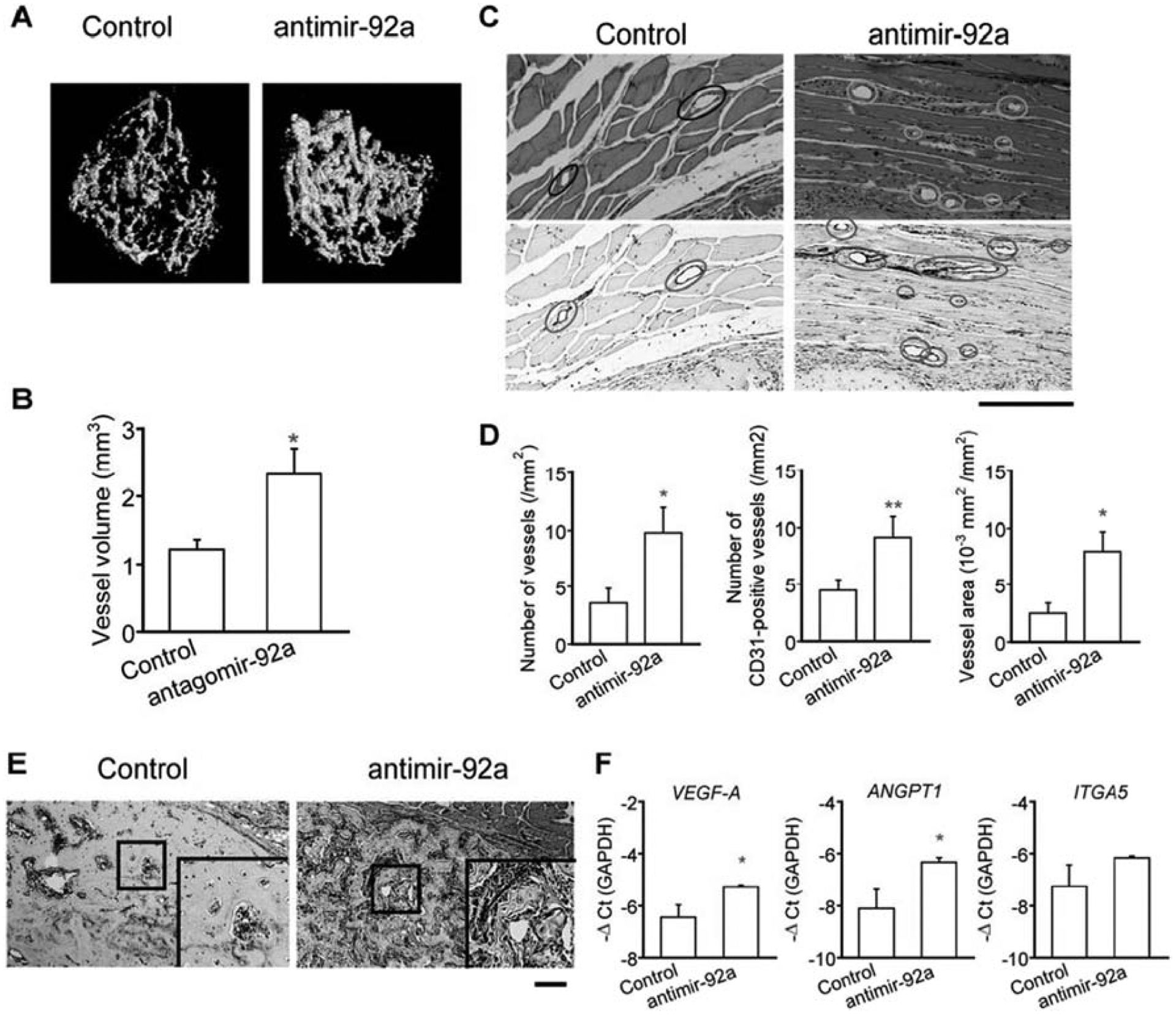
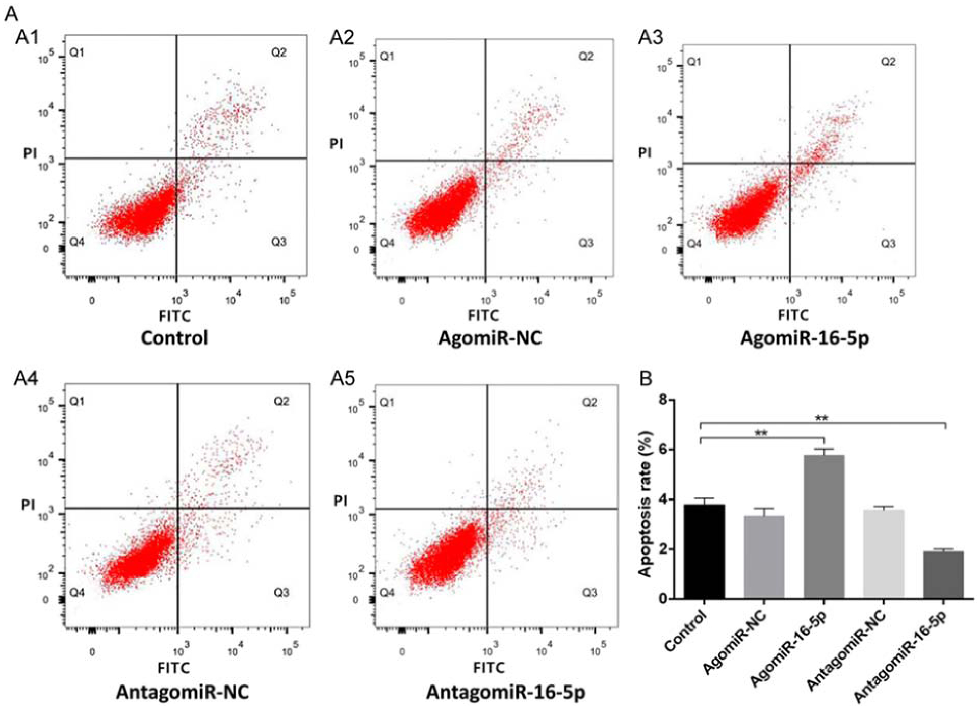
During the past several years, pre-clinical experiments have established that microRNAs (miRNAs), small non-coding RNAs, serve as key regulatory molecules of fracture healing. Their easy modulation with agonists and antagonists make them highly desirable targets for future therapeutic strategies, especially for pathophysiologic fractures that either do not heal (nonunions) or are delayed. It is now well documented that these problematic fractures lead to human suffering and impairment of life quality. Additionally, financial difficulties are also encountered as work productivity decreases and income is reduced. Moreover, targeting miRNAs may also be an avenue to enhancing normal physiological fracture healing. Herein we present the most current knowledge of the involvement of miRNAs during fracture healing in pre-clinical studies. Following a brief description on the nature of miRNAs and of the fracture healing process, we present data from studies focusing specifically, on miRNA regulation of osteoblast differentiation and osteogenesis (within the context of known signaling pathways), chondrocytes, angiogenesis, and apoptosis, all critical to successful bone repair. Further, we also discuss miRNAs and exosomes. We hope that this manuscript serves as a comprehensive review that will facilitate basic/translational scientists in the orthopaedic arena to realize and further decipher the biological and future therapeutic impact of these small regulatory RNA molecules, especially as they relate to the molecular events of each of the major phases of fracture healing.

Keywords: Chondrocytes; Exosomes; Fracture healing; Osteoblasts; miRNAs.


Figures

Figure 1.
Figure 1.
Fracture healing process. A) Fractured bone. B) Initial response showing the evelopment of a hematoma and formation of a fibrin clot at the fracture site. C) Proliferative esponse, indicated by the activation of periosteal and bone marrow stem cell progenitors and ctivation of various signaling pathways. D) Tissue regeneration, as a result of stem cell ifferentiation into osteoblasts and chondrocytes that lead to new bone directly through intramembranous ossification (hard callus) and cartilage (soft callus), respectively. E) ndochondral ossification, conversion of the soft callus into secondary bone accompanied by angiogenesis. F) Fractures healing occurs with bridging of the fracture with calcified tissue, but is completed with remodeling of the fracture which restores the bone to its normal size and shape. G) Complete healing. Adapted from [11].
Figure 2.
Figure 2.
miRNA regulation of enhanced bone repair. This schematic represents the effects of modulating various miRNAs in osteoblasts and/or mesenchymal stem cells leading to activation of signaling pathways, particularly, BMP/TGF-β, Wnt/β-catenin and PTEN/PI3K/Akt that stimulate osteogenesis and ultimately enhance bone repair. The data represented in this schematic are also summarized in Table 1.
Figure 3.
Figure 3.
In vivo bone repair as monitored by microCT. Human ADSCs were mock-transduced (Mock group), co-transduced with Bac-Cre/Bac-L148b (L148b group) or with Bac-Cre/Bac-LCBW (LCBW group), or co-transduced with Bac-Cre/Bac-LCBW/Bac-L148b (LCBW/L148b group). The cells were seeded onto gelatin-coated poly(lactic-co-glycolic acid) (PLGA) scaffolds and implanted into critical-sized (4mm in diameter) calvarial defects in nude mice. The mice were scanned by micoCT at the indicated times. Adapted from [35].
Figure 4.
Figure 4.
Newly formed bone was detected by X-ray and mechanical testing. a. miR-218 was significantly increased at day 5 post fracture. b. X-ray images at week 2 and week 4 post-fracture. c and d. Three-point bending mechanical test at week 4. *p < 0.05. Adopted from [58].
Figure 5.
Figure 5.
Suppression of miR-92a enhanced angiogenesis during fracture healing. A) Vascularity in the fractured femora of mice was visualized by microCT using a radioopaque silicon polymer medium on post fracture day 14. LNA was administered intravenously on days 0, 4, 7, 11, and 14 after the fracture. B) Vessel volume was quantified on 3D microCT images. n=5–6 mice per group. The data are shown as mean ± SEM. *p<0.05. C) Tissues surrounding the fracture callus on post fracture day 14 were stained with HE (upper panels) or immunohistochemically with anti-CD31 (lower panels). Scale bar=20μm. D) The number of vessels in HE-stained samples (left panel) and CD31-positive vessels (middle panel) were counted, and the ratio of vessel area was measured in HE-stained samples (right panel) (n=5–7 per group). E) Post fracture fay 14 calluses were stained with anti-CD31 (lower panels). Boxed areas are enlarged on right bottom. Scale bar for original images=200μm. F) The expression levels of VEGF-A, ANGPT1, and ITGA5 of callus from mice on post fracture day 14 were quantified by qRT-PCR (n=5, respectively). The data are shown as mean±SEM. *p<0.05; **p<0.01. Adopted from [93].
Figure 6.
Figure 6.
Downregulation of miR-16–5p inhibits osteoblast apoptosis. A. Apoptosis of osteoblasts in each group. B. Quantitative apoptosis rate in each group. All data were expressed as means ± SD. Significance is noted at these thresholds: *P < 0.05, **P < 0.01, ***P < 0.001. One-way ANOVA with a post hoc test was performed. Statistical differences between two groups were determined by Student’s t test. Adopted from [98].

References

    1. Initiative: USBaJ The Burden of Musculoskeletal Diseases in the United States (BMUS), Third Edition 2014.
    1. Zura R, Xiong Z, Einhorn T, Watson JT, Ostrum RF, Prayson MJ, Della Rocca GJ, Mehta S, McKinley T, Wang Z, Steen RG. Epidemiology of Fracture Nonunion in 18 Human Bones. JAMA Surg 2016;151: e162775. - PubMed
    1. Wright NC, Looker AC, Saag KG, Curtis JR, Delzell ES, Randall S, Dawson-Hughes B. The Recent Prevalence of Osteoporosis and Low Bone Mass in the United States Based on Bone Mineral Density at the Femoral Neck or Lumbar Spine. Journal of Bone and Mineral Research 2014;29: 2520–2526. - PMC - PubMed
    1. Briggs A, Woolf A, Dreinhoefer K, Homb N, Hoy D, Kopansky-Giles D, Akesson K, March L. Reducing the global burden of musculoskeletal conditions. Bulletin of the World Health Organization 2018;96: 366–368. - PMC - PubMed
    1. Force UPST. Screening for Osteoporosis to Prevent Fractures: US Preventive Services Task Force Recommendation Statement. JAMA 2018;319: 2521–2531. - PubMed

Publication types