

Comment
doi: 10.1093/brain/awaa350.
Lesion network mapping: where do we go from here?
Affiliations
- PMID: 33212509
- PMCID: PMC8453285
- DOI: 10.1093/brain/awaa350
Item in Clipboard
Comment
Lesion network mapping: where do we go from here?
Brain.
.
No abstract available
Figures
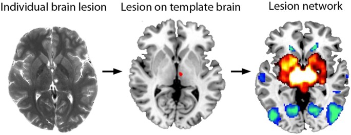
Lesion network mapping. Lesion network mapping involves three steps: (i) a brain lesion from a patient scan acquired clinically or for research is mapped onto a template brain; (ii) the lesion volume is used as a seed region of interest for a resting state functional connectivity MRI analysis that uses normative data; and (iii) the lesion-associated networks can then be analysed, such as comparing network results in relation to the presence or absence of a symptom being investigated.
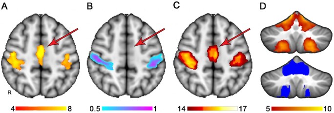
Extension of lesion localization to connected brain areas. (A) The somatomotor network derived from a previously published atlas of functional connectivity data derived from normal healthy adults (Smith et al., 2009). (B) A lesion symptom map derived from somatomotor network ‘lesion load’ values from A used as simulated behavioural data, where the success or failure of the lesion symptom map could be judged based on the similarity to A. (C) A potential use of lesion network mapping in extending the lesion symptom mapping findings in B to other functionally related brain regions, in this case identifying the medial node of the somatomotor network despite inadequate lesion coverage in this region, evident by the absence of findings in B. (D) Similarly, lesion network mapping extends the functional network to the cerebellum somatomotor network even though the cerebellum had no lesion coverage in this analysis. The results from lesion network mapping are on top and the somatomotor network of the cerebellum from a published atlas is shown below for reference (Buckner, 2011). The colour bars denote voxel-wise Z-scores for the somatomotor network (A), the strength of association of lesion location with somatomotor network lesion load based on multivariate lesion symptom mapping, which is output as arbitrary units from 0 to 1, thresholded at 0.5 to display the strongest findings (Pustina et al., 2018). And voxel-wise Z-scores reflecting strength of connectivity with the regions denoted in B.
Comment in
-
Reply: Lesion network mapping: where do we go from here?Brain. 2021 Feb 12;144(1):e6. doi: 10.1093/brain/awaa351. Brain. 2021. PMID: 33212502 Free PMC article. No abstract available.
Comment on
-
Post-stroke deficit prediction from lesion and indirect structural and functional disconnection.Brain. 2020 Jul 1;143(7):2173-2188. doi: 10.1093/brain/awaa156. Brain. 2020. PMID: 32572442 Free PMC article.
References
-
- Axer M, Amunts K, Grassel D, Palm C, Dammers J, Axer H, et al. A novel approach to the human connectome: ultra-high resolution mapping of fiber tracts in the brain. Neuroimage 2011; 54: 1091–101. - PubMed
Publication types
MeSH terms
Grants and funding
LinkOut - more resources
Full Text Sources
Medical
