Review
. 2020 Nov 17;21(22):8652.
doi: 10.3390/ijms21228652.

Peripheral Nerve Regeneration and Muscle Reinnervation

Affiliations
Review

Peripheral Nerve Regeneration and Muscle Reinnervation

Tessa Gordon. Int J Mol Sci. .

Abstract

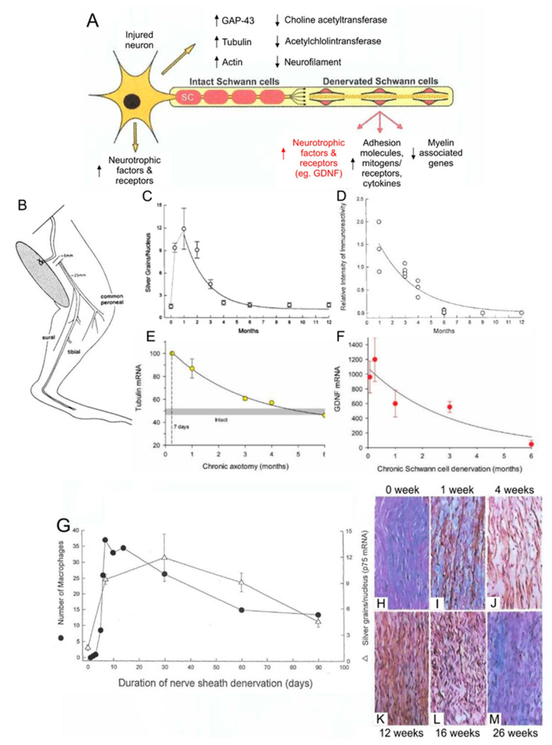
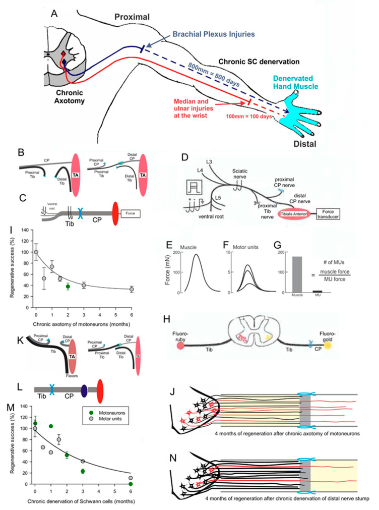
Injured peripheral nerves but not central nerves have the capacity to regenerate and reinnervate their target organs. After the two most severe peripheral nerve injuries of six types, crush and transection injuries, nerve fibers distal to the injury site undergo Wallerian degeneration. The denervated Schwann cells (SCs) proliferate, elongate and line the endoneurial tubes to guide and support regenerating axons. The axons emerge from the stump of the viable nerve attached to the neuronal soma. The SCs downregulate myelin-associated genes and concurrently, upregulate growth-associated genes that include neurotrophic factors as do the injured neurons. However, the gene expression is transient and progressively fails to support axon regeneration within the SC-containing endoneurial tubes. Moreover, despite some preference of regenerating motor and sensory axons to "find" their appropriate pathways, the axons fail to enter their original endoneurial tubes and to reinnervate original target organs, obstacles to functional recovery that confront nerve surgeons. Several surgical manipulations in clinical use, including nerve and tendon transfers, the potential for brief low-frequency electrical stimulation proximal to nerve repair, and local FK506 application to accelerate axon outgrowth, are encouraging as is the continuing research to elucidate the molecular basis of nerve regeneration.

Keywords: Schwann cells; peripheral nerve injuries; peripheral nerve regeneration; regenerating peripheral nerves; skeletal muscle reinnervation.


Conflict of interest statement

The authors declare no conflict of interest.

Figures

Figure 2
Figure 2
Outgrowth of axons after femoral nerve transection and microsurgical repair. The femoral neve was crushed 1.5 mm distal to the repair site (A) for rubyred microinjection (B) to count motoneurons that had regenerated their axons across the suture site. (C). The motoneuron count elucidated a staggering of the regenerating axons across the suture site with all axons crossing the site by 28 days, considerably longer than the calculated latent period of 2–3 days. The slow and staggered crossing the suture is seen in longitudinal sections of the nerve with axons of motoneurons immunostained for neurofilament protein, 5 (D) and 10 (E) days after the nerve repair. The complex growth of silver nitrate-stained regenerating axons (F) even turns back into the proximal nerve stump as first seen by Cajal [41]. The staggering of the regenerating axons within the endoneurial tube of the distal nerve stump is seen in (G). The cut and surgical repair is illustrated in (H,I). where regenerating axons “stagger” across the suture site in amongst the Schwann cells that move into the surgical site from both nerve stumps. The Schwann cells elongate along the basal lamina of the denervated endoneurial tubes to guide, support, and myelinate the regenerating axon. Adapted from [33].
Figure 6
Figure 6
The motor unit (MU) spatial territory that contains all the muscle fibers innervated by one motoneuron, limits the numbers of muscle fibers that each reinnervating nerve can reinnervate after complete and partial nerve injuries. Muscle force was recorded from isolated MUs in tibialis anterior (TA) muscle by all-or-none electrical stimulation of dissected ventral root filaments after a laminectomy to expose the lumbosacral spinal cord (A). The MU muscle fibers were depleted of their glycogen by repetitive stimulation at 1 and 5 Hz to fatigue the MU fibers followed by recovery during 0.1 Hz stimulation This sequence was repeated until the MU force failed to recover (B). The TA muscle was dissected rapidly. The muscle was frozen for cryosectioning and staining for glycogen with Periodic Schiff reaction to reveal and count glycogen depleted MU fibers in normally innervated (C,D) and reinnervated (F,G) TA muscle fibers after common peroneal (CP) nerve section and surgical repair and for histochemical staining of acidic mATPase to fiber type the MU muscle fibers (E,H). The MU muscle fibers are normally distributed in a mosaic pattern (CE) which changes to F, G, H. clumping of the fibers that occupy a smaller territory (FH). The branching pattern of the motor nerve is shown in normally innervated (I) and reinnervated (J) MUs. In normally innervated muscles, each motor nerve normally branches only once the nerve enters the muscle. Thereafter, the nerve branches to distribute the fibers to several fascicles to give the mosaic pattern of MU muscle fibers (I). In reinnervated muscles, the loss of the mosaic pattern reveals that the regenerating nerves “miss” some of the denervated intramuscular nerve sheaths and branch more as they approach the muscle fibers, often within single fascicles. This gives rise to the clumping of reinnervated muscle fibers (J). On the other hand, the normal mosaic distribution (I,K) is progressively lost after partial muscle denervation by transecting one of two contributing ventral roots (L). Diagrammatically, the peri-synaptic Schwann cells at the endplates (M,N) begin, after partial denervation of the muscle, to extend processes (O) some of which extend to the innervated endplate of an adjacent muscle fiber (O,P). The processes lead nerve sprouts from innervated to denervated endplates to reinnervate the denervated muscle fibers (Q,R). Normally innervated (S,T,W) and partially denervated (U,VZ) MU muscle fibers are compared to show the progressive MU fiber and muscle fiber type-grouping that occurs as the percentage of remaining MUs declines from 100% (normal; W), to 60% (X), 30% (Y), and 15% (Z) in partially denervated muscles. Adapted from [5,109,121].
Figure 7
Figure 7
Motor unit enlargement after partial denervation of muscles. Figurative illustration of muscle denervation, reinnervation after complete nerve injury, and compensatory sprouting after partial nerve injury (A). The skewed distribution of motor unit (MU) twitch forces in control tibialis anterior (TA) muscle is shifted significantly (p < 0.05) to the right to larger values after partial denervation by cutting the L5 spinal root (B). The shift is more obvious when the MU forces are plotted on a logarithmic scale (C) and as cumulative distributions (D). The cumulative distributions demonstrate that all MUs in the partially denervated muscle increase by the same factor such that the large MUs include many more muscle fibers by sprouting than the small MUs. This five-fold increase is maximum as shown in (E). by the sharp decline in muscle force recovery when partial denervation of the muscle (numbers of nerves innervating the muscle) exceeds about 80%. The sprouting in partially denervated muscles (PD) is the same whether the spinal cord is intact or hemisected (SCPD). Adapted from [109].
Figure 8
Figure 8
Size relationships in cat medial gastrocnemius (MG) muscles after complete and partial nerve injuries. At regular intervals of 10–14 days, the cat was anesthetized with fluorothane. The foot of the operated hindlimb was encased in a special boot that was attached to a force transducer to record isometric muscle and motor unit (MU) forces in response to stimulation of the MG nerve via nerve cuff electrodes (A) and in response to stimulation of single MG nerves via a needle electrode inserted through the skin into the endplate region of the MG muscle (B). The MU action potentials on the MG (C) and sciatic (D) nerves were recorded from implanted nerve cuff electrodes. The MU electromyographic action potential (EMG; E) was recorded from EMG pad electrodes on the muscle surface and the twitch tension (F) was recorded with the force transducer attached to the boot. MU action potential amplitude was directly correlated with the twitch tension of the innervated muscle fibers (G) and inversely correlated with the contraction times of the twitch contractions (H). These correlations are in accordance with Henneman’s size principle (see text). The inverse relationship demonstrates that force increases as contraction times become shorter with the fast MUs being the most forceful and the slow MUs being the least. Early during reinnervation, the size relationships were lost when each nerve reinnervated muscles fibers of different histochemical types(I,J) but they returned with time (KN) as the heterogenous muscle fibers were respecified by their new motoneuron innervation (not shown). In normal (O,P) and partially denervated (RU) MG muscles after transecting the L7 root, the size relationships were retained as the forces increased when the partial denervation removed 50% and 80% of the nerve supply to the muscles. All correlations in which lines are drawn were statistically significant at the 0.05 level. Adapted from [112,125,133].
Figure 1
Figure 1
Expression of growth-associated genes in axotomized motoneurons after loss of target connections and in chronically denervated Schwann cells (SCs) in the distal nerve stumps. An illustrated motoneuron showing expression of growth-associated genes in motoneurons and in Schwann cells in the distal nerve stumps after a nerve injury (A). Experiments were performed in which we cut the sciatic nerve and sutured both proximal and distal nerve stumps to innervated muscle to prevent regeneration (B). p75NTFR in situ hybridization showing an early rise in expression in the denervated SCs in the distal nerve stump that declined within 6 months (C). Immunocytochemical evidence of a concomitant decline in p75 protein in the chronically denervated Schwann cells. (D). In situ hybridization showing a progressive decline to baseline values in the upregulation of the mRNA of the cytoskeletal protein tubulin in axotomized motoneurons (E). Semi-quantitation of the gene expression of Glial derived neurotrophic factor (GDNF) in the denervated distal nerve stump with rt-PCR, showing an exponential decline (F). The parallel expression of p75 and infiltration of macrophages into the denervated distal nerve stump (G). The slow rate of Wallerian degeneration of the isolated nerve in the denervated distal nerve stump is illustrated in the longitudinal micrographs of the denervated distal nerve stump (HJ) leaving parallel endothelial tubes with immunologically stained p75 expressing SCs by 12 weeks (K) that declines within 6-months (L,M). Adapted from [19].
Figure 3
Figure 3
A. Motoneurons remain without targets (chronic axotomy) as they regenerate after proximal nerve injuries such as brachial plexus injuries and Schwann cells (SCs) in the denervated distal nerve stumps remain chronically denervated. At the slow rate of 1 mm/day regeneration rate in human, a year or more will pass for regenerating nerves to reach hand muscles, which are generally believed to atrophy and be replaced by fat (A). To isolate the effects of chronic axotomy from chronic denervation, the tibial (TIB) nerve was cut and the stumps sutured to innervated muscle prior to a cross-suture of TIB nerve to freshly denervated common peroneal (CP) distal nerve stump (B) and tibialis anterior (TA) muscle reinnervation was assessed by recording muscle and motor unit (MU) isometric forces at least 4 months later (C), as shown in more detail in (D). After cutting all nerves other than the CP nerve to the TA muscle, the sciatic nerve was stimulated at 2x threshold to evoke and record isometric muscle twitch (and tetanic—not shown) force (E). Thereafter, ventral root filaments were dissected to stimulate, at 2x threshold, single CP nerve fibers and record all-or-none MU twitch forces (F). The number of MUs was determined by dividing TA muscle force by average TA MU forces to give the number of TIB nerves that reinnervated the TA muscle (G). Numbers of axotomized motoneurons regenerating axons into the freshly denervated CP nerve stumps were also counted after their labelling by applying retrograde dyes, either fluororuby or fluorogold, to the distal nerve stump 10 mm from the cross-suture site (H). Plots of the numbers of reinnervated MUs and motoneurons regenerating their axons as a function of the days of axotomy prior to TIB-CP cross-suture, demonstrated an exponential decline to 33% of the numbers after immediate cross-suture at 0 days (I). The outgrowth of axons across the suture site is shown diagrammatically in (J). In order to examine the effects of chronic Schwann cell denervation, the CP nerve was transected and the proximal stump sutured to innervated muscle before the TIB-CP cross-suture of the freshly cut TIB nerve to the chronically denervated CP nerve stump to record muscle and MU forces (K) or to apply retrograde dye to the regenerated nerve 10 mm from the suture site (L). The numbers of reinnervated MUs and TIB motoneurons that regenerated their axons into the CP nerve stump, declined exponentially to <5% within 6-months of CP distal nerve chronic denervation (M), as illustrated figuratively in (N). Adapted from [35,36].
Figure 4
Figure 4
The expression of neurotrophic factors in Schwann cells in denervated nerve stumps and their role in preferential reinnervation of appropriate pathways by regenerating femoral nerves. The time course of the gene expression measured with rtPCR in the cutaneous sensory nerve branch of the femoral nerve (A) and in ventral root motor nerves (B). The mRNA normalized to values in innervated nerve stumps increased to a peak at 15 days and receded thereafter. Two different retrograde dyes (fluorogold and rubyred) were applied for retrograde labelling and counting of the motoneurons that regenerated their axons into appropriate and inappropriate motor (muscle) and sensory (cutaneous) nerve branches (C), and 1.5 mm across the site of microsurgical repair of the transected femoral nerve (D). The regeneration of the motor axons is non-selective at 2 and 3 weeks for the appropriate and inappropriate motor and sensory femoral nerve branches (E). However, as also shown by the data in (F,G), motoneurons progressively regenerate their axons preferentially into the motor branch in the time frame in which Schwann cells in the sensory and motor nerve branches show selective expression of growth factors (A). The number of motoneurons that had regenerated their axons non-selectively into both branches did not change over time (F,G). Adapted from [33,98,99].
Figure 5
Figure 5
Non-selective reinnervation of muscles after peripheral nerve surgery. The diagrammatic representation of the innervation of the intrinsic abductor (AB) and adductor (AB) muscles of the vocal cords by inspiration and phonation (speaking) in normal (A), denervated (B), and reinnervated (C) intrinsic muscles. The large and small motoneurons innervating the AB and AD muscles, respectively, represents the 4:1 ratio of the number of the motoneurons innervating the AB and AD muscles. After bilateral denervation, the vocal cords do not close during phonation so that speech is severely impaired (B). After surgical repair of the laryngeal nerve, the axotomized motoneurons randomly reinnervate the AB and AD muscles (C) with the result that the vocal cords are effectively paralyzed in a position of openness that disallows effective speech. Adapted from [104].

References

    1. Lieberman A. The axon reaction: A review of the principal features of perikaryal responses to axon injury. Int. Rev. Neurobiol. 1971;14:49–124. doi: 10.1016/s0074-7742(08)60183-x. - DOI - PubMed
    1. Fu S.Y., Gordon T. The cellular and molecular basis of peripheral nerve regeneration. Mol. Neurobiol. 1997;14:67–116. doi: 10.1007/BF02740621. - DOI - PubMed
    1. Gaudet A.D., Popovich P.G., Ramer M.S. Wallerian degeneration: Gaining perspective on inflammatory events after peripheral nerve injury. J. Neuroinflammation. 2011;8:110. doi: 10.1186/1742-2094-8-110. - DOI - PMC - PubMed
    1. Gordon T. Nerve regeneration Understanding the biology and its influence on return of function after nerve transfers. Hand Clin. 2016;32:103–117. doi: 10.1016/j.hcl.2015.12.001. - DOI - PubMed
    1. Gordon T., Borschel G.H. The use of the rat as a model for studying peripheral nerve regeneration and sprouting after complete and partial nerve injuries. Exp. Neurol. 2017;287:331–347. doi: 10.1016/j.expneurol.2016.01.014. - DOI - PubMed

MeSH terms