. 2020 Nov 17;25(22):5361.
doi: 10.3390/molecules25225361.

Quantum Chemical Study Aimed at Modeling Efficient Aza-BODIPY NIR Dyes: Molecular and Electronic Structure, Absorption, and Emission Spectra

Affiliations

Quantum Chemical Study Aimed at Modeling Efficient Aza-BODIPY NIR Dyes: Molecular and Electronic Structure, Absorption, and Emission Spectra

Alexander E Pogonin et al. Molecules. .

Abstract

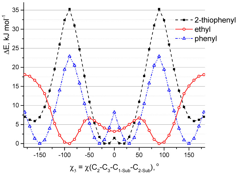
A comprehensive study of the molecular structure of aza-BODIPY and its derivatives, obtained by introduction of one or more substituents, was carried out. We considered the changes in the characteristics of the electronic and geometric structure of the unsubstituted aza-BODIPY introducing the following substituents into the dipyrrin core; phenyl, 2-thiophenyl, 2-furanyl, 3-pyridinyl, 4-pyridinyl, 2-pyridinyl, and ethyl groups. The ground-state geometries of the unsubstituted Aza-BODIPY and 27 derivatives were computed at the PBE/6-31G(d) and CAM-B3LYP/6-31+G(d,p) levels of theory. The time-dependent density-functional theory (TDDFT) together with FC vibronic couplings was used to investigate their absorption and emission spectra.

Keywords: absorption spectra; aza-BODIPY; intramolecular rotation; molecular structure; quantum chemical calculations; vibronic spectra.


Conflict of interest statement

The authors declare no conflict of interest.

Figures

Figure 1
Figure 1
Atom numbering, structures, and naming scheme of the investigated molecules used throughout the paper. Color denotes dihedral angles C2-C1-C1-Sub-X2-Sub1), C6-C7-C1-Sub-X2-Sub2), C2-C3-C1-Sub-X2-Sub3), and C6-C5-C1-Sub-X2-Sub4). Molecules are named according to the symbols of substituents R1–R4: A1—1,7-diphenyl-3,5-di(2-thiophenyl)-aza-BODIPY, A2—1,3,5,7-tetra(2-thiophenyl)-aza-BODIPY, A3—1,7-di(2-furanyl)-3,5-di(2-thiophenyl)-aza-BODIPY, A4—1,7-di(3-pyridinyl)-3,5-di(2-thiophenyl)-aza-BODIPY, A5—1,7-di(4-pyridinyl)-3,5-di(2-thiophenyl)-aza-BODIPY, A6—1,7-di(2-pyridinyl)-3,5-di(2-thiophenyl)-aza-BODIPY, A7—3,5-di(2-thiophenyl)-aza-BODIPY, B1—1,7-diphenyl-3,5-diethyl-aza-BODIPY, B2—1,7-di(2-thiophenyl)-3,5-diethyl-aza-BODIPY, B3—1,7-di(2-furanyl)-3,5-diethyl-aza-BODIPY, B4—1,7-di(3-pyridinyl)-3,5-diethyl-aza-BODIPY, B5—1,7-di(4-pyridinyl)-3,5-diethyl-aza-BODIPY, B6—1,7-di(2-pyridinyl)-3,5-diethyl-aza-BODIPY, B7—3,5-diethyl-aza-BODIPY, C1—1,3,5,7-tetraphenyl-aza-BODIPY, C2—1,7-di(2-thiophenyl)-3,5-diphenyl-aza-BODIPY, C3—1,7-di(2-furanyl)-3,5-diphenyl-aza-BODIPY, C4—1,7-di(3-pyridinyl)-3,5-diphenyl-aza-BODIPY, C5—1,7-di(4-pyridinyl)-3,5-diphenyl-aza-BODIPY, C6—1,7-di(2-pyridinyl)-3,5-diphenyl-aza-BODIPY, C7—3,5-diphenyl-aza-BODIPY, D1—1,7-diphenyl-aza-BODIPY, D2—1,7-di(2-thiophenyl)-aza-BODIPY, D3—1,7-di(2-furanyl)-aza-BODIPY, D4—1,7-di(3-pyridinyl)-aza-BODIPY, D5—1,7-di(4-pyridinyl)-aza-BODIPY, D6—1,7-di(2-pyridinyl)-aza-BODIPY, D7—aza-BODIPY.
Figure 2
Figure 2
Conformer models of aza-BODIPY derivatives with substituent groups at C1, C7 (or C3, C5) positions. Depiction of conformational multiformity coupled to different mutual orientations of neighboring cyclic groups: (a) structure of C2 symmetry; (b) structure of Cs symmetry; to different arrangement of heteroatoms with respect to the center of the molecule: (c) heteroatoms in cyclic groups are oriented by “outer” direction with respect to center of molecule; (d) heteroatoms in cyclic groups are directed by “inner” direction with respect to center of molecule; (e) one heteroatom in one cyclic group is directed by “inner” direction, another heteroatom in another group—by “outer” direction; different orientations of ethyl groups: (f) model of Cs symmetry according to which ethyl groups are oriented in the same direction relating to aza-BODIPY core; (g) model of C2 symmetry according to which ethyl groups are oriented in the opposite direction relating to aza-BODIPY core; (h) two ethyl groups are in plane of molecular core and the groups are oriented by “outer” direction with respect to center of molecule; (i) two ethyl groups are in plane of molecular core with direction of the ethyl group toward each other.
Figure 3
Figure 3
Conformation models of aza-BODIPY analogues substituted by cyclic groups in positions 1, 3, 5, and 7. Depiction of conformational multiformity coupled to different mutual orientations of cyclic groups: I—all four cyclic substituents are quasi-parallel to each other; II—neighboring cyclic groups R1 and R2, R3 and R4 are quasi-parallel to each other in pairs, however R1 and R3, R2 and R4 are not quasi-parallel to each other in pairs; III—neighboring cyclic groups R3 and R4 are quasi-parallel, R1 and R2 are mirrored relative to each other; IV—neighboring cyclic groups R3 and R4 are mirrored relative to each other, R1 and R2 are quasi-parallel; V—neighboring cyclic groups R1 and R2, R3 and R4 are mirrored relative to each other in pairs, R1 and R3, R2 and R4 are quasi-parallel to each other in pairs; VI—neighboring cyclic groups R1 and R2 are mirrored relative to each other in pairs, R1 and R3, R2 and R4 are not quasi-parallel to each other in pairs.
Figure 4
Figure 4
Relaxed potential energy function (PBE/6-31G(d)) of internal rotation of one group R1 in the derivatives of aza-BODIPY molecules around the C1-C1-Sub bond. Positions C3, C5, C7 (Figure 1) in these molecules are occupied by hydrogen atoms.
Figure 5
Figure 5
Relaxed potential energy function (PBE/6-31G(d)) of internal rotation of one group R3 in the derivatives of aza-BODIPY molecules around the C3-C1-Sub bond. Positions C1, C5, and C7 (Figure 1) in these molecules are occupied by hydrogen atoms.
Figure 6
Figure 6
Potential energy surface (PES) obtained at the PBE/6-31G(d) level of theory showing potential energy as a function of rotations of two substituents (R1 and R2) around the bonds C1-C1-Sub and C7-C1-Sub: (a) phenyl groups in molecule D1; (b) 2-thiophenyl groups in molecule D2; (c) 2-furanyl groups in molecule D3; (d) 3-pyridinyl groups in molecule D4; (e) 4-pyridinyl groups in molecule D5; (f) 2-pyridinyl groups in molecule D6. Black circles indicate the calculations performed for the corresponding values of the angles. White letters indicate conformations which are described in Figure 2.
Figure 6
Figure 6
Potential energy surface (PES) obtained at the PBE/6-31G(d) level of theory showing potential energy as a function of rotations of two substituents (R1 and R2) around the bonds C1-C1-Sub and C7-C1-Sub: (a) phenyl groups in molecule D1; (b) 2-thiophenyl groups in molecule D2; (c) 2-furanyl groups in molecule D3; (d) 3-pyridinyl groups in molecule D4; (e) 4-pyridinyl groups in molecule D5; (f) 2-pyridinyl groups in molecule D6. Black circles indicate the calculations performed for the corresponding values of the angles. White letters indicate conformations which are described in Figure 2.
Figure 7
Figure 7
PES obtained at the PBE/6-31G* level of theory showing potential energy as a function of rotations of two substituents (R3, R4) around the bonds C3-C1-Sub and C5-C1-Sub: (a) 2-thiophenyl groups in molecule A7, (b) ethyl groups in molecule B7, and (c) phenyl groups in molecule C7. Black circles indicate the calculations performed for the corresponding values of the angles. White letters indicate conformations which are described in the Figure 2.
Figure 8
Figure 8
Visual representation of the first three transitions (nm) for molecules A1A7 and D7 calculated at CAM-B3LYP/6-31+G(d,p) level. Isosurface cutoff is 0.03. Visual representation of transitions for molecules B1B7, C1C7, and C1D7 is presented in Figures S5–S7.
Figure 9
Figure 9
The CAM-B3LYP/6-31+G(d,p) MO energies and HOMO–LUMO energy gaps for A1A7, B1B7, C1C7, and D1D7.
Figure 10
Figure 10
Calculated TDDFT (CAM-B3LYP/6-31+G(d,p)) electronic absorption spectra for aza-BODIPY derivatives.
Figure 11
Figure 11
Calculated vibronic (FC, CAM-B3LYP/6-31G(d)) electronic absorption and emission spectra for A1A7 and D7 aza-BODIPY derivatives. Solid lines correspond to absorption and dotted lines correspond to emission.

References

    1. Wang C., Wang Z., Zhao T., Li Y., Huang G., Sumer B.D., Gao J. Optical molecular imaging for tumor detection and image-guided surgery. Biomaterials. 2018;157:62–75. doi: 10.1016/j.biomaterials.2017.12.002. - DOI - PMC - PubMed
    1. Mezzanotte L., van’t Root M., Karatas H., Goun E.A., Löwik C.W.G.M. In Vivo Molecular Bioluminescence Imaging: New Tools and Applications. Trends Biotechnol. 2017;35:640–652. doi: 10.1016/j.tibtech.2017.03.012. - DOI - PubMed
    1. Park G.K., Hoseok I., Kim G.S., Hwang N.S., Choi H.S. Optical spectroscopic imaging for cell therapy and tissue engineering. Appl. Spectrosc. Rev. 2018;53:360–375. doi: 10.1080/05704928.2017.1328428. - DOI - PMC - PubMed
    1. Ziessel R., Ulrich G., Harriman A. The chemistry of Bodipy: A new El Dorado for fluorescence tools. New J. Chem. 2007;31:496–501. doi: 10.1039/b617972j. - DOI
    1. Vodyanova O.S., Kochergin B.A., Usoltsev S.D., Marfin Y.S., Rumyantsev E.V., Aleksakhina E.L., Tomilova I.K. BODIPY dyes in bio environment: Spectral characteristics and possibilities for practical application. J. Photochem. Photobiol. A Chem. 2018;350:44–51. doi: 10.1016/j.jphotochem.2017.09.049. - DOI

LinkOut - more resources