

Monte Carlo Modeling of Shortwave-Infrared Fluorescence Photon Migration in Voxelized Media for the Detection of Breast Cancer
- PMID: 33212890
- PMCID: PMC7698463
- DOI: 10.3390/diagnostics10110961
Monte Carlo Modeling of Shortwave-Infrared Fluorescence Photon Migration in Voxelized Media for the Detection of Breast Cancer
Abstract
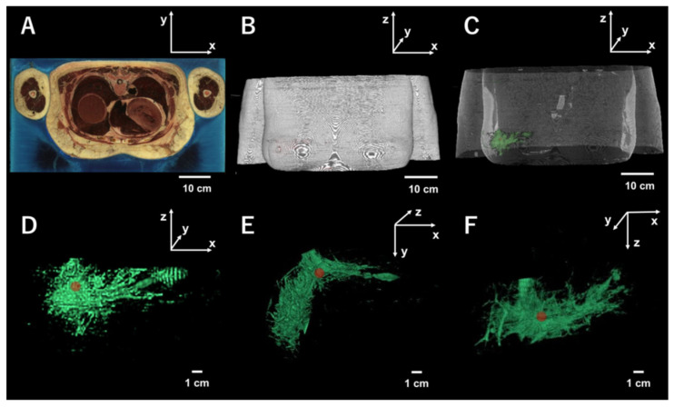
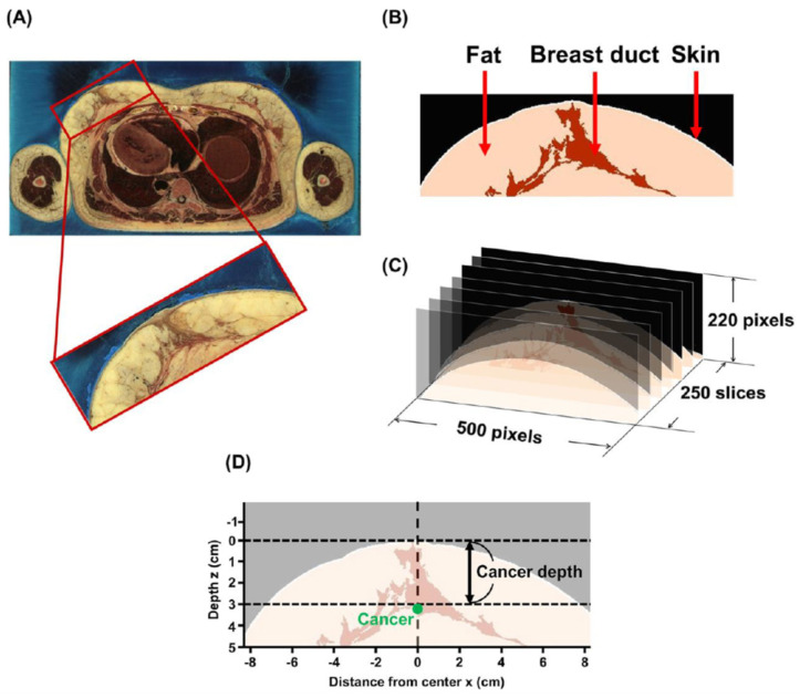
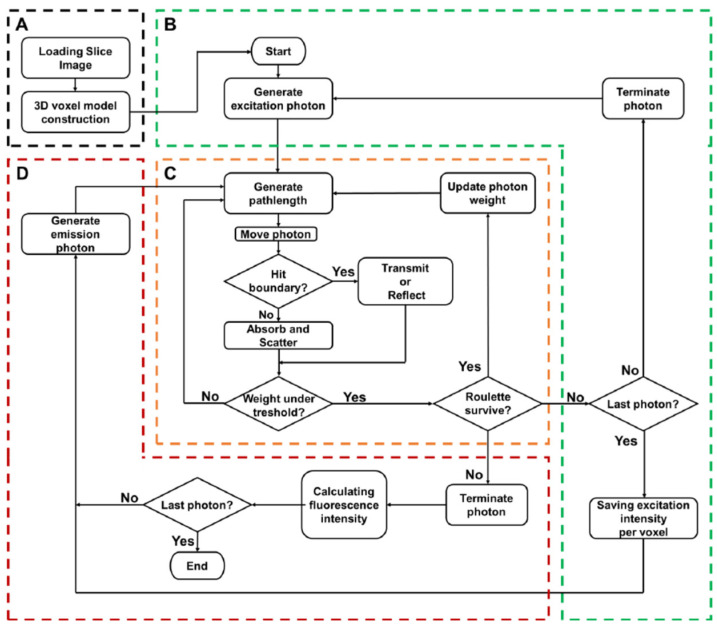
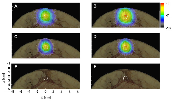
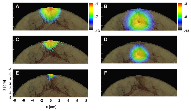
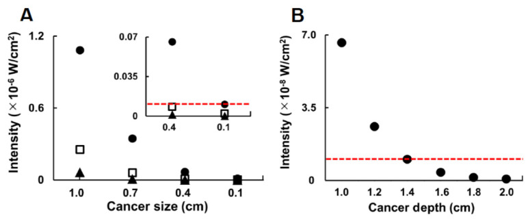
Recent progress regarding shortwave-infrared (SWIR) molecular imaging technology has inspired another modality of noninvasive diagnosis for early breast cancer detection in which previous mammography or sonography would be compensated. Although a SWIR fluorescence image of a small breast cancer of several millimeters was obtained from experiments with small animals, detailed numerical analyses before clinical application were required, since various parameters such as size as well as body hair differed between humans and small experimental animals. In this study, the feasibility of SWIR was compared against visible (VIS) and near-infrared (NIR) region, using the Monte Carlo simulation in voxelized media. In this model, due to the implementation of the excitation gradient, fluorescence is based on rational mechanisms, whereas fluorescence within breast cancer is spatially proportional to excitation intensity. The fluence map of SWIR simulation with excitation gradient indicated signals near the upper surface of the cancer, and stronger than those of the NIR. Furthermore, there was a dependency on the fluence signal distribution on the contour of the breast tissue, as well as the internal structure, due to the implementation of digital anatomical data for the Visible Human Project. The fluorescence signal was observed to become weaker in all regions including the VIS, the NIR, and the SWIR region, when fluorescence-labeled cancer either became smaller or was embedded in a deeper area. However, fluorescence in SWIR alone from a cancer of 4 mm diameter was judged to be detectable at a depth of 1.4 cm.
Keywords: Monte Carlo simulation; breast cancer; duct; fluorescence; near-infrared light; shortwave-infrared light; visible human project; visible light; voxelized media.
Conflict of interest statement
The authors have no conflicts of interest with any company or commercial organization.
Figures
Similar articles
-
Shortwave infrared fluorescence imaging with the clinically approved near-infrared dye indocyanine green.Proc Natl Acad Sci U S A. 2018 Apr 24;115(17):4465-4470. doi: 10.1073/pnas.1718917115. Epub 2018 Apr 6. Proc Natl Acad Sci U S A. 2018. PMID: 29626132 Free PMC article.
-
Shortwave-infrared (SWIR) fluorescence molecular imaging using indocyanine green-antibody conjugates for the optical diagnostics of cancerous tumours.RSC Adv. 2020 Jul 28;10(47):28171-28179. doi: 10.1039/d0ra04710d. eCollection 2020 Jul 27. RSC Adv. 2020. PMID: 35519107 Free PMC article.
-
Shortwave Infrared Imaging Enables High-Contrast Fluorescence-Guided Surgery in Neuroblastoma.Cancer Res. 2023 Jun 15;83(12):2077-2089. doi: 10.1158/0008-5472.CAN-22-2918. Cancer Res. 2023. PMID: 36934744 Free PMC article.
-
A Review of Image Sensors Used in Near-Infrared and Shortwave Infrared Fluorescence Imaging.Sensors (Basel). 2024 May 30;24(11):3539. doi: 10.3390/s24113539. Sensors (Basel). 2024. PMID: 38894330 Free PMC article. Review.
-
In vivo near-infrared fluorescent optical imaging for CNS drug discovery.Expert Opin Drug Discov. 2020 Aug;15(8):903-915. doi: 10.1080/17460441.2020.1759549. Epub 2020 May 12. Expert Opin Drug Discov. 2020. PMID: 32396023 Review.
Cited by
-
Optical Diagnostics in Human Diseases.Diagnostics (Basel). 2021 May 12;11(5):873. doi: 10.3390/diagnostics11050873. Diagnostics (Basel). 2021. PMID: 34066215 Free PMC article.
References
-
- van Dongen J.A., Voogd A.C., Fentiman I.S., Legrand C., Sylvester R.J., Tong D., van der Schueren E., Helle P.A., van Zijl K., Bartelink H. Long-Term Results of a Randomized Trial Comparing Breast-Conserving Therapy with Mastectomy: European Organization for Research and Treatment of Cancer 10801 Trial. J. Natl. Cancer Inst. 2000;92:1143–1150. doi: 10.1093/jnci/92.14.1143. - DOI - PubMed
-
- Tsukasaki Y., Morimatsu M., Nishimura G., Sakata T., Yasuda H., Komatsuzaki A., Watanabe T.M., Jin T. Synthesis and Optical Properties of Emission-Tunable PbS/CdS Core/Shell Quantum Dots for In Vivo Fluorescence Imaging in the Second Near-Infrared Window. RSC. Adv. 2014;4:41164–41171. doi: 10.1039/C4RA06098A. - DOI
LinkOut - more resources
Full Text Sources
Miscellaneous
