. 2020 Nov 17;11(11):1361.
doi: 10.3390/genes11111361.

Segregation Analysis of Rare NRP1 and NRP2 Variants in Families with Lymphedema

Affiliations

Segregation Analysis of Rare NRP1 and NRP2 Variants in Families with Lymphedema

Sandro Michelini et al. Genes (Basel). .

Abstract

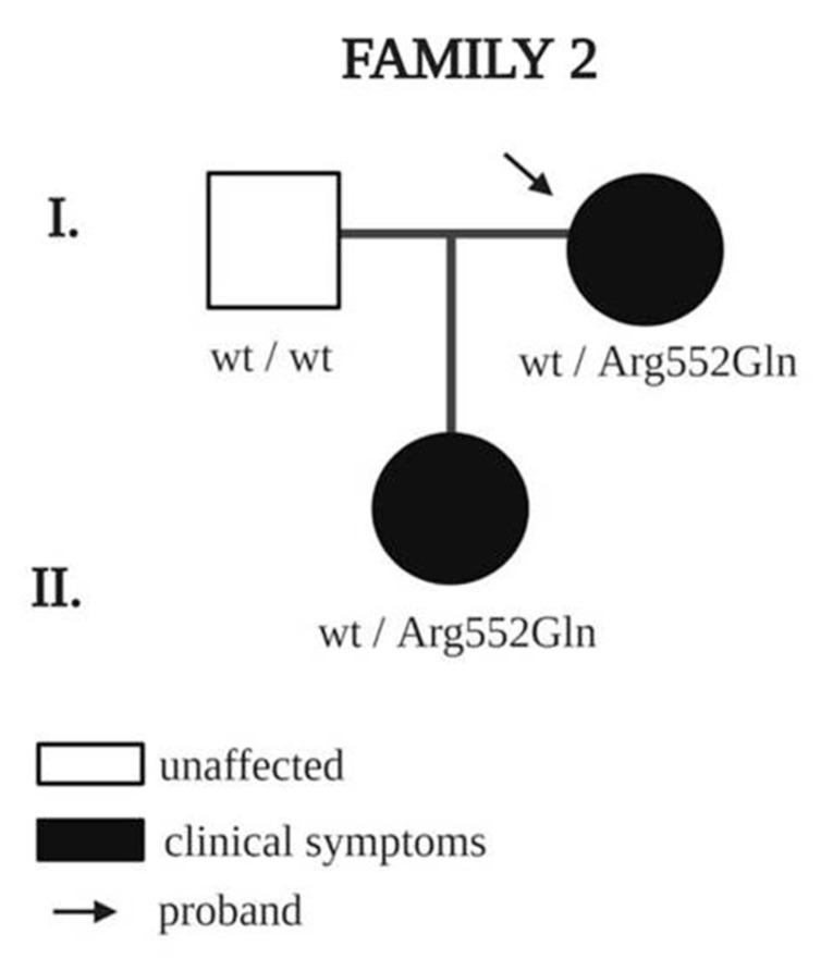
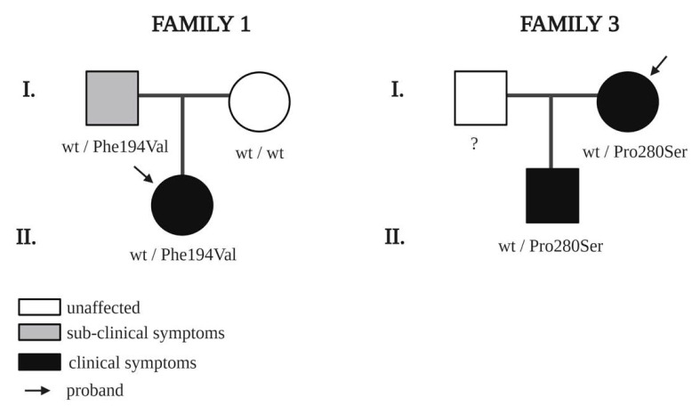
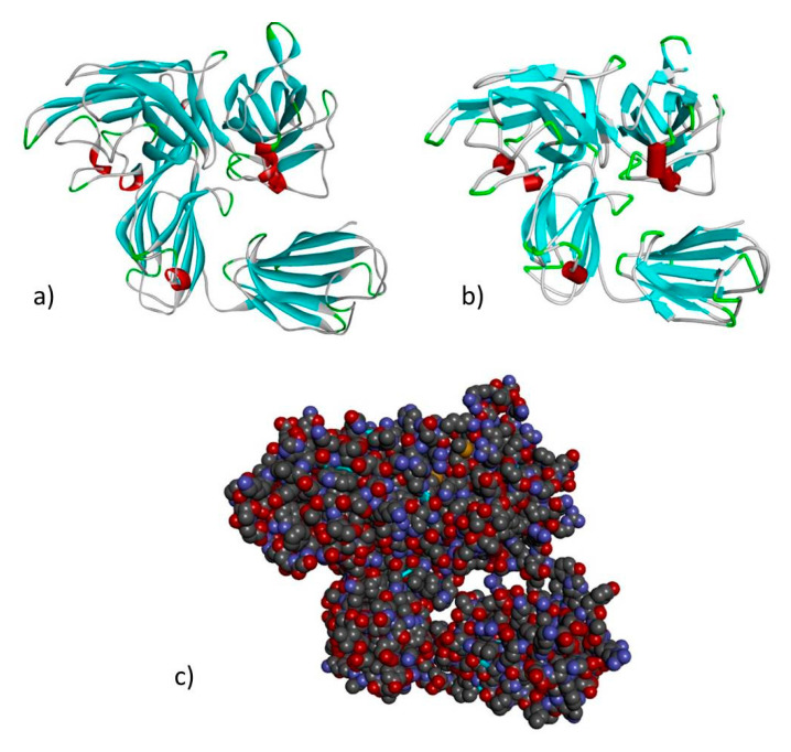
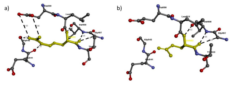
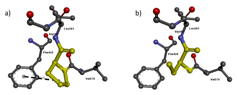
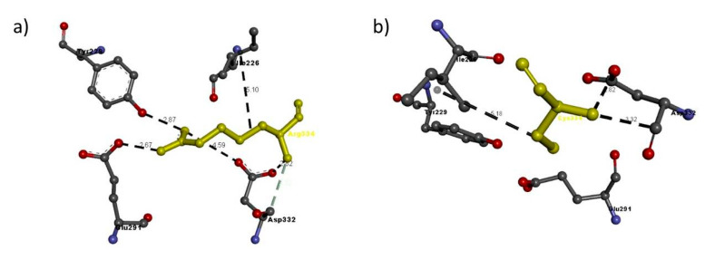
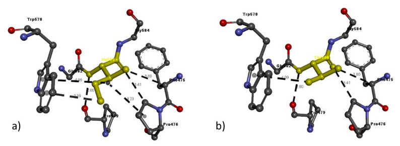
Neuropilins are transmembrane coreceptors expressed by endothelial cells and neurons. NRP1 and NRP2 bind a variety of ligands, by which they trigger cell signaling, and are important in the development of lymphatic valves and lymphatic capillaries, respectively. This study focuses on identifying rare variants in the NRP1 and NRP2 genes that could be linked to the development of lymphatic malformations in patients diagnosed with lymphedema. Two hundred and thirty-five Italian lymphedema patients, who tested negative for variants in known lymphedema genes, were screened for variants in NRP1 and NRP2. Two probands carried variants in NRP1 and four in NRP2. The variants of both genes segregated with lymphedema in familial cases. Although further functional and biochemical studies are needed to clarify their involvement with lymphedema and to associate NRP1 and NRP2 with lymphedema, we suggest that it is worthwhile also screening lymphedema patients for these two new candidate genes.

Keywords: NGS; NRP1; NRP2; genetic diagnostics; lymphedema.


Conflict of interest statement

The authors declare that they have no conflicts of interest.

Figures

Figure 1
Figure 1
Pedigree of family 2 with a rare NRP1 variant. Square indicates male, and circles indicate females; black indicates clinical symptoms of lymphedema. Arrows indicate probands, and wt indicates wildtype amino acid.
Figure 2
Figure 2
Pedigrees of families with rare NRP2 variants. Squares indicate males and circles, females; black indicates clinical symptoms of lymphedema; grey indicates subclinical symptoms confirmed by lymphoscintigraphy. Arrows indicate probands. ? means “not screened”, and wt indicates wildtype amino acid.
Figure 3
Figure 3
Lymphoscintigraphy of father of the proband with c.580T > C variants in NRP2 (family 1). Although phenotypically healthy, lymphoscintigraphy highlighted subclinical anomalies (arrows): radiotracer accumulates in multiple lymph nodes of the left (panel 4) and right arm (panel 5). After 90 min of tracer administration, there is reduced tardive visualization of the right axillary lymph nodes (right axillary packet hypogenesis) and even more reduced tardive visualization of the left axillary lymph nodes (left axillary packet hypogenesis) (arrows in panel 6).
Figure 4
Figure 4
Modeled structure of the NRP1 gene in (a) ribbon (b) schematic and (c) CPK views. Cyan regions are β sheets, white represents loops and red represents α helices.
Figure 5
Figure 5
Modeled structure of the NRP2 gene in (a) ribbon (b) schematic and (c) CPK views. Cyan regions are β sheets, white represents loops and red represents α helices.
Figure 6
Figure 6
Molecular interactions of (a) Arg552 and (b) Gln552 (in yellow) of the modeled NRP1 protein with adjacent residues.
Figure 7
Figure 7
Molecular interactions of (a) Phe194 and (b) Val194 (in yellow) of the modeled NRP2 protein with adjacent residues.
Figure 8
Figure 8
Molecular interactions of (a) Pro280 and (b) Ser280 (in yellow) of the modeled NRP2 protein with adjacent residues.
Figure 9
Figure 9
Molecular interactions of (a) Arg334 and (b) Cys334 (in yellow) of the modeled NRP2 protein with adjacent residues.
Figure 10
Figure 10
Molecular interactions of (a) Ile583 and (b) Thr583 (in yellow) of the modeled NRP2 protein with adjacent residues.

Similar articles

Cited by

References

    1. Kolodkin A.L., Levengood D.V., Rowe E.G., Tai Y.T., Giger R.J., Ginty D.D. Neuropilin is a semaphorin III receptor. Cell. 1997;90:753–762. doi: 10.1016/S0092-8674(00)80535-8. - DOI - PubMed
    1. He Z., Tessier-lavigne M. Neuropilin Is a Receptor for the Axonal Chemorepellent Semaphorin III. Cell. 1997;90:739–751. doi: 10.1016/S0092-8674(00)80534-6. - DOI - PubMed
    1. Giger R.J., Urquhart E.R., Gillespie S.K.H., Levengood D.V., Ginty D.D., Kolodkin A.L. Neuropilin-2 is a receptor for semaphorin IV: Insight into the structural basis of receptor function and specificity. Neuron. 1998;21:1079–1092. doi: 10.1016/S0896-6273(00)80625-X. - DOI - PubMed
    1. Gluzman-Poltorak Z., Cohen T., Herzog Y., Neufeld G. Neuropilin-2 and neuropilin-1 are receptors for the 165-amino acid form of vascular endothelial growth factor (VEGF) and of placenta growth factor-2, but only neuropilin-2 functions as a receptor for the 145-amino acid form of VEGF. J. Biol. Chem. 2000;275:18040–18045. doi: 10.1074/jbc.M909259199. - DOI - PubMed
    1. Raimondi C., Ruhrberg C. Neuropilin signalling in vessels, neurons and tumours. Semin. Cell Dev. Biol. 2013;24:172–178. doi: 10.1016/j.semcdb.2013.01.001. - DOI - PubMed

Publication types