Review
. 2020 Nov 17;9(11):2496.
doi: 10.3390/cells9112496.

Extracellular ATP: A Feasible Target for Cancer Therapy

Affiliations
Review

Extracellular ATP: A Feasible Target for Cancer Therapy

Valentina Vultaggio-Poma et al. Cells. .

Abstract

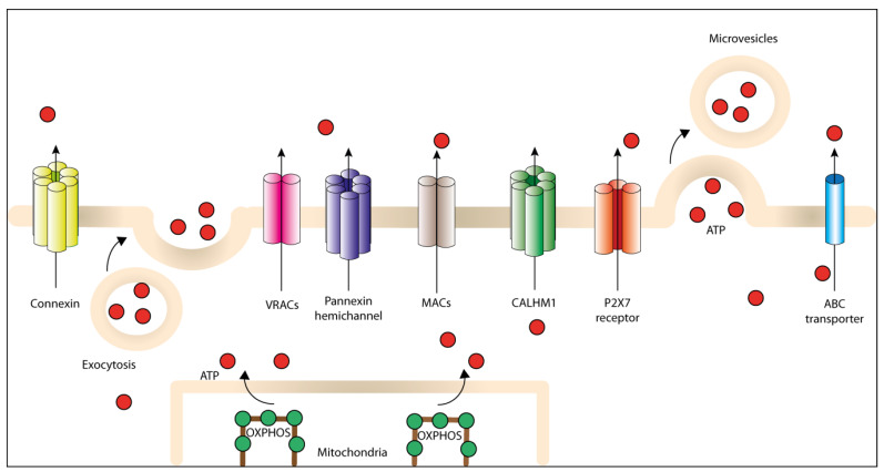
Adenosine triphosphate (ATP) is one of the main biochemical components of the tumor microenvironment (TME), where it can promote tumor progression or tumor suppression depending on its concentration and on the specific ecto-nucleotidases and receptors expressed by immune and cancer cells. ATP can be released from cells via both specific and nonspecific pathways. A non-regulated release occurs from dying and damaged cells, whereas active release involves exocytotic granules, plasma membrane-derived microvesicles, specific ATP-binding cassette (ABC) transporters and membrane channels (connexin hemichannels, pannexin 1 (PANX1), calcium homeostasis modulator 1 (CALHM1), volume-regulated anion channels (VRACs) and maxi-anion channels (MACs)). Extracellular ATP acts at P2 purinergic receptors, among which P2X7R is a key mediator of the final ATP-dependent biological effects. Over the years, P2 receptor- or ecto-nucleotidase-targeting for cancer therapy has been proposed and actively investigated, while comparatively fewer studies have explored the suitability of TME ATP as a target. In this review, we briefly summarize the available evidence suggesting that TME ATP has a central role in determining tumor fate and is, therefore, a suitable target for cancer therapy.

Keywords: cancer; extracellular ATP; purinergic signaling; tumor microenvironment.


Conflict of interest statement

Francesco Di Virgilio is a member of the Scientific Advisory Board of Biosceptre Ltd., a UK-based company involved in the development of P2X7-targeted antibodies. The other authors declare no conflicts of interest.

Figures

Figure 1
Figure 1
Different pathways for regulated ATP release into the tumor microenvironment (TME). ATP generated inside the cell can be actively released through plasma membrane-derived microvesicles, vesicular exocytosis or different non-exocytotic conductive pathways, including specific ATP-binding cassette (ABC) transporters, the P2X7R, connexin and pannexin channels, calcium homeostasis modulator 1 (CALHM1) channel, volume-regulated ion channels (VRACs) and maxi-anion channels (MACs).
Figure 2
Figure 2
Extracellular ATP shapes the TME. ATP is released into the tumor microenvironment (TME) in a non-regulated or regulated fashion. Extracellular ATP can promote immunosuppression or support antitumor immunity depending on its concentration and specific receptors expressed by immune and cancer cells. ATP is degraded by ecto-nucleotidases (CD39 and CD73) to generate ADP, AMP and adenosine (ADO), which promote immunosuppression via adenosine receptors P1 (P1Rs) (mainly A2AR and A2BR). A2AR stimulation inhibits antigen presentation by dendritic cells (DCs) and impairs cytotoxic T lymphocytes functions. Extracellular ATP acting at P2Y receptors (P2Y1R, P2Y2R or P2Y6R) and P2X receptors (mainly P2X7R) supports tumor cell survival and proliferation, but at the same time drives recruitment and activation of immune cells such as CD8+ and CD4+ T lymphocytes, Tregs, tumor-associated macrophages (TAMs) and myeloid-derived suppressor cells (MDSCs). In addition, ATP activates DCs to promote the release of pro-inflammatory cytokines, such as IL-1β and tumor necrosis factor (TNF), and potentiate tumor antigen presentation. ATP-activated DCs increase CD8+ T cell responses, thus supporting antitumor immunity.In the TME, eATP triggers via P2X7R the release of metalloproteinases (MMPs) and cathepsin from tumor cells, T lymphocytes and macrophages. MMPs degrade the extracellular matrix and support tumor cell invasion and metastatic spreading. ATP-mediated activation of P2YR, e.g., P2Y2R, drives the formation of pseudopodia and facilitates tumor cell migration across vessel endothelium.
Figure 3
Figure 3
Stimulation of cancer cell metabolism. Extracellular ATP acting at P2X7R causes an increase in the mitochondrial Ca2+ level, stimulates oxidative phosphorylation (OXPHOS) and promotes ATP generation. At the same time, P2X7R activation upregulates via PI3K-AKT expression of the plasma membrane glucose transporter GLUT1 and of several enzymes of the glycolytic cascade. This generates ATP and, at the same time, increases the production of lactate, which causes acidification of TME.
Figure 4
Figure 4
Strategies to exploit the elevated eATP concentration in the TME for antitumor therapy. A novel anti-CD137 monoclonal antibody was generated by Igawa and co-workers [171] that binds the antigen only in the presence of ATP levels close to those found in the TME (left panel). Under these conditions, and only under these conditions, this antibody binds and activates the costimulatory receptor CD137 expressed by CD8+ T cells, thus promoting T cell proliferation and cytokines release (e.g., IFN-γ), which boosts antitumor immune response (right panel). In the same way, we can hypothesize the engineering of an antibody that binds and activates suicide receptors on tumor cells, such as the FAS receptor, that is selectively activated by high ATP in the TME (right panel).

References

    1. Langen P., Hucho F. Karl Lohmann and the discovery of ATP. Angew Chem. Int. Ed. Engl. 2008;47:1824–1827. doi: 10.1002/anie.200702929. - DOI - PubMed
    1. Burnstock G. Purinergic signalling: From discovery to current developments. Exp. Physiol. 2014;99:16–34. doi: 10.1113/expphysiol.2013.071951. - DOI - PMC - PubMed
    1. Burnstock G., Campbell G., Satchell D., Smythe A. Evidence that adenosine triphosphate or a related nucleotide is the transmitter substance released by non-adrenergic inhibitory nerves in the gut. Br. J. Pharmacol. 1970;40:668–688. doi: 10.1111/j.1476-5381.1970.tb10646.x. - DOI - PMC - PubMed
    1. Burnstock G. Purinergic nerves. Pharmacol. Rev. 1972;24:509–581. - PubMed
    1. Webb T.E., Simon J., Krishek B.J., Bateson A.N., Smart T.G., King B.F., Burnstock G., Barnard E.A. Cloning and functional expression of a brain G-protein-coupled ATP receptor. FEBS Lett. 1993;324:219–225. doi: 10.1016/0014-5793(93)81397-I. - DOI - PubMed

Publication types

Substances