

Proteomic Insights into Senescence of Testicular Peritubular Cells from a Nonhuman Primate Model
- PMID: 33213088
- PMCID: PMC7698562
- DOI: 10.3390/cells9112498
Proteomic Insights into Senescence of Testicular Peritubular Cells from a Nonhuman Primate Model
Abstract
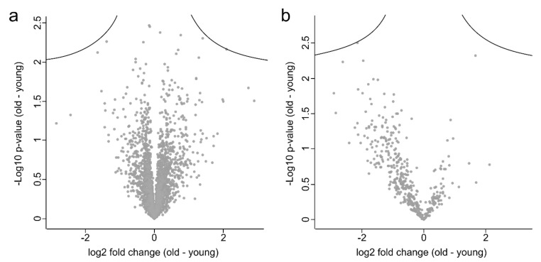
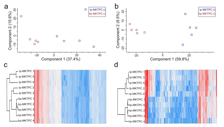
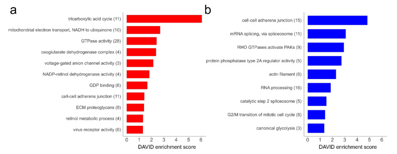
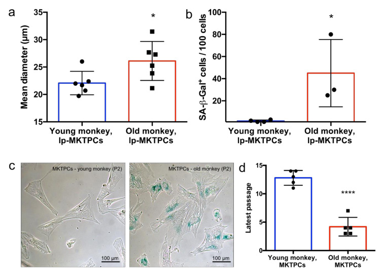
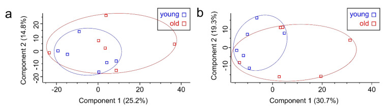
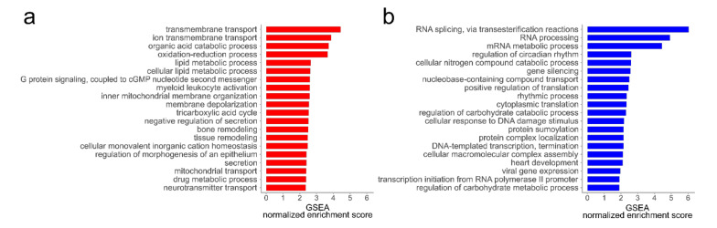
Age-related changes in the human testis may include morphological alterations, disturbed steroidogenesis, and impaired spermatogenesis. However, the specific impact of cell age remains poorly understood and difficult to assess. Testicular peritubular cells fulfill essential functions, including sperm transport, contributions to the spermatogonial stem cell niche, and paracrine interactions within the testis. To study their role in age-associated decline of testicular functions, we performed comprehensive proteome and secretome analyses of repeatedly passaged peritubular cells from Callithrix jacchus. This nonhuman primate model better reflects the human testicular biology than rodents and further gives access to young donors unavailable from humans. Among 5095 identified proteins, 583 were differentially abundant between samples with low and high passage numbers. The alterations indicate a reduced ability of senescent peritubular cells to contract and secrete proteins, as well as disturbances in nuclear factor (NF)-κB signaling and a reduced capacity to handle reactive oxygen species. Since this in vitro model may not exactly mirror all molecular aspects of in vivo aging, we investigated the proteomes and secretomes of testicular peritubular cells from young and old donors. Even though the age-related alterations at the protein level were less pronounced, we found evidence for impaired protein secretion, altered NF-κB signaling, and reduced contractility of these in vivo aged peritubular cells.
Keywords: aging; cellular model; marmoset monkey; nonhuman primate; proteome; secretome; senescence; testis.
Conflict of interest statement
The authors declare no conflict of interest.
Figures


Similar articles
-
Characterization of a non-human primate model for the study of testicular peritubular cells-comparison with human testicular peritubular cells.Mol Hum Reprod. 2018 Aug 1;24(8):401-410. doi: 10.1093/molehr/gay025. Mol Hum Reprod. 2018. PMID: 29846669
-
Age-Related Alterations in the Testicular Proteome of a Non-Human Primate.Cells. 2021 May 24;10(6):1306. doi: 10.3390/cells10061306. Cells. 2021. PMID: 34074003 Free PMC article.
-
A translational cellular model for the study of peritubular cells of the testis.Reproduction. 2020 Aug;160(2):259-268. doi: 10.1530/REP-20-0100. Reproduction. 2020. PMID: 32449695
-
Peritubular cells of the human testis: prostaglandin E2 and more.Andrology. 2020 Jul;8(4):898-902. doi: 10.1111/andr.12669. Epub 2019 Jun 24. Andrology. 2020. PMID: 31237067 Review.
-
Human testicular peritubular cells: more than meets the eye.Reproduction. 2013 Apr 29;145(5):R107-16. doi: 10.1530/REP-12-0497. Print 2013 May. Reproduction. 2013. PMID: 23431272 Review.
Cited by
-
The Molecular Signature of Human Testicular Peritubular Cells Revealed by Single-Cell Analysis.Cells. 2022 Nov 19;11(22):3685. doi: 10.3390/cells11223685. Cells. 2022. PMID: 36429113 Free PMC article.
-
Hallmarks of Testicular Aging: The Challenge of Anti-Inflammatory and Antioxidant Therapies Using Natural and/or Pharmacological Compounds to Improve the Physiopathological Status of the Aged Male Gonad.Cells. 2021 Nov 10;10(11):3114. doi: 10.3390/cells10113114. Cells. 2021. PMID: 34831334 Free PMC article. Review.
-
Systems approaches to investigate the role of NF-κB signaling in aging.Biochem J. 2022 Jan 28;479(2):161-183. doi: 10.1042/BCJ20210547. Biochem J. 2022. PMID: 35098992 Free PMC article. Review.
-
Cannabidiol-induced transcriptomic changes and cellular senescence in human Sertoli cells.Toxicol Sci. 2023 Feb 17;191(2):227-238. doi: 10.1093/toxsci/kfac131. Toxicol Sci. 2023. PMID: 36519830 Free PMC article.
-
Testicular aging, male fertility and beyond.Front Endocrinol (Lausanne). 2022 Oct 13;13:1012119. doi: 10.3389/fendo.2022.1012119. eCollection 2022. Front Endocrinol (Lausanne). 2022. PMID: 36313743 Free PMC article. Review.
References
-
- Wu F.C., Tajar A., Pye S.R., Silman A.J., Finn J.D., O’Neill T.W., Bartfai G., Casanueva F., Forti G., Giwercman A., et al. Hypothalamic-pituitary-testicular axis disruptions in older men are differentially linked to age and modifiable risk factors: The European male aging study. J. Clin. Endocrinol. Metab. 2008;93:2737–2745. doi: 10.1210/jc.2007-1972. - DOI - PubMed
Publication types
MeSH terms
LinkOut - more resources
Full Text Sources
