. 2021 Oct;17(10):3068-3081.
doi: 10.1080/15548627.2020.1852725. Epub 2020 Dec 7.

Pancreas-specific SNAP23 depletion prevents pancreatitis by attenuating pathological basolateral exocytosis and formation of trypsin-activating autolysosomes

Affiliations

Pancreas-specific SNAP23 depletion prevents pancreatitis by attenuating pathological basolateral exocytosis and formation of trypsin-activating autolysosomes

Subhankar Dolai et al. Autophagy. 2021 Oct.

Abstract

Intrapancreatic trypsin activation by dysregulated macroautophagy/autophagy and pathological exocytosis of zymogen granules (ZGs), along with activation of inhibitor of NFKB/NF-κB kinase (IKK) are necessary early cellular events in pancreatitis. How these three pancreatitis events are linked is unclear. We investigated how SNAP23 orchestrates these events leading to pancreatic acinar injury. SNAP23 depletion was by knockdown (SNAP23-KD) effected by adenovirus-shRNA (Ad-SNAP23-shRNA/mCherry) treatment of rodent and human pancreatic slices and in vivo by infusion into rat pancreatic duct. In vitro pancreatitis induction by supraphysiological cholecystokinin (CCK) or ethanol plus low-dose CCK were used to assess SNAP23-KD effects on exocytosis and autophagy. Pancreatitis stimuli resulted in SNAP23 translocation from its native location at the plasma membrane to autophagosomes, where SNAP23 would bind and regulate STX17 (syntaxin17) SNARE complex-mediated autophagosome-lysosome fusion. This SNAP23 relocation was attributed to IKBKB/IKKβ-mediated SNAP23 phosphorylation at Ser95 Ser120 in rat and Ser120 in human, which was blocked by IKBKB/IKKβ inhibitors, and confirmed by the inability of IKBKB/IKKβ phosphorylation-disabled SNAP23 mutant (Ser95A Ser120A) to bind STX17 SNARE complex. SNAP23-KD impaired the assembly of STX4-driven basolateral exocytotic SNARE complex and STX17-driven SNARE complex, causing respective reduction of basolateral exocytosis of ZGs and autolysosome formation, with consequent reduction in trypsinogen activation in both compartments. Consequently, pancreatic SNAP23-KD rats were protected from caerulein and alcoholic pancreatitis. This study revealed the roles of SNAP23 in mediating pathological basolateral exocytosis and IKBKB/IKKβ's involvement in autolysosome formation, both where trypsinogen activation would occur to cause pancreatitis. SNAP23 is a strong candidate to target for pancreatitis therapy.Abbreviations: AL: autolysosome; AP: acute pancreatitis; AV: autophagic vacuole; CCK: cholecystokinin; IKBKB/IKKβ: inhibitor of nuclear factor kappa B kinase subunit beta; SNAP23: synaptosome associated protein 23; SNARE: soluble NSF (N-ethylmaleimide-sensitive factor) attachment protein receptor; STX: syntaxin; TAP: trypsinogen activation peptide; VAMP: vesicle associated membrane protein; ZG: zymogen granule.

Keywords: Autophagy; IKKβ; SNAREs; caerulein; experimental pancreatitis; pancreatic acinar cell.


Conflict of interest statement

All authors declare no competing financial interests.

Figures

Figure 1.
Figure 1.
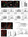
SNAP23 depletion in acini inhibits amylase secretion and perturbs pancreatitis-induced autophagy. (A,B) Rat (A) and human (B) pancreas slices were treated with Ad-SNAP23-shRNA/mCherry (SNAP23-KD/KD) vs Ad-sc(scramble)-shRNA/RFP (control/WT) for 48 h. Western blot analysis (left) and densitometry analyses (right, N = 3, normalized to loading control TUBA4A/tubulin) showed reduced SNAP23 expression (see Results). (C-F) SNAP23-KD reduced AMY/amylase secretion in rat (C,D) and human (E,F) pancreatic slices as assessed by a colorimetric assay (C,E; secreted AMY/amylase expressed as percentage of total pancreatic slice AMY/amylase, n = 8 from 3 independent experiments); and as detected by western blots (D,F; left) of AMY/amylase released into the buffer or retained in the slices, expressed as a densitometry ratio (D,F; right) of WT (100 pM for buffer and 0 pM for slices) as 100. (G,H) Western blot detection of autophagic vacuole (AV) marker protein LC3B-II and autophagy substrate protein SQSTM1/p62 (left- representative western blots; right- densitometry analyses (N = 3) by the ratio of LC3B-II or SQSTM1/p62 to loading control ACTB/actin) in human pancreatic slices exposed to pancreatitis stimuli (G: 10 nM CCK; H: 100 pM CCK plus 20 mM EtOH) vs starvation (G). (I) Increased accumulation of GFP-LC3B (by Ad-GFP-LC3B co-infection) puncta (which are AVs) in SNAP23-KD human acini (left) induced by pancreatitis stimuli. Quantification of GFP-LC3B puncta is shown on right from 30 acinar cells from 3 independent experiments. AMY/amylase secretion, densitometry and quantification values are expressed as mean±S.E.M; NS = Not significant. *, P< 0.05
Figure 2.
Figure 2.
SNAP23-KD of human acini reduces pathological basolateral exocytosis by impairing the formation of its exocytotic SNARE complexes. (A) Analysis of exocytosis. 36 h post-infected WT and SNAP23-KD pancreatic slices were co-infected with Ad-syncollin-pHluorin for another 12 h, then dispersed into acini and plated on coverslips for imaging. Acini were subjected to pancreatitis (10 nM CCK-8) stimulation. Representative images from the indicated time points are shown in left. ZG fusion events observed as pHluorin hotspots were assigned to “apical” and “basolateral” areas [15,43,44,57], and normalized to cell area and recording time (right). Data (N = 6) expressed as mean±SEM, *P< 0.05, Scale bars: 10 μm. (B,C) Analysis of STX3 (B) and STX4 (C) exocytotic SNARE complex formation in WT vs SNAP23-KD pancreatic slices after pancreatitis (10 nM CCK-8) stimulation. Degree of association are expressed as band intensity ratios of indicated co-IPed proteins that immunoprecipitated (IP) with STX3 (B) or STX4 (C) in right and expressed as mean±SEM (N = 3). Set-1 and Set-2 are 2 independent sets of experiments. Red asterisks showing reduced levels of co-IPed VAMP8 in SNAP23-KD pancreas. “Input” controls (10% or 20 μg total acini lysates) with densitometric analyses are shown in Figure S2A,B
Figure 3.
Figure 3.
SNAP23-KD in human acini attenuates pancreatitis-induced autolysosome formation by blocking autophagosome-lysosome fusion. (A) WT and SNAP23-KD pancreatic slices were kept under starvation condition (Starv; 2 h in EBSS) or treated with Baf A1 (2 h), or subjected to pancreatitis stimulus (10 nM CCK-8, 1 h) alone or in presence of salubrinol, Baf A1 or 3-MA. Level of LC3B-I conversion to LC3B-II was assessed by western (top) and quantified and expressed as densitometric ratio to loading control ACTB/actin (bottom). (B) WT and SNAP23-KD slices were cultured as in Figure 2 and co-infected with Ad-GFP-mRFP-LC3B for 12 h, then dispersed into acini that were plated and fixed immediately as Control (top panel), or exposed to starvation condition (middle panel) or stimulation with 10 nM CCK-8 (bottom panel), then fixed and imaged. Yellow puncta indicate autophagosomes, red puncta indicate autolysosomes. Scale bars: 10 µm. (C) Analyses of the two AV populations (autophagosomes: yellow bars and autolysosomes: red bars) from 42 WT and 61 KD acinar cells (at least 10 cells from each experiment) from 3 independent experiments. (D) Immunofluorescence images (enlarged) of transiently expressed GFP-LC3B with native LAMP1 (lysosome marker protein) in Control (top panel), or 10 nM CCK-8-stimulated (lower panel) WT and KD acini. Scale bars: 10 µm. Complete sequences of images demarcated with enlarged regions are shown in Figure S3. Quantification of autolysosomes (colocalized yellow puncta) from 17 acini (WT) and 22 acini (KD) from 2 independent experiments is shown at the bottom. (E,F) Defect in CTSL (E) and CTSB (F) processing and their activities during 10 nM CCK-8 pancreatitis stimulation were similar in the lysosomes from WT and SNAP23-KD pancreatic slices. I: intermediate form and SC: single-chain form. (G) Representative images showing increased appearance of TAP-positive puncta in WT acini during pancreatitis stimulation compared to SNAP23-KD acini. (H) Quantification of TAP-positive puncta from experiments related to Figure G. Control (unstimulated) acini displayed very low numbers of puncta (data not shown). (I) Detection of total cellular trypsin activity in WT vs SNAP23-KD acini. Scale bars: 10 μm. Data shown as mean±S.E.M. *, P < 0.05; NS, not significant
Figure 4.
Figure 4.
Pancreatitis stimuli promote SNAP23 association with the STX17 SNARE complex required for autolysosome formation, which is disabled by SNAP23-KD. (A-D) Assessment of autophagosome-lysosome fusion SNARE complexes (STX17-SNAP29-VAMP8 and STX7-SNAP29-YKT6) were analyzed on WT and SNAP23-KD human pancreatic slices by co-IP. Pancreatitis stimulation (10 nM CCK-8 or ethanol pancreatitis stimulation) of human pancreatic slices induced SNAP23 association with STX17-SNAP29-VAMP8 complex (A) but not with STX7-SNAP29-VAMP8 complex (B); and for both not in slices exposed to starvation condition (C,D). STX17 co-IPed less amounts of VAMP8 from SNAP23-KD slices (A) whereas SNAP23-KD had no effect on STX7-SNAP29-YKT6 complex formation (B). Association of proteins are expressed in the corresponding right panels as band intensity ratios of indicated co-IPed proteins to IPed STX17 (A,C) or STX7 (B,D). “Input” controls and their densitometric analyses are shown in Figure S4. Data shown as mean±S.E.M (N = 3). *, P < 0.05; NS, not significant
Figure 5.
Figure 5.
IKBKB/IKKβ-mediated phosphorylation of SNAP23 is required for SNAP23 to translocate to STX17-containing autophagosomes. (A) Representative images showing translocation of SNAP23 from PM to intracellular STX17-positive GFP-LC3B-puncta (autophagosomes) after pancreatitis stimulation (middle: 10 nM CCK-8; right: ethanol pancreatitis stimulation). Individual channels are shown in magnified boxed regions at the bottom. Scale bars: 10 µm. (B) Quantification of SNAP23-positive STX17-containing autophagosomes. 30 acinar cells were quantified from 3 independent experiments. (C-F) Rat (C,D) and human (E,F) pancreatic SNAP23 are a substrate of IKBKB/IKKβ-induced phosphorylation during pancreatitis stimulation. Quantifications of phosphorylation in the specified conditions are shown as densitometric ratio of phosphorylated SNAP23 (p-Ser95 in C; p-Ser120 in D-F) to total SNAP23 in the corresponding right panels. TUBA4A/tubulin is the loading control. (G) IKBKB/IKKβ inhibition prevented the translocation of SNAP23 to the autophagosomes (- CCK-8, + CCK-8, scale bar: 10 µm; + CCK-8/IKK-In, scale bar: 9 µm). (H) Quantification of GFP-LC3B puncta associated with SNAP23. (I) Quantification of GFP-LC3B puncta in the indicated conditions. 25 acinar cells were analyzed from 3 independent experiments for H and I. (J) Immunoprecipitation assessment of STX17 association with overexpressed WT vs IKBKB/IKKβ-phosphorylation-disabled SNAP23 in AR42J cells after pancreatitis stimuli. Co-IPed proteins within the complexes were analyzed as in the IP analyses in Figures 2 and Figures 4, and expressed as mean±S.E.M at the bottom. Input controls and their densitometry analyses are in Figure S5D. *, P < 0.05; NS, not significant
Figure 6.
Figure 6.
In vivo SNAP23 depletion of the rat pancreas can provide protection against acute pancreatitis. (A) Representative western blots showing pancreatic duct infusion of Ad-SNAP23-shRNA-mCherry (KD) can reduce SNAP23 expression in rat pancreas, preferentially in the pancreatic head (N = 8), less so in the pancreatic body, and leaving unaltered expression of SNAP23 in the pancreatic tail; the latter having similar level of SNAP23 as those found in all three portions of the pancreatic head, body and tail of Ad-sc(scramble)-shRNA/RFP-infused WT (control) pancreas (N = 8). Densitometry analyses (bottom, normalized to loading control TUBA4A/tubulin) showing effective SNAP23-KD in the head (H) > body (B) >tail (T), but equal in all three portions in WT control. (B,F) Representative H&E-stained images (14X magnification) of WT (top panels, n = 3) vs KD (bottom panels, n = 3) pancreatic “H”, “B” and “T” sections (left to right) after caerulein pancreatitis (B) and alcoholic pancreatitis (F), scale bar: 200 μm. (C,G) Individual (to the left of red dotted line) and combined (Total, to the right of red dotted line) histology scores of acinar injury: edema (Ed), inflammatory cell infiltration (Inf), necrosis (Nec) and vacuolization (Vac), on a scale of 0–4 from “H” (top), “B” (middle) and “T” (bottom) portions of pancreases of caerulein (C) and ethanol (G) pancreatitis-induced rats. H&E images of pancreases of controls (n = 2 each) that were injected saline or only-ethanol, and their histology injury score analyses are shown in Figure S6G-I (for saline) and Figure S6J-L (for only-ethanol). (D,H,E,I) Activities of pancreatic tissue trypsin (left; D,H), MPO (right; D,H), serum amylase (left; E,I) and lipase (right; E,I) for caerulein (D,E) and ethanol (H,I)-induced pancreatitis. Data shown as mean±S.E.M. *, P < 0.05; NS, not significant

References

    1. Habtezion A, Gukovskaya AS, Pandol SJ.. Acute pancreatitis: a multifaceted set of organelle and cellular interactions. Gastroenterology. 2019;156:1941–1950. - PMC - PubMed
    1. Gukovskaya AS, Gorelick FS, Groblewski GE, et al. Recent insights into the pathogenic mechanism of pancreatitis: role of acinar cell organelle disorders. Pancreas. 2019;48:459–470. - PMC - PubMed
    1. Williams JA, Yule DI. Stimulus-secretion coupling in pancreatic acinar cells. In: JL R, editor. Physiology of the gastrointestinal tract. Amsterdam: Elsevier; 2012. p. 1361–1398.
    1. Dolai S, Liang T, Cosen-Binker LI, Lam PL, et al.Regulation of physiologic and pathologic exocytosis in pancreatic acinar cells. The Pancreapedia: Exocrine Pancreas Knowledge Base Available from: https://wwwpancreapediaorg/reviews/regulation-of-physiologic-and-patholo...; DOI: 10.3998/panc.2012.12. - DOI
    1. Antonucci L, Fagman JB, Kim JY, et al. Basal autophagy maintains pancreatic acinar cell homeostasis and protein synthesis and prevents ER stress. Proc Natl Acad Sci U S A. 2015;112:E6166–74. - PMC - PubMed

Publication types

Grants and funding