. 2020 Nov 20;18(1):350.
doi: 10.1186/s12916-020-01812-6.

Sleep in disorders of consciousness: behavioral and polysomnographic recording

Affiliations

Sleep in disorders of consciousness: behavioral and polysomnographic recording

Isabella Mertel et al. BMC Med. .

Abstract

Background: Sleep-wakefulness cycles are an essential diagnostic criterion for disorders of consciousness (DOC), differentiating prolonged DOC from coma. Specific sleep features, like the presence of sleep spindles, are an important marker for the prognosis of recovery from DOC. Based on increasing evidence for a link between sleep and neuronal plasticity, understanding sleep in DOC might facilitate the development of novel methods for rehabilitation. Yet, well-controlled studies of sleep in DOC are lacking. Here, we aimed to quantify, on a reliable evaluation basis, the distribution of behavioral and neurophysiological sleep patterns in DOC over a 24-h period while controlling for environmental factors (by recruiting a group of conscious tetraplegic patients who resided in the same hospital).

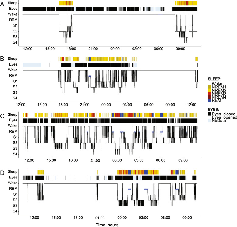
Methods: We evaluated the distribution of sleep and wakefulness by means of polysomnography (EEG, EOG, EMG) and video recordings in 32 DOC patients (16 unresponsive wakefulness syndrome [UWS], 16 minimally conscious state [MCS]), and 10 clinical control patients with severe tetraplegia. Three independent raters scored the patients' polysomnographic recordings.

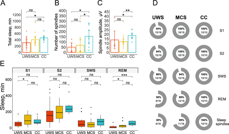
Results: All but one patient (UWS) showed behavioral and electrophysiological signs of sleep. Control and MCS patients spent significantly more time in sleep during the night than during daytime, a pattern that was not evident in UWS. DOC patients (particularly UWS) exhibited less REM sleep than control patients. Forty-four percent of UWS patients and 12% of MCS patients did not have any REM sleep, while all control patients (100%) showed signs of all sleep stages and sleep spindles. Furthermore, no sleep spindles were found in 62% of UWS patients and 21% of MCS patients. In the remaining DOC patients who had spindles, their number and amplitude were significantly lower than in controls.

Conclusions: The distribution of sleep signs in DOC over 24 h differs significantly from the normal sleep-wakefulness pattern. These abnormalities of sleep in DOC are independent of external factors such as severe immobility and hospital environment.

Keywords: EEG; Minimally conscious state; Polysomnography; Sleep; Unresponsive wakefulness; Vegetative state.


Conflict of interest statement

The authors declare that they have no competing interests.

Figures

Fig. 1
Fig. 1
Behavioral and electrophysiological sleep. UWS, unresponsive wakefulness syndrome; MCS, minimally conscious state; CC, clinical control. a The amount of behavioral sleep (time in minutes with eyes closed) during the night and at daytime, for each group. b The amount of electrophysiological sleep evaluated by polysomnographic recording during the night and at daytime, for each group. c The distribution of sleep probability (percentage of patients who slept during the respective epoch) across all 2880 epochs. Solid lines show the results of smoothing according to the LOESS algorithm with the smoothing span of 0.2 by means of the ggplot2 R package. d Scatterplots relating the probabilities (in %) that a particular epoch was a sleep epoch as scored with electrophysiological and behavioral measures (each dot represents one epoch). Note that the whole graph is “larger” for CC than for UWS and MCS, indicating a higher behavioral-electrophysiological correspondence among CC patients than DOC patients. There were epochs when all CC patients slept, and epochs when all of them where awake, but there were no such epochs in the two DOC groups. e Behavioral-electrophysiological sleep Kendall correlations calculated within each subject (dots) across epochs and averaged for each group (columns). *p < .05, **p < .01, ***p < .001; ns, not significant. Error bars are 95% confidence intervals
Fig. 2
Fig. 2
Exemplary hypnograms of four patients. a An UWS patient who remained awake at night but slept during the day. b An MCS patient with close-to-normal sleep distribution. c An UWS patient with uniformly distributed sleep over the 24-h period. d A CC patient with a pattern of well-structured sleep during the night and an afternoon nap. Notes: sleep—electrophysiological sleep; eyes—behavioral sleep
Fig. 3
Fig. 3
Sleep stage distribution. a The total amount of sleep by group. Mean ± SD 311 ± 184, 405 ± 185, and 464 ± 98 min in the UWS, MCS, and CC groups, respectively. b The number of sleep spindles by group. Mean ± SD 50.3 ± 50.6, 68.6 ± 101, and 155 ± 97.2 spindles in S2 in the UWS, MCS, and CC groups, respectively. Only patients with present spindles (defined by visual screening) were included (see Table 2). c The amplitude of sleep spindles by group. Mean ± SD 13.8 ± 12.2, 11.8 ± 4.23, and 18.7 ± 4.52 μV in the UWS, MCS, and CC groups, respectively. Only patients with present spindles (defined by visual screening) were included (see Table 2). d The percentage of patients in each group, showing signs of the respective sleep stage during the 24-h recording period. e Time spent in single sleep stages by group. UWS, unresponsive wakefulness syndrome; MCS, minimally conscious state; CC, clinical control; SWS, slow-wave sleep; REM, rapid eye movement sleep; S1, sleep stage 1; S2, sleep stage 2; *p < .05, **p < .01, ***p < .001; ns, not significant. Error bars are 95% confidence intervals

References

    1. The Multi-Society Task Force on PVS Medical aspects of the persistent vegetative state. N Engl J Med. 1994;330:1499–1508. doi: 10.1056/NEJM199405263302107. - DOI - PubMed
    1. Landsness E, Bruno M-A, Noirhomme Q, Riedner B, Gosseries O, Schnakers C, et al. Electrophysiological correlates of behavioural changes in vigilance in vegetative state and minimally conscious state. Brain. 2011;134:2222–2232. doi: 10.1093/brain/awr152. - DOI - PMC - PubMed
    1. Yang X, Song C, Yuan F, Zhao J, Jiang Y, Yang F, et al. Prognostic roles of sleep electroencephalography pattern and circadian rhythm biomarkers in the recovery of consciousness in patients with coma: a prospective cohort study. Sleep Medicine. 2020;69:204–12. - PubMed
    1. Sutter R, Barnes B, Leyva A, Kaplan PW, Geocadin RG. Electroencephalographic sleep elements and outcome in acute encephalopathic patients: a 4-year cohort study. Eur J Neurol. 2014;21:1268–1275. doi: 10.1111/ene.12436. - DOI - PubMed
    1. Schnakers C, Vanhaudenhuyse A, Giacino J, Ventura M, Boly M, Majerus S, et al. Diagnostic accuracy of the vegetative and minimally conscious state: clinical consensus versus standardized neurobehavioral assessment. BMC Neurol. 2009;9:1. doi: 10.1186/1471-2377-9-35. - DOI - PMC - PubMed

Publication types