. 2020 Nov 19;13(1):156.
doi: 10.1186/s13045-020-00991-2.

Tumor-derived exosomal miR-934 induces macrophage M2 polarization to promote liver metastasis of colorectal cancer

Affiliations

Tumor-derived exosomal miR-934 induces macrophage M2 polarization to promote liver metastasis of colorectal cancer

Senlin Zhao et al. J Hematol Oncol. .

Erratum in

Abstract

Background: Mounting evidence has demonstrated the vital importance of tumor-associated macrophages (TAMs) and exosomes in the formation of the premetastatic niche. However, the molecular mechanisms by which tumor-derived exosomal miRNAs interact with TAMs underlying premetastatic niche formation and colorectal cancer liver metastasis (CRLM) remain largely unknown.

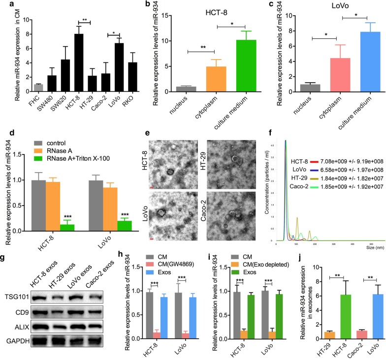
Methods: Transmission electron microscopy and differential ultracentrifugation were used to verify the existence of exosomes. In vivo and in vitro assays were used to identify roles of exosomal miR-934. RNA pull-down assay, dual-luciferase reporter assay, etc. were applied to clarify the mechanism of exosomal miR-934 regulated the crosstalk between CRC cells and M2 macrophages.

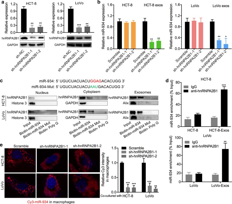
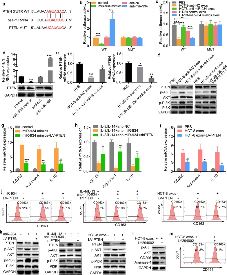
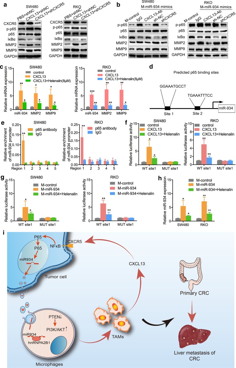
Results: In the present study, we first demonstrated the aberrant overexpression of miR-934 in colorectal cancer (CRC), especially in CRLM, and its correlation with the poor prognosis of CRC patients. Then, we verified that CRC cell-derived exosomal miR-934 induced M2 macrophage polarization by downregulating PTEN expression and activating the PI3K/AKT signaling pathway. Moreover, we revealed that hnRNPA2B1 mediated miR-934 packaging into exosomes of CRC cells and then transferred exosomal miR-934 into macrophages. Interestingly, polarized M2 macrophages could induce premetastatic niche formation and promote CRLM by secreting CXCL13, which activated a CXCL13/CXCR5/NFκB/p65/miR-934 positive feedback loop in CRC cells.

Conclusions: These findings indicate that tumor-derived exosomal miR-934 can promote CRLM by regulating the crosstalk between CRC cells and TAMs. These findings reveal a tumor and TAM interaction in the metastatic microenvironment mediated by tumor-derived exosomes that affects CRLM. The present study also provides a theoretical basis for secondary liver cancer.

Keywords: Colorectal cancer liver metastasis; Exosome; M2 macrophage polarization; Premetastatic niche; miR-934.


Conflict of interest statement

The authors declare that they have no conflicts of interest.

Figures

Fig. 1
Fig. 1
Aberrant expression of miR-934 significantly correlates with liver metastasis and poor prognosis of CRC patients. a Volcano plot comparing the miRNA-Seq data between stage I and stage IV CRC patients from the TCGA CC dataset. Each dot represents a microRNA. Dots in red represent microRNAs that are significantly upregulated in stage IV CRC patients (p < 0.05 based on DESeq). b miR-934 expression in stage I and stage IV CRC patients. Each dot represents a sample. c, d Expression levels of miR-934 in tissues and serum of two groups of CRC patients classified on the basis of the presence (n = 50) or absence (n = 60) of liver metastasis. e Representative in situ hybridization assay for the detection of miR-934 in normal mucosa (score = 0), CRC tissue without liver metastasis (score = 1), and CRC tissue with liver metastasis (score = 3). f, g Kaplan–Meier survival analysis with the log-rank test was used to determine the association of miR-934 with OS (e) and DFS (f) in 308 CRC patients (*p < 0.05; **p < 0.01; ***p < 0.001)
Fig. 2
Fig. 2
miR-934 is encapsulated within CRC cell-derived exosomes. a qPCR analysis of the expression levels of miR-934 in the culture medium (CM) of seven different CRC cell lines and one normal colon cell line. b, c qPCR analysis of the expression levels of miR-934 in the nucleus, cytoplasm, and culture medium (CM) of the HCT-8 and LoVo cell lines. These cell lines showed relatively higher miR-934 expression than other cell lines. d qPCR analysis of the expression levels of miR-934 in the HCT-8 and LoVo cell lines treated with control medium or RNase A (2 mg/mL) alone or in combination with Triton X-100 (0.1%), for 0.5 h. e, f Phenotype analysis of exosomes derived from HCT-8, HT29, LoVo, and Caco-2 cells using electron microscopy (e) and Nano Sight nanoparticle tracking analysis (f). g Western blot analysis was performed to detect typical exosomal biomarkers (TSG101, CD9, and ALIX) in exosomes derived from the above four CRC cell lines. h, i qPCR analysis of miR-934 expression in the CM of HCT-8/LoVo cells depleted of exosomes by GW4869 (an inhibitor of exosome secretion) (h) or by ultracentrifugation (i). j qPCR analysis of the expression levels of miR-934 in HT-29/HCT-8/LoVo/Caco-2-derived exosomes (*p < 0.05; **p < 0.01; ***p < 0.001)
Fig. 3
Fig. 3
CRC cell-derived exosomal miR-934 induces M2 polarization of macrophages. a ClueGO analysis of the 191 genes showing the highest correlation with miR-934 using Cytoscape software. Enriched pathways are shown as nodes interconnected based on the κ score. b Correlation between miR-934 and specific gene signatures of different immune cells. The node size represents the association p value between the neighbor gene and miR-934. c IHC staining of TAMs (for the M2 macrophage marker CD163) in primary human CRC tissues and liver-metastatic tissues, n50. The red arrows indicate TAMs; the black arrows indicate tumor cells. Scale bar, 200 μm. The correlation between TAM infiltration and miR-934 expression is also shown. d Representative image of macrophages derived from THP-1 cells treated with phorbol 12-myristate 13-acetate (PMA) for 24 h. qPCR analysis of the expression of the macrophage marker CD68 was also performed. e Representative immunofluorescence image showing the internalization of DiO-labeled HT-29/HCT-8/Caco-2/LoVo-derived exosomes (green) by PMA-treated THP-1 cells. f qPCR analysis of the expression of typical M2 markers (CD206, arginase-1, and IL10) and M1 markers (iNOS and IL-1β) in PMA-pretreated THP-1 cells treated with HT-29/HCT-8/Caco-2/LoVo-derived exosomes or PBS (control) g Flow cytometry was performed to analyze the effect of CRC cell-derived exosomes on the expression of the typical M2 marker CD163. qPCR (h) and flow cytometry (i) were used to determine the effect of exogenous miR-934 on the expression of typical M2 markers in PMA-treated THP-1 cells. qPCR (j) and flow cytometry (k) were used to determine the effect of exosomes derived from HCT-8 and HT-29 cells transfected with anti-miR-934 on the expression of CD206, arginase-1, IL10, and CD163 (*p < 0.05; **p < 0.01; ***p < 0.001)
Fig. 4
Fig. 4
hnRNPA2B1 plays a role in the transfer of CRC cell-derived exosomal miR-934 to macrophages a qPCR and western blot analysis of hnRNPA2B1 expression in HCT-8 and LoVo cells after hnRNPA2B1 knockdown. b qPCR analysis of miR-934 expression in cell lysates and exosomes derived from hnRNPA2B1-knockdown HCT-8 and LoVo cells. c Western blot analysis was performed to determine the association between biotinylated wild-type or mutant miR-934 and hnRNPA2B1 expression in samples derived by miRNA pull-downs performed with nuclear, cytoplasmic, or exosomal extracts from the HCT-8 and LoVo cell lines. Biotinylated poly(G) served as the negative control. d RIP assays with anti-hnRNPA2B1 antibody were performed on HCT-8/LoVo-CM and HCT-8/LoVo-derived exosomes. IgG served as the negative control. qPCR was then used to analyze the expression levels of miR-934 in the immunoprecipitated samples, and the expression levels of miR-934 were expressed as percentages with respect to the input sample (% input) in HCT-8 and LoVo cell lines. e HCT-8 cells and LoVo cells cotransfected with 100 nM Cy3-labeled miR-934 and sh-hnRNPA2B1 or negative control and then cocultured with PMA-pretreated THP-1 cells for 12 h. An immunofluorescence assay was performed to analyze the effects of hnRNPA2B1 knockdown on the transfer of exosomal (Cy3-labeled) miR-934 from HCT-8 and LoVo cells to macrophages (*p < 0.05; **p < 0.01; ***p < 0.001)
Fig. 5
Fig. 5
Exosomal miR-934 induces M2 macrophage polarization via downregulation of PTEN expression and activation of the PI3K/AKT signaling pathway. a Sequences of the predicted binding sites between miR-934 and the 3′-UTR of the wild-type (WT)/mutant (MUT) PTEN gene. b A luciferase reporter gene activity assay was performed to determine the effects of miR-934 mimics or anti-miR-934 on the luciferase activity of the 3′-UTR of the WT/MUT PTEN gene. c A luciferase reporter gene activity assay was performed to determine the effect of exosomal miR-934 from HCT-8 and HT-29 cells transfected with miR-934 mimics or anti-miR-934 on the luciferase activity of the 3′-UTR of the WT/MUT PTEN gene in THP-1 cells. d The effects of exogenous miR-934 mimics and anti-miR-934 on the expression of PTEN in PMA-treated THP-1 cells were tested using qPCR and western blotting. e, f The effects of CRC cell-derived exosomal miR-934 on PTEN expression and the expression of PTEN, AKT, p-AKT, PI3k, and p-PI3K in THP-1 cells pretreated with PMA and the aforementioned exosomes were determined using qPCR (e) and WB (f). g, j PMA-treated THP-1 cells were transfected with miR-934 mimics, IL3/IL4–anti-miR-934, or HCT-8-derived exosomes and further cocultured with LV-PTEN or shPTEN. The combined effects of exogenous miR-934/LV-PTEN, anti-miR-934/shPTEN, and exosomes/LV-PTEN on the expression of typical M2 markers (CD206, arginase-1, and IL-10) were determined using qPCR (gi) and flow cytometry (j). k Western blot analysis was performed to detect the expression of PI3K/AKT pathway components in PMA-pretreated THP-1 cells transfected with miR-934 mimics, IL3/IL4–anti-miR-934, or HCT-8-derived exosomes. lm Western blotting (l) and flow cytometry (m) were performed to determine the expression of AKT, p-AKT, and M2 markers (CD206, arginase-1, and CD163) in PMA-treated THP-1 cells cocultured with HCT-8 cell-derived exosomes and an inhibitor of PI3K (LY294002) (*p < 0.05; **p < 0.01; ***p < 0.001; scale bar, 200 μm)
Fig. 6
Fig. 6
CRC cell-derived exosomal miR-934 induces M2 polarization of macrophages to promote invasion and liver metastasis of CRC cells in vitro and in vivo. a, b The CRC cell lines SW480 and RKO were cocultured with CM from PMA-treated THP-1 cells for 12 h (THP-1 cells were pretreated with exogenous miR-934 mimics). The effects of exogenous miR-934 on the migratory and invasive abilities of SW480 (a) and RKO (b) cells were tested using transwell assays. HCT-8 and HT-29 cells were transfected with anti-miR-934 or miR-934 mimics or their controls; exosomes were isolated from all groups. THP-1 cells prestimulated with PMA were treated with these exosomes at a concentration of 25 μg/mL or PBS; then, the CM were obtained and incubated with SW480 or RKO cells for 12 h. Migration and invasion in vitro (c, d) and liver metastasis in vivo (e–g) were evaluated using transwell assays and live imaging combined with HE staining, respectively (*p < 0.05; **p < 0.01; ***p < 0.001)
Fig. 7
Fig. 7
M2 macrophage polarization by exosomal miR-934 promotes invasion and liver metastasis of CRC cells by activating the CXCL13/CXCR5 axis. a A human chemokine screening test kit was applied to analyze the levels of chemokines involved in M2 macrophage polarization in PMA-pretreated THP-1 cells treated with HCT-8 cell-derived exosomes. b The effect of exogenous miR-934 on the expression of CXCL13 in PMA-treated THP-1 cells was determined using ELISA. c ELISAs were used to determine the expression of CXCL13 in PMA-treated THP-1 cells cocultured with exosomes derived from HCT-8 and HT-29 cells, which were transfected with miR-934 mimics or anti-miR-934. d Representative IHC staining of CXCR5 in CRC tissues and paired normal tissues. ei PMA-treated THP-1 cells were cotransfected with miR-934 mimics and anti-CXCL13 antibody, and then the CM were obtained and cocultured with SW480 or RKO cells for 12 h. SW480/RKO cells were transfected with shCXCR5 and negative control vectors; we obtained CM from PMA-treated THP-1 cells cotransfected with miR-934 mimics and cocultured CXCR5 knockdown or control SW480 or RKO cells with the CM for 12 h. Migration and invasion in vitro (e, f) and liver metastasis in vivo (gi) were evaluated using a transwell assay and live imaging combined with HE staining, respectively (*p < 0.05; **p < 0.01; ***p < 0.001)
Fig. 8
Fig. 8
CXCL13/CXCR5/NFκB/p65/miR-934 positive feedback loop mediates the interaction between M2 macrophages and CRC cells during CRLM. a Western blot assays were performed to evaluate the expression levels of total p65, phosphorylated p65, IκBα, MMP2, and MMP9 in SW480 and RKO cells pretransfected with exogenous CXCL13 or shCXCR5. b PMA-treated THP-1 cells were transfected with miR-934 mimics and treated with anti-CXCL13 antibody or transfected with shCXCR5. The CM of these THP-1 cells was added to SW480 and RKO cells, and the expression of p65, phosphorylated p65, IκBα, MMP2, and MMP9 was determined again using western blot assay. c SW480 and RKO cells were transfected with exogenous CXCL13 and cocultured with or without an inhibitor of NFκB/p65 signaling (helenalin), and the expression of miR-934, MMP2, and MMP9 was examined using qPCR. d Schematic diagram representing the two specific binding sites between p65 and the promoter of miR-934. E. A ChIP assay was used to evaluate five potential binding regions between p65 and the miR-934 promoter in SW480 and RKO cells via LASAGNA-Search 2.0 and the human genomic databases of the National Center for Biotechnology Information (NCBI). f Mutants with truncated binding sites and their control vectors were cloned into pGL3-luciferase reporter plasmids and then transfected into SW480 and RKO cells. g A luciferase reporter gene activity assay was used to analyze the effects of helenalin (a p65 inhibitor) as well as the CM of miR-934-overexpressing THP-1 cells (PMA pretreatment) on the luciferase activity of the WT or MUT p65 binding site 1 of miR-934 promoter constructs in SW480 and RKO cells. h qPCR assays were performed to analyze the effects of helenalin on the RNA expression levels of miR-934 in SW480 and RKO cells cocultured with the culture medium of PMA-treated THP-1 cells treated with miR-934. i Graphical illustration of the interaction between TAMs and CRC cells (*p < 0.05; **p < 0.01; ***p < 0.001)

References

    1. Wolf AMD, Fontham ETH, Church TR, Flowers CR, Guerra CE, LaMonte SJ, et al. Colorectal cancer screening for average-risk adults: 2018 guideline update from the American Cancer Society. CA Cancer J Clin. 2018;68:250–281. doi: 10.3322/caac.21457. - DOI - PubMed
    1. Bray F, Ferlay J, Soerjomataram I, Siegel RL, Torre LA, Jemal A. Global cancer statistics 2018: GLOBOCAN estimates of incidence and mortality worldwide for 36 cancers in 185 countries. CA Cancer J Clin. 2018;68:394–424. doi: 10.3322/caac.21492. - DOI - PubMed
    1. Beckers RCJ, Lambregts DMJ, Lahaye MJ, Rao SX, Kleinen K, Grootscholten C, et al. Advanced imaging to predict response to chemotherapy in colorectal liver metastases—a systematic review. HPB (Oxford) 2018;20:120–127. doi: 10.1016/j.hpb.2017.10.013. - DOI - PubMed
    1. Christofori G. New signals from the invasive front. Nature. 2006;441:444–450. doi: 10.1038/nature04872. - DOI - PubMed
    1. Liu Y, Cao X. Characteristics and significance of the pre-metastatic niche. Cancer Cell. 2016;30:668–681. doi: 10.1016/j.ccell.2016.09.011. - DOI - PubMed

Publication types