. 2021 Feb;70(2):342-356.
doi: 10.1136/gutjnl-2020-322540. Epub 2020 Nov 19.

Neutrophils interact with cholangiocytes to cause cholestatic changes in alcoholic hepatitis

Affiliations

Neutrophils interact with cholangiocytes to cause cholestatic changes in alcoholic hepatitis

Masahiro Takeuchi et al. Gut. 2021 Feb.

Abstract

Background & objectives: Alcoholic hepatitis (AH) is a common but life-threatening disease with limited treatment options. It is thought to result from hepatocellular damage, but the presence of cholestasis worsens prognosis, so we examined whether bile ducts participate in the pathogenesis of this disease.

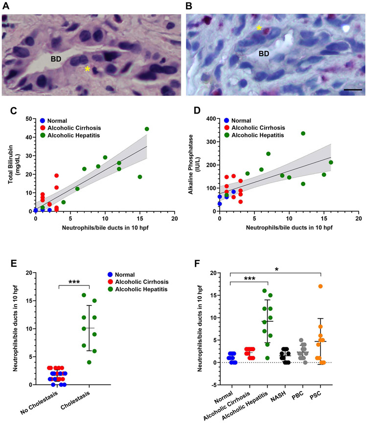
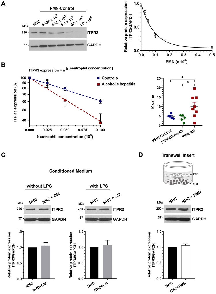
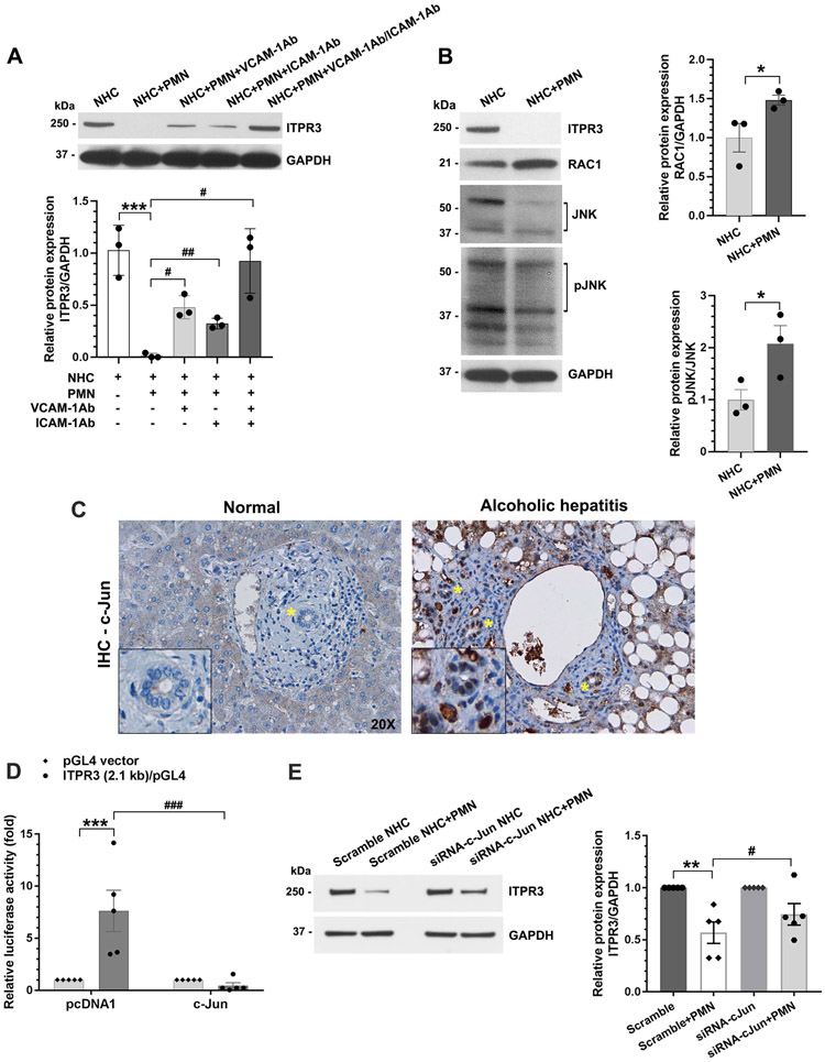
Design: Cholangiocytes derived from human bile ducts were co-cultured with neutrophils from patients with AH or controls. Loss of type 3 inositol 1,4,5-trisphosphate receptor (ITPR3), an apical intracellular calcium channel necessary for cholangiocyte secretion, was used to reflect cholestatic changes. Neutrophils in contact with bile ducts were quantified in liver biopsies from patients with AH and controls and correlated with clinical and pathological findings.

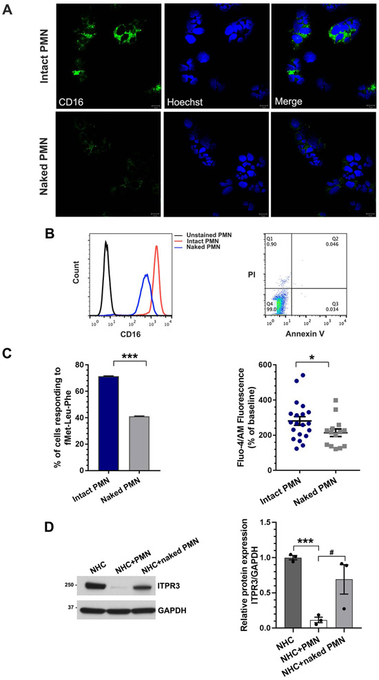
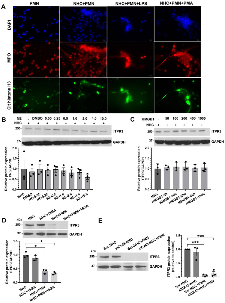
Results: Liver biopsies from patients with AH revealed neutrophils in contact with bile ducts, which correlated with biochemical and histological parameters of cholestasis. Cholangiocytes co-cultured with neutrophils lost ITPR3, and neutrophils from patients with AH were more potent than control neutrophils. Biochemical and histological findings were recapitulated in an AH animal model. Loss of ITPR3 was attenuated by neutrophils in which surface membrane proteins were removed. RNA-seq analysis implicated integrin β1 (ITGB1) in neutrophil-cholangiocyte interactions and interference with ITGB1 on cholangiocytes blocked the ability of neutrophils to reduce cholangiocyte ITPR3 expression. Cell adhesion molecules on neutrophils interacted with ITGB1 to trigger RAC1-induced JNK activation, causing a c-Jun-mediated decrease in ITPR3 in cholangiocytes.

Conclusions: Neutrophils bind to ITGB1 on cholangiocytes to contribute to cholestasis in AH. This previously unrecognised role for cholangiocytes in this disease alters our understanding of its pathogenesis and identifies new therapeutic targets.

Keywords: alcoholic liver disease; bilirubin; chemokines; cholestasis.


Conflict of interest statement

Competing interests: None declared.

Figures

Figure 1.
Figure 1.. Neutrophils are in contact with bile ducts in alcoholic hepatitis and this correlates with disease severity.
(A) Representative micrograph of a liver biopsy specimen from a patient with alcoholic hepatitis stained with H&E shows a neutrophil (asterisk) adjacent to a bile duct (BD). Scale bar: 20 μm. (B) Representative image of a chloroacetate esterase (CAE)-stained neutrophil (asterisk) adjacent to a bile duct in a liver biopsy from a patient with alcoholic hepatitis. Scale bar: 20 μm. (C) Degree of hyperbilirubinemia correlates with the number of neutrophils adjacent to bile ducts. There is a positive correlation between serum total bilirubin levels and the frequency of neutrophil-bile duct contacts (R2=0.753, p<0.0001). (D) The cholestatic marker serum alkaline phosphatase correlates with the numbers of neutrophils adjacent to bile ducts (R2=0.4477, p<0.001). Data were analysed using the Pearson correlation coefficient. (E) Histological evidence for cholestasis in the hepatic lobule (including bile pigments in hepatocytes and canalicular bile plugs) was only observed when neutrophils were adjacent to bile ducts. Data reflect blinded observations from liver biopsies of patients with alcoholic hepatitis (n=10), abstinent patients with alcoholic cirrhosis (n=10), and histologically normal control subjects (n=10). Data represent mean ± SEM, ***p<0.0001 compared with non-cholestatic biopsies. (F) Analysis of the number of neutrophils adjacent to bile ducts among patients with a spectrum of liver diseases. Neutrophils adjacent to bile ducts were rarely observed in alcoholic cirrhosis (n=10), non-alcoholic steatohepatitis (NASH; n=10), and primary biliary cholangitis (PBC; n=10) but were common in alcoholic hepatitis (n=10) and primary sclerosing cholangitis (PSC; n=10). Data represent mean ± SEM, *p<0.05, ***p<0.001 compared with histologically normal liver biopsies.
Figure 2.
Figure 2.. Neutrophils inhibit ITPR3 expression when they are co-cultured with cholangiocytes.
(A) Human neutrophils decrease ITPR3 expression in the normal human cholangiocyte (NHC) cell line in a concentration-dependent fashion. Cells were co-cultured with freshly isolated human neutrophils at given concentrations for 18 hours. Following co-culture, cells were harvested and assessed for ITPR3 expression by immunoblotting. Representative blot of ITPR3 expression in the cells in co-culture with human neutrophils from healthy controls at given neutrophil concentrations (left panel) and quantification (n=5, right panel). The relationship between neutrophil concentration (x-axis) and ITPR3 expression in the NHC cell line (y-axis) is closely described by a mono-exponential decay curve (correlation coefficient R2=0.991 in this example and ranged from 0.988 to 0.992). (B) Neutrophils from alcoholic hepatitis patients are more potent than neutrophils from normal controls or from abstinent patients with alcoholic cirrhosis in inhibiting ITPR3 expression in the NHC cell line. Logarithmic scale of the neutrophil concentration versus ITPR3 expression in NHCs illustrates a steeper slope for neutrophils from alcoholic hepatitis patients, reflecting more potent inhibition (left panel). The individual value for the rate constant k for each patient from the three groups is shown (n=5–8, right panel), *p<0.05. (C) Conditioned medium (CM) from human neutrophils does not decrease ITPR3 expression in the NHC cell line. Neutrophils were stimulated either with or without lipopolysaccharides (LPS) (0.2 μg/mL) for 8 hours prior to obtaining CM that was used for the cells. After 18-24 hours of co-culture, cells were harvested and assessed for ITPR3 expression by immunoblotting. Representative blot (top panel), and quantitative analysis (bottom panel) reflect no effect of conditioned medium on ITPR3 expression in the NHC cell line (n=6). (D) Neutrophils do not inhibit ITPR3 expression in the NHC cell line when they are separated by a semipermeable membrane. Neutrophils were placed in the upper compartment of 3 μM pore Transwell system and co-cultured with NHCs cultured in the lower compartment. After 18-24 hours of co-culture, NHCs were harvested and assessed for ITPR3 expression by immunoblotting. Representative blot (top panel) and quantitative analysis (bottom panel) reflect no significant change in NHC ITPR3 expression in the Transwell co-culture system (n= 4).
Figure 3.
Figure 3.. Direct contact with neutrophils is required to decrease ITPR3 expression in cholangiocytes.
Generation of ‘naked’ neutrophils. (A) Representative immunofluorescence images of untreated neutrophils (intact polymorphonuclear neutrophil (PMN)) or phosphatidylinositol-specific phospholipase C (PI-PLC)-treated neutrophils (naked PMN) stained for the neutrophil surface marker CD16 (green) and the nuclear marker Hoechst (blue). Scale bar: 10 μm. Naked neutrophils demonstrate a marked reduction in surface expression of CD16 relative to intact neutrophils. (B) Flow cytometry analysis of intact neutrophils or naked neutrophils stained with CD16 (left panel). Surface expression of CD16 was markedly reduced in naked neutrophils compared with intact neutrophils. Flow cytometry analysis of intact and naked neutrophils stained with propidium iodide (PI) and annexin V-FITC indicates that nearly all naked neutrophils remain viable (rightpanel). (C) Transmembrane signalling is impaired in naked neutrophils. Cells were stimulated with fMet-Leu-Phe (fMLP), which activates the G protein-coupled FPR1 receptor in the plasma membrane. Both the percentage of neutrophils that responded to fMLP (left panel), and the amplitude of the fMLP-induced Ca2+ signal in responding cells (right panel) were significantly reduced. Data represent mean ± SEM (n=28 intact neutrophils and n=34 naked neutrophils from three independent experiments); *p<0.05, ***p<0.0001, relative to intact neutrophils. (D) Naked neutrophils lose their ability to inhibit ITPR3 expression in the NHC cell line. Representative immunoblot (left panel) and quantitative blot analysis (right panel) of ITPR3 expression in cells co-cultured with control or naked neutrophils for 18 hours. Data represent mean ± SEM (n=4); ***p<0.0001 and #p<0.05.
Figure 4.
Figure 4.. Neutrophil extracellular trap (NET) formation and gap junction communication do not participate in the downregulation of ITPR3 in cholangiocytes.
(A) Representative confocal images of neutrophils alone and co-cultured with the normal human cholangiocyte (NHC) cell line. Neutrophils were stained with the NET markers myeloperoxidase (MPO, red) and citrullinated histone H3 (green). Nuclei were stained with DAPI (blue). Lipopolysaccharides (LPS) and phorbol 12-myristate 13-acetate (PMA) were used as positive controls to induce NET formation. Scale bar: 10 μm. (B) The NET constituent neutrophil elastase (NE) does not inhibit ITPR3 expression in the NHC cell line. Cells were co-incubated with varying concentrations of NE for 18 hours and then assessed for ITPR3 protein expression. Representative immunoblot (top panel) and quantitative blot analysis (bottom panel) of ITPR3 expression in NHCs after treatment with NE. Data represent mean ± SEM (n=3). (C) High mobility group box-1 (HMGB1), a mediator released during NET formation, does not inhibit ITPR3 expression in the NHC cell line. Representative immunoblot (top panel) and quantitative blot analysis (bottom panel) of ITPR3 expression in NHCs after treatment with HMGB1 for 18 hours. Data represent mean ± SEM (n=3). (D) Treatment of the NHC cell line with the gap junction inhibitor, 18α-glycyrrhetinic acid (18α-GA), does not prevent the neutrophil-induced decrease in ITPR3 expression. (Top) Representative immunoblot and (bottom) quantitative blot analysis of ITPR3 in NHCs treated either with vehicle or 18α-GA, either alone or in co-culture for 18 hours with neutrophils. Data represent mean ± SEM (n=3); *p<0.05. (E) Knockdown of CX43 in the NHC cell line does not prevent the neutrophil-induced reduction in ITPR3 expression. Representative immunoblot (left) and (right) quantitative blot analysis of ITPR3 expression in cells after CX43 knockdown and co-cultured with neutrophils. GAPDH is used as an internal loading control. Data represent mean ± SEM (n=3); ***p<0.0001.
Figure 5.
Figure 5.. RNA-seq analysis implicates integrin β1 in neutrophil-cholangiocyte interactions and integrin β1 is abundantly expressed in the normal human cholangiocyte (NHC) cell line.
(A) Analysis of RNA-seq transcriptome data with ingenuity pathways analysis (IPA) software identifies the six signalling pathways that are significantly altered in the NHC cell line when co-cultured with neutrophils. Bars represent −log(p value) of significance level for each pathway; orange indicates upregulated and blue indicates downregulated pathways. (B) Volcano plot of RNA-seq analysis of NHC proteins that are affected by exposure of the cells to neutrophils. RNA was extracted from the NHC cell line alone (n=3) and co-cultured with neutrophils (n=4), and both groups were analysed by Affymetrix High-Throughput Transcriptomics Array. A total of 5818 mRNA genes were differentially expressed (grey circles), including plasma membrane proteins (orange circles) and integrin β1 (ITGB1) (blue circle). (C) RT-PCR demonstrates that ITGB1 is more heavily expressed than ITGB3 or ITGA5 in the NHC cell line (n=4). mRNA expression levels were normalised to ACTB. (D) Representative immunofluorescence images of NHCs stained for ITGB1 (green). Hoechst 33342 is used to stain the nuclei (blue). Scale bar: 10 μm.
Figure 6.
Figure 6.. Neutrophils inhibit ITPR3 expression in cholangiocytes via integrin β1 (ITGB1) signalling.
(A) ITGB1 expression is increased in the normal human cholangiocyte (NHC) cell line when it is co-cultured with neutrophils. Representative immunoblot (left panel) and quantitative blot analysis (right panel) of ITGB1 expression in cells in the absence or presence of neutrophils. GAPDH is used as an internal loading control. Data represent mean ± SEM (n=5); **p<0.01 relative to NHCs alone. (B) Representative immunohistochemical staining of bile ducts for ITGB1 in liver biopsy specimens from patients with alcoholic hepatitis and normal controls (left panel). ITGB1-positive cells are brown. Scale bars: 20 μm. Quantitative analysis of ITGB1 staining (right panel) demonstrates that ITGB1 labelling is significantly increased in bile ducts of patients with alcoholic hepatitis (n=8) relative to what is seen in normal controls (n=9). Values are mean ± SEM, ***p<0.0001. (C) An anti-ITGB1 neutralising antibody (ITGB1Ab) mitigates the inhibitory effect of neutrophils on ITPR3 expression in the NHC cell line. Representative immunoblot (top panel) and quantitative blot analysis (bottom panel) of ITPR3 expression in NHCs after blocking of ITGB1 and co-cultured for 18 hours with neutrophils. GAPDH is used as an internal loading control. Data represent mean ± SEM (n=3); **p<0.01 relative to NHCs alone, #p<0.05 relative to NHC + polymorphonuclear neutrophil (PMN). (D) Knockdown of ITGB1 in the NHC cell line by small interfering RNA (siRNA) protects against neutrophil-induced down-regulation of ITPR3 expression. Cells were transfected with human ITGB1-specific siRNA for 24 hours and then co-cultured with neutrophils for an additional 18 hours. Representative immunoblot (top panel) and quantitative blot analysis (bottom panel) of ITPR3 expression in NHCs after treatment with scrambled or ITGB1 siRNA and co-culture with neutrophils. GAPDH is used as an internal loading control. Data represent mean ± SEM (n=4); *p<0.05 relative to NHCs treated with scrambled siRNA and #p<0.05 relative to cells treated with scrambled siRNA+PMN.
Figure 7.
Figure 7.. Interaction of vascular cell adhesion molecule-1 (VCAM-1)/intercellular adhesion molecule-1 (ICAM-1) on neutrophils with integrin β1 (ITGB1) on cholangiocytes activates a RAC1/JNK/c-Jun cascade to suppress ITPR3 expression.
(A) An anti-VCAM-1 antibody (VCAM-1Ab) and anti-ICAM-1 antibody (ICAM-1Ab) each attenuate the inhibitory effect of neutrophils on ITPR3 expression in the normal human cholangiocyte (NHC) cell line. Representative immunoblot (top panel) and quantitative blot analysis (bottom panel) of ITPR3 expression in NHCs after blocking of VCAM-1 and/or ICAM-1 on surface of neutrophils and co-cultured with neutrophils. GAPDH is used as an internal loading control. Data represent mean ± SEM (n=3); ***p<0.0001 relative to the NHC cell line alone, #p<0.05, ##p<0.001 relative to NHC+ polymorphonuclear neutrophil (PMN). (B) Co-culture of neutrophils and NHCs activates RAC1-induced JNK activation and decreases ITPR3 expression. Representative immunoblot (left panel) and quantitative blot analysis of RAC1 (right top panel) and JNK activation (pJNK/JNK) (right bottom panel) in the NHC cell line alone and co-cultured with neutrophils. GAPDH is used as an internal loading control. Data represent mean ± SEM (n=3); *p<0.05 relative to the NHC cell line alone. (C) c-Jun expression is increased in bile ducts of patients with alcoholic hepatitis. Representative images of immunohistochemistry staining of c-Jun in human liver samples. In each panel, a bile duct is indicated by the asterisk and shown under higher magnification in the inset in the lower left corner. Images are representative of what was observed in four to six patients in each category. Original magnification, ×20. (D) c-Jun inhibits ITPR3 promoter activity. A human ITPR3 promoter construct (p-1812/+326-Luc) was co-transfected with a c-Jun expression plasmid in the NHC cell line. Data represent mean ± SEM (n=5); ***p<0.0001 relative to cells transfected with empty vector, and ###p<0.0001 relative to cells co-transfected with c-Jun. (E) c-Jun mediates the neutrophil-induced-downregulation of ITPR3 expression in the NHC cell line. Quantitative blot analysis of ITPR3 expression in NHCs after treatment with scrambled or c-Jun small interfering RNA (siRNA) and co-culture with neutrophils. GAPDH is used as an internal loading control. Data represent mean ± SEM (n=6); **p<0.01 relative to cells treated with scrambled siRNA, #p<0.05 relative to cells treated with scrambled siRNA+PMN.
Figure 8.
Figure 8.. Neutrophils are recruited to cholangiocytes by CXCL8.
Human neutrophils increase the expression of (A) the pro-inflammatory cytokine interleukin-6 (IL-6), (B) the pro-inflammatory chemokines CXCL1 and (C) the pro-inflammatory chemokine CXCL8 in the normal human cholangiocyte (NHC) cell line. IL-6, CXCL1 and CXCL8 expression each is significantly increased in the NHC cell line co-cultured with neutrophils from healthy control subjects, and the increase is even greater when co-cultured with neutrophils from patients with AH. Data represent mean ± SEM (n=4), *p<0.05. Lipopolysaccharides (LPS) increases the expression of (D) IL-6, (E) CXCL1 and (F) CXCL8 in the NHC cell line in a dose-dependent manner. Data represent mean ± SEM (n=4), (**p<0.01; ***p<0.0001). Note that the increase in CXCL8 expression is much more than that of IL-6 or CXCL1 in response to either neutrophils or LPS. (G) CXCL8 is responsible for recruiting neutrophils to NHCs. Left: representative differential interference contrast (DIC) and CMTMR fluorescence images of neutrophil migration assay (left). Neutrophils (polymorphonuclear neutrophil (PMN)) were stained with CMTMR CellTracker (red) and co-cultured with NHCs in the presence of either LPS or CXCL8 antagonist. Recombinant CXCL8 was used as a positive control for neutrophil migration. LPS and recombinant CXCL8 each stimulate migration of neutrophils through Transwell membranes, and the CXCL8 antagonist inhibits LPS-induced neutrophil migration. Original magnification ×20. Right: quantitative assessment of neutrophil migration across a permeable Transwell chamber. Data represent mean ± SEM (n=5), ***p<0.0001. (H) Proposed mechanism by which neutrophils interact with cholangiocytes to cause cholestasis in alcoholic hepatitis. Endotoxin (LPS) stimulates cholangiocytes to produce IL-6, CXCL1 and CXCL8, thus recruiting neutrophils to bile ducts. Vascular cell adhesion molecule-1 (VCAM-1) and intercellular adhesion molecule-1 (ICAM-1) on the neutrophils interact with integrin β1 (ITGB1) on cholangiocytes, which triggers RAC1 signalling to phosphorylate JNK. Phospho-JNK (pJNK) then enters the nucleus to phosphorylate the transcription factor c-Jun/AP-1, which in turn binds to AP1 sites on the promoter of the ITPR3 gene to suppress expression of ITPR3. Loss of ITPR3 expression results in loss of Ca2+-mediated biliary bicarbonate secretion, which contributes to cholestasis.

References

    1. Mokdad AH, Marks JS, Stroup DF, et al. Actual causes of death in the United States, 2000. JAMA 2004;291:1238–45. - PubMed
    1. Basra S, Anand BS. Definition, epidemiology and magnitude of alcoholic hepatitis. World J Hepatol 2011;3:108–13. - PMC - PubMed
    1. GBD 2016 Alcohol Collaborators. Alcohol use and burden for 195 countries and territories, 1990-2016: a systematic analysis for the Global Burden of Disease Study 2016. Lancet 2018;392:1015–35. - PMC - PubMed
    1. Tapper EB, Parikh ND. Mortality due to cirrhosis and liver cancer in the United States, 1999-2016: observational study. BMJ 2018;362:k2817. - PMC - PubMed
    1. Maddrey WC, Boitnott JK, Bedine MS, et al. Corticosteroid therapy of alcoholic hepatitis. Gastroenterology 1978;75:193–9. - PubMed

Publication types

Substances