Case Reports
. 2021 Jan;32(1):33-40.
doi: 10.1681/ASN.2020060804. Epub 2020 Nov 19.

COVID-19-Associated Glomerular Disease

Affiliations
Case Reports

COVID-19-Associated Glomerular Disease

Aneesha A Shetty et al. J Am Soc Nephrol. 2021 Jan.

Abstract

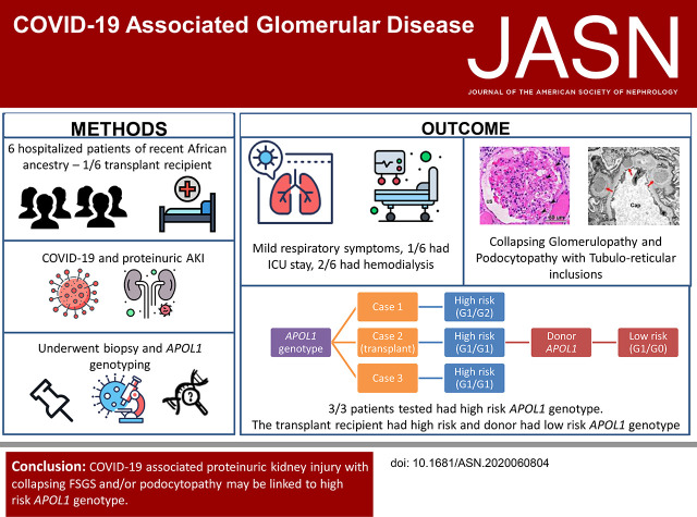
Background: Studies have documented AKI with high-grade proteinuria in patients with severe acute respiratory syndrome coronavirus 2 (SARS-CoV-2) infection. In some patients, biopsies have revealed collapsing glomerulopathy, a distinct form of glomerular injury that has been associated with other viruses, including HIV. Previous patient reports have described patients of African ancestry who developed nephrotic-range proteinuria and AKI early in the course of disease.

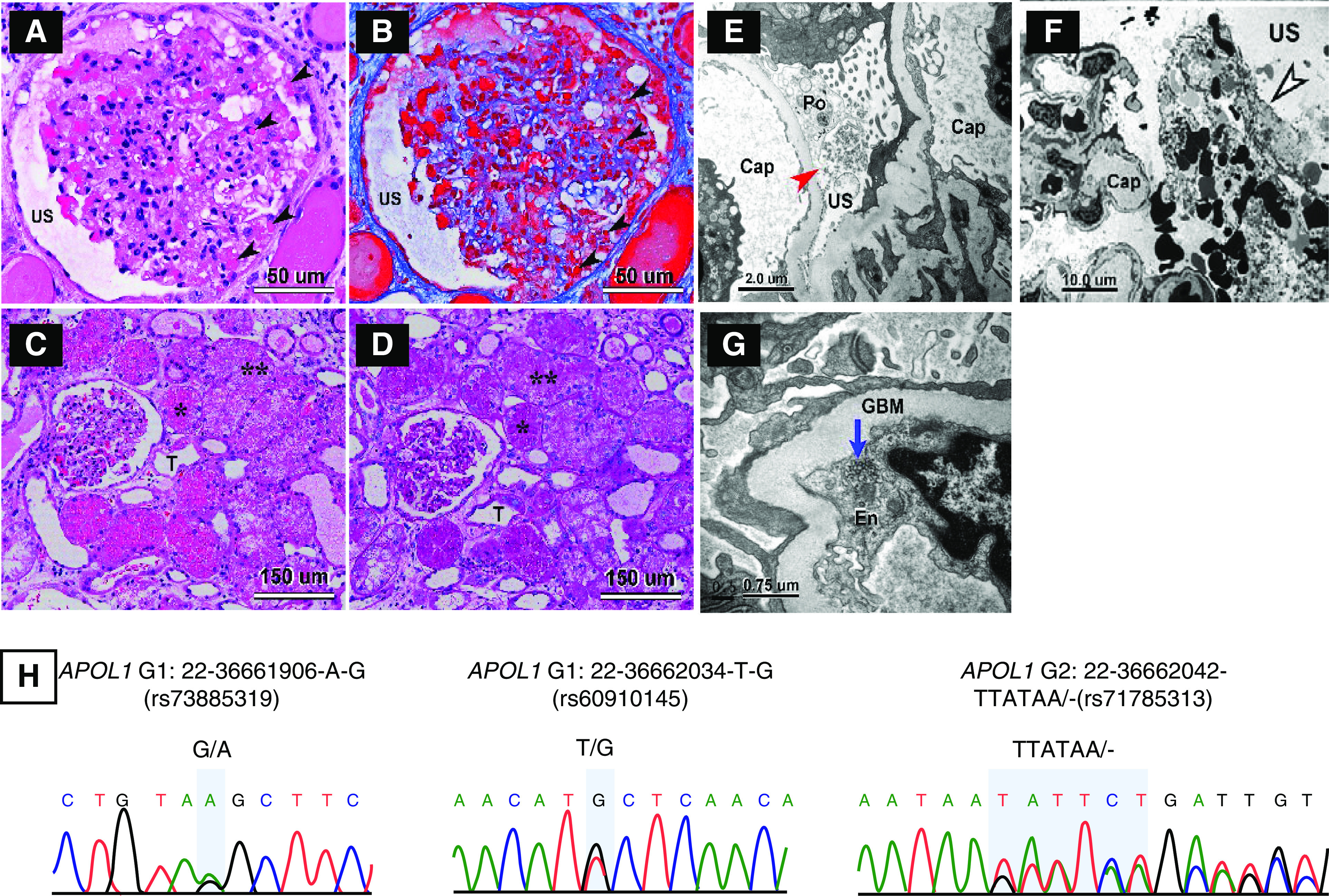
Methods: In this patient series, we identified six patients with coronavirus disease 2019 (COVID-19), AKI, and nephrotic-range proteinuria. COVID-19 was diagnosed by a positive nasopharyngeal swab RT-PCR for SARS-CoV-2 infection. We examined biopsy specimens from one transplanted kidney and five native kidneys. Three of the six patients underwent genetic analysis of APOL1, the gene encoding the APOL1 protein, from DNA extracted from peripheral blood. In addition, we purified genomic DNA from paraffin-embedded tissue and performed APOL1 genotype analysis of one of the native biopsies and the donor kidney graft.

Results: All six patients were of recent African ancestry. They developed COVID-19-associated AKI with podocytopathy, collapsing glomerulopathy, or both. Patients exhibited generally mild respiratory symptoms, and no patient required ventilator support. Genetic testing performed in three patients confirmed high-risk APOL1 genotypes. One APOL1 high-risk patient developed collapsing glomerulopathy in the engrafted kidney, which was transplanted from a donor who carried a low-risk APOL1 genotype; this contradicts current models of APOL1-mediated kidney injury, and suggests that intrinsic renal expression of APOL1 may not be the driver of nephrotoxicity and specifically, of podocyte injury.

Conclusions: Glomerular disease presenting as proteinuria with or without AKI is an important presentation of COVID-19 infection and may be associated with a high-risk APOL1 genotype.

Keywords: APOL1; COVID-19; collapsing glomerulopathy.


Figures

None
Graphical abstract
Figure 1.
Figure 1.
Biopsy findings showing CGN and APOL1 genotyping showing high risk genotype for patient 1. (A–D) Light micrographs of glomerular and tubular morphologic features in patient’s renal biopsy. (A and B) Mild to moderate focal glomerular hypercellularity with shrinkage of the capillary tuft is seen. The lumina of many capillary loops are effaced. The urinary space (US) is expanded. Many protein absorption lysosomal droplets are seen in the glomerular podocytes and parietal epithelial cells (black arrowheads). (A and C) Hematoxylin and eosin stain. (B) Trichrome stain. (D) Periodic acid–Schiff (PAS) stain. T, tubule. *In (C and D), numerous protein absorption lysosomal PAS-positive droplets are seen in the tubular epithelia. **Many tubules have undergone severe vacuolar degeneration. (E–G) Electron micrographs of glomerular and tubular morphologic features in patient’s renal biopsy. (E and F) Podocyte effacement (Po) and dilation of USs (red arrow) with cellular debris are present in the US (black and white arrowhead), which is likely made up of deformed red blood cells and fibrin tactoids. (G) shows tubular reticular inclusions (TRIs) in the rough endoplasmic reticulum of glomerular endothelium (En; blue arrow). GBM, glomerular basement membrane. (H) Sequencing for APOL1 was performed on DNA obtained from blood and native kidney biopsy. Genotypes were concordant and revealed a high–renal risk APOL1 G1/G2 genotype.
Figure 2.
Figure 2.
Biopsy findings showing CGN and APOL1 genotyping showing discordance between donor and recipient for patient 2. (A–D) Light micrographs of glomerular and tubular morphologic features in renal biopsy. (A–C) Moderate diffuse glomerular hypercellularity with compression of the capillary tuft is seen. The lumina of some capillary loops (Cap) are not readily discernible. (C) A portion of the glomerular tuft with intact Cap is seen only with silver stain sections. There is marked proliferation of visceral epithelial cells/podocytes (Po). Many protein absorption lysosomal (Ly) droplets are seen in the glomerular Po (black arrowheads in A and B). (D) Numerous protein absorption Ly periodic acid–Schiff (PAS)-positive droplets are seen in the tubular epithelia (T). Some tubules have undergone vacuolar degeneration, as seen in lower right corner of (D). (A) Hematoxylin and eosin stain. (B) Trichrome stain. (C) Silver stain. (D) PAS stain. (E–H) Electron micrographs showing glomerular morphologic features in renal biopsy. (E) shows a Po containing numerous Ly droplets and focal effacement of the foot processes. Electron-dense immune complex deposits are seen in the subepithelial (red arrows) and paramesangial (black and green arrow) regions of the glomerulus. (F) shows disrupted Po with dilated urinary space (US; red arrowheads) flanked by glomerular basement membrane (GBM) of two Cap. Tubuloreticular inclusions (black and white arrowhead) are present in the glomerular endothelium (En). (G) shows the presence of subepithelial and intramembranous electron-dense deposits (red arrows). (H) shows an intramembranous electron-dense deposit (red arrow) flanked by striated collagen fibers (black and green arrowheads). (I) Sequencing of APOL1 from DNA obtained from renal allograft demonstrates a G1/G0 genotype, whereas (J) analysis of DNA obtained from peripheral blood shows a G1/G1 genotype, which was discordant from the renal allograft.

Comment in

References

    1. Cheng Y, Luo R, Wang K, Zhang M, Wang Z, Dong L, et al. .: Kidney disease is associated with in-hospital death of patients with COVID-19. Kidney Int 97: 829–838, 2020 - PMC - PubMed
    1. Pei G, Zhang Z, Peng J, Liu L, Zhang C, Yu C, et al. .: Renal involvement and early prognosis in patients with COVID-19 pneumonia. J Am Soc Nephrol 31: 1157–1165, 2020 - PMC - PubMed
    1. Kissling S, Rotman S, Fakhouri F: The authors reply. Kidney Int 98: 232, 2020 - PMC - PubMed
    1. Larsen CP, Bourne TD, Wilson JD, Saqqa O, Sharshir MA: Collapsing glomerulopathy in a patient with COVID-19. Kidney Int Rep 5: P935–P939, 2020 - PMC - PubMed
    1. Peleg Y, Kudose S, D’Agati V, Siddall E, Ahmad S, Kisselev S, et al. .: Acute kidney injury due to collapsing glomerulopathy following COVID-19 infection. Kidney Int Rep 5: 940–945, 2020 - PMC - PubMed

Publication types

MeSH terms