. 2021 Jan 8;371(6525):eabd2638.
doi: 10.1126/science.abd2638. Epub 2020 Nov 19.

Recapitulation of HIV-1 Env-antibody coevolution in macaques leading to neutralization breadth

Affiliations

Recapitulation of HIV-1 Env-antibody coevolution in macaques leading to neutralization breadth

Ryan S Roark et al. Science. .

Abstract

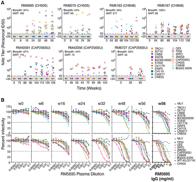
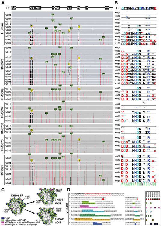
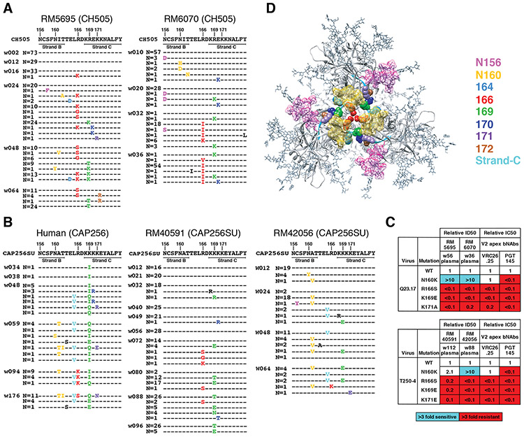
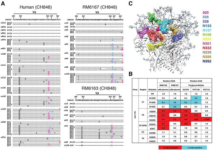
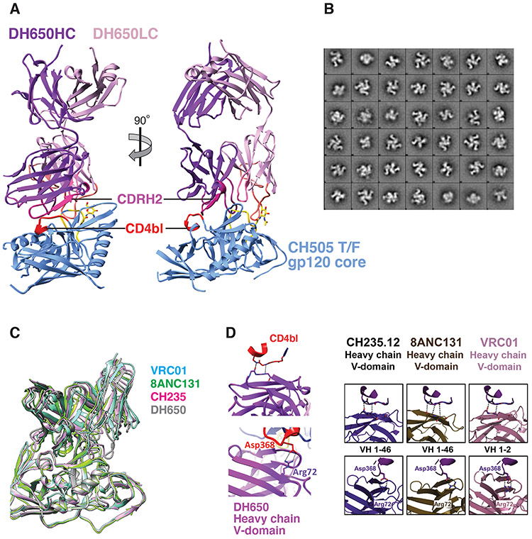
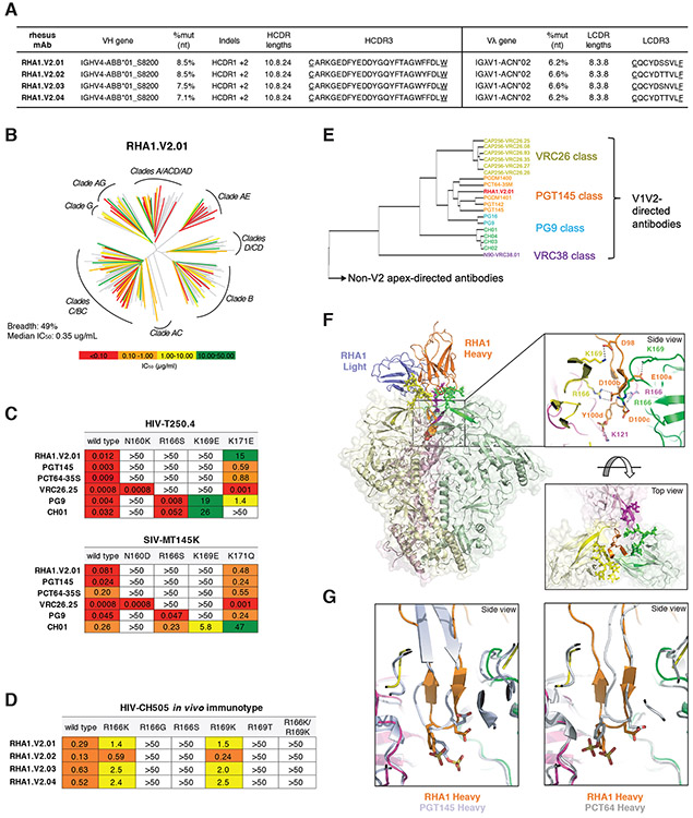
Neutralizing antibodies elicited by HIV-1 coevolve with viral envelope proteins (Env) in distinctive patterns, in some cases acquiring substantial breadth. We report that primary HIV-1 envelope proteins-when expressed by simian-human immunodeficiency viruses in rhesus macaques-elicited patterns of Env-antibody coevolution very similar to those in humans, including conserved immunogenetic, structural, and chemical solutions to epitope recognition and precise Env-amino acid substitutions, insertions, and deletions leading to virus persistence. The structure of one rhesus antibody, capable of neutralizing 49% of a 208-strain panel, revealed a V2 apex mode of recognition like that of human broadly neutralizing antibodies (bNAbs) PGT145 and PCT64-35S. Another rhesus antibody bound the CD4 binding site by CD4 mimicry, mirroring human bNAbs 8ANC131, CH235, and VRC01. Virus-antibody coevolution in macaques can thus recapitulate developmental features of human bNAbs, thereby guiding HIV-1 immunogen design.


Figures

Fig. 1.
Fig. 1.. Broadly neutralizing antibodies in seven rhesus macaques.
(A) RMs 5695 and 6070 were infected by SHIV.CH505, RMs 6163 and 6167 by SHIV.CH848, and RMs 40591, 42056 and 6727 by SHIV.CAP256SU. Neutralizing antibody titers (ID50, 50% inhibitory dilution) from longitudinal plasma specimens against 19 global tier 2 HIV-1 strains (26, 27, 86) are depicted. Full designations of target viruses are provided in Fig. S22 and Table S3. Maximum neutralization breadth across all time points and maximum geometric mean titer (GMT) of neutralization at any one time point are indicated for each animal. (B) Neutralization curves for longitudinal plasma specimens and purified plasma IgG (highlighted bold) from RM5695 show the development of broad and potent neutralization. MT145K.Q171K is a chimpanzee-derived SIVcpz strain that shares antigenic cross reactivity with HIV-1 in the V2 apex (35, 134). Dashed lines indicate 50% reduction in virus infectivity. MLV, murine leukemia virus.
Fig. 2.
Fig. 2.. Env evolution in SHIV.CH505 infected rhesus macaques recapitulates HIV-1 Env evolution in the human subject CH505.
(A) A PIXEL plot (https://www.hiv.lanl.gov/content/sequence/pixel/pixel.html) (https://www.hiv.lanl.gov/content/sequence/HIV/HIVTools.html) of amino acid alignments of longitudinal HIV-1 Env sequences obtained by single genome sequencing of plasma virion RNA. Between 15-60 individual sequences are grouped by time point and amino acids are colored red to highlight mutations relative to the infecting CH505 transmitted/founder strain illustrated schematically at the top. The image is highly compressed. Each row within a time block represents a single sequence, and each column represents an amino acid position in the alignment; thus each pixel represents a single amino acid that is colored grey if it matches the T/F sequence, red if it is a mutated residue, and black if it is an insertion or deletion (Indel) relative to the T/F virus. All SHIV sequences differ from the T/F at position 375, reflecting the SHIV design strategy that enables replication in rhesus macaques (9). Green tags indicate amino positions (HXB2 numbering) that are mutated in both human and rhesus. Yellow tags indicate three sites of identical Indels observed in both human and rhesus. (B) LASSIE analysis (44) of the same longitudinal Env sequences was used to characterize mutations under positive selection. If a T/F amino acid was retained in <25% of the sequences in the human CH505 infection, the site was considered to be under selective pressure and tracked in all hosts. The height of each amino acid mutation is proportional to its frequency at the respective time point. Red, dark blue and black indicate acidic, basic and neutral residues. “O” indicates asparagine (N) embedded in an N-linked glycosylation motif. Numbers at the bottom indicate residue positions (HXB2 numbering). Green numbers indicate mutations that reached 75% frequency in the human and in at least one animal. (C) Side projection of the CH505 Env trimer with potential N-linked glycans (PNGS) indicated in blue, 10Å neighbors of PNGs shown in green, and “glycan holes” that are typically covered by glycans in >80% and 50-80% of group M HIV-1 strains in magenta and pink, respectively. The light grey area in the central region of the trimer is the inter-protomer surface that forms a cleft with low glycan coverage (45). Two glycan holes in the CH505 T/F at positions 234 and 332 were filled by the addition of NXS/T motifs over time in the human subject CH505 and in RM6072 as well as in all other monkeys (see Fig. S3). (D) The V1 sequence of CH505 env is shown at the top of the panel with the hypervariable region indicated in red. Indels in V1 that arose in the first year of infection in the human host are illustrated in the sequences below the reference TF sequence. Indels occurred in nucleotide lengths divisible by three so as to maintain a viable Env open reading frame, and in the case of insertions, consisted of direct repeats. Indels that were replicated identically in one or more rhesus macaques are indicated by color-matched boxes to the right. Identical Indels in human and rhesus CH505 sequences were also found in V4 and V5 (Fig. S9).
Fig. 3.
Fig. 3.. bNAb responses in four rhesus macaques map to the V2 apex.
(A) Single genome sequencing (N = number of sequences; w = weeks post-SHIV infection) in SHIV.CH505 infected animals RM5695 and RM6070 reveals selection and fixation of mutations in and immediately proximal to strand C and additional mutations that eliminate PNG sites at 156 and 160. (B) Sequential Env sequences from human subject CAP256 and SHIV.CAP256SU infected RMs 40591 and 42056 showed selection and fixation of mutations in and proximal to strand C, with fewer mutations eliminating the PNG site at 160. (C) Heterologous neutralization of Q23.17 and T250-4 Env-pseudotyped viruses by rhesus plasma is drastically reduced (expressed as fold-change from wildtype) by mutations at residues 166, 169 and 171 similar to human V2 apex bNAbs. Enhanced neutralization against the N160K mutants is illustrated in Fig. S13 and further described in the Supplement. (D) Escape mutations from rhesus V2 apex bNAbs are displayed on the 5FYL structure of BG505.N332 SOSIP (top view).
Fig. 4.
Fig. 4.. bNAb responses in two rhesus macaques map to the V3 glycan high mannose patch.
(A) Sequential Env sequences from human subject CH848 and SHIV.CH848 infected RMs 6167 and 6163 showed selection and fixation of mutations in the GDIR motif. Ser to Asn substitutions at position 334 shift the PNG from residue 332 to 334 in the human subject and in RM6167. (B) Heterologous neutralization of Ce1176 Env-pseudotyped virus by RMs 6163 and 6167 plasma is reduced (expressed as fold-change from wildtype) by mutations at N332D and V295N, consistent with human V3 glycan bNAbs. Neutralization of GDIR mutants is reduced by 2-5-fold. Elimination of a glycan at position N138 enhances neutralization in RMs 6163 and 6167 and for some human V3 glycan bNAbs. (C) V3 glycan bNAb escape mutations are displayed on the 5FYL structure of BG505.N332 SOSIP (side view) highlighting their close spatial proximity.
Fig. 5.
Fig. 5.. Structure of Fab DH650 bound to CH505 gp120 core mimics human CD4bs antibodies.
(A) Ribbon representations of Fab DH650 heavy chain (purple) and light chain (pink) and CH505 gp120 core (blue). The heavy-chain CDRH2 is in magenta, the light-chain CDRL1 is in orange, and the CD4 binding loop (CD4bl) is in red. Loop D of gp120, which shifts from its position in other complexes to accommodate the long CDRL1 of DH650, is in yellow; the first N-acetyl glucosamine of glycan 276, which also shifts to accommodate CDRL1, is in stick representation (carbon, yellow; nitrogen, blue; oxygen, red). (B) 2D class averages after negative stain EM analysis of DH650-CH505 DS-SOSIP trimer showing three Fab bound per trimer. (C) Superposition of Fab DH650-CH505 gp120 complex with other CD4 binding site Fab-gp120 complexes, with the essentially identical structures of gp120 as the common reference. The view corresponds to the orientation in the right-hand part of panel A. Only the Fv modules of the Fabs are shown. Complex of DH650 (this study) is shown in grey, VRC01 (PDB-3NGB) in cyan, CH235.12 (PDB-5F96) in magenta, and 8ANC131 (PDB-4RWY) in green. DH650-bound gp120 core Cαs have rmsds of 0.78Å, 0.82 Å and 0.71Å from those of the VRC01-, CH235.12- and 8AC131-bound gp120 structures, respectively. (D) Left panel: Recognition of CD4 binding loop of CH505 T/F gp120 by variable heavy domain of DH650 (top). Coordination of Asp368 by Arg72 of DH650 (bottom). Right panel: Comparison with other CD4 binding site antibodies (6).
Fig. 6.
Fig. 6.. Rhesus bNAb lineage RHA1 targets the V2 apex, is broadly reactive, and contains a sulfated tyrosine in HCDR3 that shows precise chemical mimicry to human bNAbs PCT64-35S and PGT145.
(A) Immunogenetic characteristics of four RHA1 lineage broadly neutralizing monoclonal antibodies. A key feature is the 24 amino acid long CDRH3 that contains an acidic EDDY core motif. (B) Neutralization breadth and potency of RHA1.V2.01 against a 208 strain global virus panel. The dendrogram depicts phylogenetic relatedness of the HIV-1 Envs tested. (C) Neutralization expressed as IC50 (μg/ml) of wildtype heterologous viruses and their V2 apex mutants by RHA1.V2.01 and by prototypic human V2 apex bNAbs. Like most human V2 apex bNAbs, RHA1.V2.01 is strictly dependent on N160 and positively charged residues at 166 and 169. (D) Neutralization of CH505 T/F (wildtype) virus and C-strand variants, or “immunotypes,” that evolved in vivo in RM5695. Predominant mutations at 24 wks post-SHIV infection in RM5695 were R166K or R169K; at 48 wks R166S, R166G, R169T, and R166K plus R169K were prevalent; at 64 wks R166S or R169T became fixed (see Fig. 3A). Panel D shows progressive loss in neutralization sensitivity to RHA1 bNAbs by the evolving CH505 Envs, beginning with CH505 T/F wild type (most sensitive), CH505.R166K or R169K (intermediately sensitive), and ending with CH505.R166G, R166S, R169T or R166K+R169K (all resistant). Results are expressed as IC50 (μg/ml). (E) Neutralization fingerprint for RHA1.V2.01 shows it to cluster within the PGT145 class. (F) Cryo-EM structure (side view) of RHA1.V2.01 in complex with BG505 DS-SOSIP at 4-Å resolution. Inset (top) highlights electrostatic contacts of the HCDR3 with Env protomers, including interactions of the tyrosine-sulfated 100d residue with Env K121. Inset (bottom) shows the trimer apex cavity highlighting glycans at N160 and the C-strands. (G) Alignment of gp120 from the complex trimer structure with RHA1.V2.01 Fab to trimer complexes with human Fabs PGT145 (PDB-5V8L) and PCT64-35S (modeled with PDB-6CA6 fit to EMD-7865) reveals alignment of tyrosine sulfated residues within the respective HCDR3 tips, which insert into the hole at the V2 apex of the Env trimer.

References

    1. Keele BF, Giorgi EE, Salazar-Gonzalez JF, Decker JM, Pham KT, Salazar MG, Sun C, Grayson T, Wang S, Li H, Wei X, Jiang C, Kirchherr JL, Gao F, Anderson JA, Ping LH, Swanstrom R, Tomaras GD, Blattner WA, Goepfert PA, Kilby JM, Saag MS, Delwart EL, Busch MP, Cohen MS, Montefiori DC, Haynes BF, Gaschen B, Athreya GS, Lee HY, Wood N, Seoighe C, Perelson AS, Bhattacharya T, Korber BT, Hahn BH, Shaw GM, Identification and characterization of transmitted and early founder virus envelopes in primary HIV-1 infection. Proc Natl Acad Sci U S A 105, 7552–7557 (2008). 10.1073/pnas.0802203105. - DOI - PMC - PubMed
    1. Liao HX, Lynch R, Zhou T, Gao F, Alam SM, Boyd SD, Fire AZ, Roskin KM, Schramm CA, Zhang Z, Zhu J, Shapiro L, N. C. S. Program, Mullikin JC, Gnanakaran S, Hraber P, Wiehe K, Kelsoe G, Yang G, Xia SM, Montefiori DC, Parks R, Lloyd KE, Scearce RM, Soderberg KA, Cohen M, Kamanga G, Louder MK, Tran LM, Chen Y, Cai F, Chen S, Moquin S, Du X, Joyce MG, Srivatsan S, Zhang B, Zheng A, Shaw GM, Hahn BH, Kepler TB, Korber BT, Kwong PD, Mascola JR, Haynes BF, Co-evolution of a broadly neutralizing HIV-1 antibody and founder virus. Nature 496, 469–476 (2013). 10.1038/nature12053. - DOI - PMC - PubMed
    1. Doria-Rose NA, Schramm CA, Gorman J, Moore PL, Bhiman JN, DeKosky BJ, Ernandes MJ, Georgiev IS, Kim HJ, Pancera M, Staupe RP, Altae-Tran HR, Bailer RT, Crooks ET, Cupo A, Druz A, Garrett NJ, Hoi KH, Kong R, Louder MK, Longo NS, McKee K, Nonyane M, O'Dell S, Roark RS, Rudicell RS, Schmidt SD, Sheward DJ, Soto C, Wibmer CK, Yang Y, Zhang Z, N. C. S. Program, Mullikin JC, Binley JM, Sanders RW, Wilson IA, Moore JP, Ward AB, Georgiou G, Williamson C, Abdool Karim SS, Morris L, Kwong PD, Shapiro L, Mascola JR, Developmental pathway for potent V1V2-directed HIV-neutralizing antibodies. Nature 509, 55–62 (2014). 10.1038/nature13036. - DOI - PMC - PubMed
    1. Gao F, Bonsignori M, Liao HX, Kumar A, Xia SM, Lu X, Cai F, Hwang KK, Song H, Zhou T, Lynch RM, Alam SM, Moody MA, Ferrari G, Berrong M, Kelsoe G, Shaw GM, Hahn BH, Montefiori DC, Kamanga G, Cohen MS, Hraber P, Kwong PD, Korber BT, Mascola JR, Kepler TB, Haynes BF, Cooperation of B cell lineages in induction of HIV-1-broadly neutralizing antibodies. Cell 158, 481–491 (2014). 10.1016/j.cell.2014.06.022. - DOI - PMC - PubMed
    1. Bhiman JN, Anthony C, Doria-Rose NA, Karimanzira O, Schramm CA, Khoza T, Kitchin D, Botha G, Gorman J, Garrett NJ, Abdool Karim SS, Shapiro L, Williamson C, Kwong PD, Mascola JR, Morris L, Moore PL, Viral variants that initiate and drive maturation of V1V2-directed HIV-1 broadly neutralizing antibodies. Nat Med 21, 1332–1336 (2015). 10.1038/nm.3963. - DOI - PMC - PubMed

Publication types

MeSH terms