. 2020 Nov 19;10(1):20220.
doi: 10.1038/s41598-020-77001-5.

Characterization of Plasmodium falciparum NEDD8 and identification of cullins as its substrates

Affiliations

Characterization of Plasmodium falciparum NEDD8 and identification of cullins as its substrates

Manish Bhattacharjee et al. Sci Rep. .

Abstract

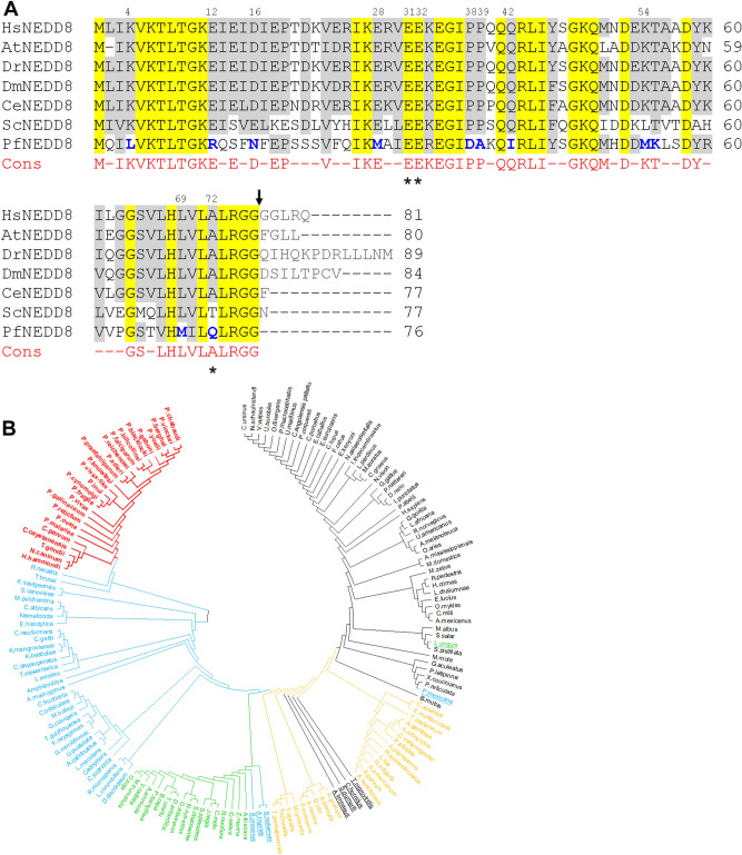
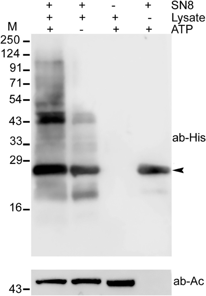
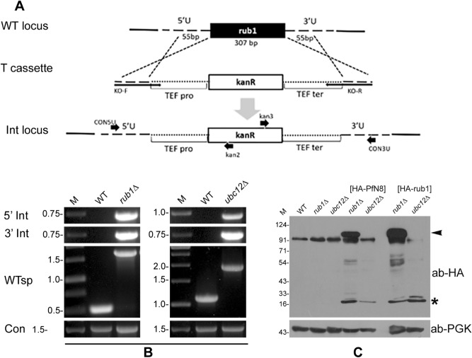
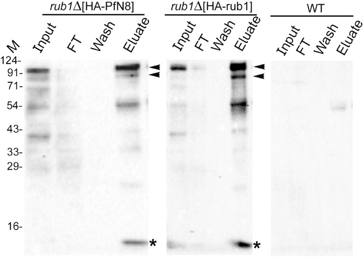
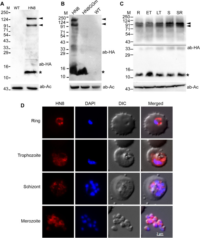
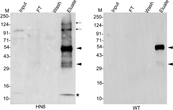
A variety of post-translational modifications of Plasmodium falciparum proteins, including phosphorylation and ubiquitination, are shown to have key regulatory roles during parasite development. NEDD8 is a ubiquitin-like modifier of cullin-RING E3 ubiquitin ligases, which regulates diverse cellular processes. Although neddylation is conserved in eukaryotes, it is yet to be characterized in Plasmodium and related apicomplexan parasites. We characterized P. falciparum NEDD8 (PfNEDD8) and identified cullins as its physiological substrates. PfNEDD8 is a 76 amino acid residue protein without the C-terminal tail, indicating that it can be readily conjugated. The wild type and mutant (Gly75Ala/Gly76Ala) PfNEDD8 were expressed in P. falciparum. Western blot of wild type PfNEDD8-expressing parasites indicated multiple high molecular weight conjugates, which were absent in the parasites expressing the mutant, indicating conjugation of NEDD8 through Gly76. Immunoprecipitation followed by mass spectrometry of wild type PfNEDD8-expressing parasites identified two putative cullins. Furthermore, we expressed PfNEDD8 in mutant S. cerevisiae strains that lacked endogenous NEDD8 (rub1Δ) or NEDD8 conjugating E2 enzyme (ubc12Δ). The PfNEDD8 immunoprecipitate also contained S. cerevisiae cullin cdc53, further substantiating cullins as physiological substrates of PfNEDD8. Our findings lay ground for investigation of specific roles and drug target potential of neddylation in malaria parasites.


Conflict of interest statement

The authors declare no competing interests.

Figures

Figure 1
Figure 1
(A) Sequence alignment of PfNEDD8. The amino acid sequences of 160 NEDD8 proteins from various organisms representing protozoans, metazoans, plants and fungi were aligned. Shown is the alignment of P. falciparum NEDD8 (PfNEDD8) with homologs from the indicated model organisms (H. sapiens: HsNEDD8, A. thaliana: AtNEDD8, D. rerio: DrNEDD8, D. melanogaster: DmNEDD8, C. elegans: CeNEDD8, S. cerevisiae: ScNEDD8). Also shown is the consensus (Cons) sequence for at least 75% of the 160 NEDD8 proteins, with hyphens representing variation at these positions. The conserved residues are yellow shaded, and grey shaded are physicochemically similar residues in at least 5 of the 7 sequences. The PfNEDD8 amino acid residues that are different from the consensus in physicochemical properties are in bold blue font. Amino acid residues marked with asterisks differentiate ubiquitin and NEDD8 for being correctly recognized by their respective enzymatic machinery. Numbers on the top of the alignment indicate position of that amino acid residue in PfNEDD8. The black arrow represents the processing site to remove the C-terminal tail. (B) Phylogenetic analysis of NEDD8 homologs. 160 NEDD8 protein sequences of the indicated organisms representing metazoans, plants, fungi and protozoans were aligned using the MUSCLE algorithm, and subjected to phylogenetic analysis using the maximum likelihood method. The NEDD8 homologs of apicomplexans are in red, non-apicomplexan protists and fungi are in blue, plants are in green, invertebrates are in yellow and vertebrates are in black. The outlier NEDD8 homologs are underlined.
Figure 2
Figure 2
Western blot analysis of in vitro neddylation. Conjugation of recombinant SUMO-PfNEDD8 (SN8) was carried out using parasite lysate and ATP at 37 °C for 1 h. The reactions were stopped and subjected to immunoblotting using anti-His antibody. The presence (+) or absence (−) of reaction component is indicated. The arrowhead indicates signal corresponding to unconjugated SN8. β-actin was used as a loading control for reactions containing the parasite lysate. The positions of proteins size markers are shown in kDa (M).
Figure 3
Figure 3
Complementation of ScRub1 by PfNEDD8. (A) Schematic for generation of rub1Δ or ubc12Δ. The wild type locus (WT locus) was replaced with a linear kanamycin cassette flanked by the homology regions of target locus (T cassette) via a double crossover homologous recombination, resulting in the integration locus (Int locus). The kanamycin coding sequence (kanR) is under the control of translation elongation factor promoter (TEF pro) and terminator (TEF ter). The horizontal arrows indicate positions of primers used in the analysis of knockout strains. (B) Confirmation of knockout. The knockout was confirmed by PCR of the gDNAs of wild type (WT), rub1Δ and ubc12Δ strains using locus-specific primers (5′ Int for 5′ integration locus, 3′ Int for 3′ integration locus, WTsp for wild type locus and Con for ScAtg18). The ethidium bromide stained agarose gel shows PCR products, with DNA markers in kbp (M). (C) Western blot analysis of complemented strains. HA-PfNEDD8 (HA-PfN8) or HA-ScRub1 (HA-rub1) were episomally expressed in rub1Δ and ubc12Δ strains. The lysates of wild type (WT), rub1Δ, ubc12Δ, HA-PfNEDD8-complemented rub1Δ or ubc12Δ (HA-PfN8), and HA-ScNEDD8-complemented rub1Δ or ubc12Δ (HA-rub1) strains were processed for western blotting using anti-HA antibodies (ab-HA). The lanes containing lysates of complemented strains show free NEDD8/Rub1 protein (marked with an asterisk), whereas the lanes with rub1Δ[HA-PfN8] and rub1Δ[HA-rub1] strains also contain a high molecular weight band (marked with an arrowhead) and some lower molecular weight conjugates, which are absent in other lanes. Phosphoglycerate kinase (anti-PGK) was used as a loading control. The protein size markers are in kDa (M).
Figure 4
Figure 4
Immunoprecipitation of PfNEDD8 and Rub1 from complemented strains. The lysates of rub1∆[HA-PfN8], rub1∆[HA-rub1] and wild type (WT) strains were immunoprecipitated with mouse anti-HA antibody. The eluates (Eluate) along with the extract (Input), flow-through (FT) and wash (Wash) were analyzed by western blotting using rabbit anti-HA antibodies. The eluate lanes contain the respective target protein (indicated by an asterisk) and two prominent high molecular weight bands (indicated by arrowheads). The sizes of protein size markers are in kDa (M).
Figure 5
Figure 5
Expression, conjugation and localization of P. falciparum NEDD8. (A) Lysates of the wild type (WT) and HA-PfNEDD8–expressing (HN8) P. falciparum parasites were processed for western blotting using anti-HA antibodies (ab-HA). β-actin was used as a loading control (ab-Ac). The blot shows a prominent band close to the predicted size of HA-PfHANEDD8 (indicated with an asterisk) and two high molecular bands (indicated with arrowheads) in the HN8 lane only. (B) The HA-PfNEDD8-expressing (HN8), mutant HA-PfNEDD8 (HN8GGm) and wild type (WT) parasites were processed for western blotting using anti-HA antibodies (ab-HA), and β-actin was used as a loading control (ab-Ac). Note that the prominent high molecular bands (indicated with arrowheads) are present in HN8 lane only, whereas a band of the predicted size of HA-PfHANEDD8 or mutant HA-PfNEDD8 (indicated with an asterisk) is present in both HN8 and HN8GGm lanes. (C) A synchronized culture of HA-PfNEDD8–expressing P. falciparum parasites was harvested at ring (R), early trophozoite (ET), late trophozoite (LT) and schizont/ring (SR) stages, and the parasites (1 × 108 parasites/lane) were processed for western blotting using anti-HA antibodies (ab-HA). β-actin was used as a loading control (ab-Ac). The blot shows two prominent high molecular weight bands (indicated by arrowheads) along with the free HA-NEDD8 (indicated by an asterisk), indicating neddylation throughout the erythrocytic cycle. The double line in the top panel marks two different blots, which were developed separately to minimize over saturation of the blot due to the band around 12 kDa. The protein size markers in A, B and C are in kDa (M). (D) Fixed HA-NEDD8-expressing P. falciparum parasites of the indicated stages were evaluated for localization of HA-NEDD8 using anti-HA antibodies. The images show HA-NEDD8 specific signal (HN8), nucleic acid staining (DAPI), the parasite and the erythrocyte boundaries (DIC), and the merged of all three images (Merged). The HA-NEDD8 signal is present throughout the parasite in all the stages shown, except the food vacuole. The scale bar shown is identical for all the images.
Figure 6
Figure 6
The effect of MLN4924 on neddylation. (A) Wild type P. falciparum D10 ring stage parasites were incubated with various concentrations of MLN4924 for 48–50 h, and the % growth (y-axis) at different MLN4924 concentrations (x-axis) was plotted to determine 50% inhibitory concentration (IC50) as described in the method section. The graph shows data from three independent experiments, with each point representing mean of two replicates. The IC50 value is average of three experiments with SD. (B) The HA-PfNEDD8-expressing P. falciparum trophozoite stage parasites were grown with MLN4924 (+) or DMSO (−) for 4 or 8 h, and then processed for western blotting using anti-HA antibodies (ab-HA). β-actin was used as a loading control (ab-Ac). The blot shows multiple high molecular weight bands along a band of the predicted size of free HA-PfNEDD8 (indicated with an asterisk). The intensity of high molecular weight bands in MNL4924-treated parasites for both the time points appears to be comparable to that of DMSO-treated ones. The protein size markers are in kDa (M).
Figure 7
Figure 7
Western blot of parasite immunoprecipitates. The lysates of HA-PfNEDD8-expressing (HN8) and wild type (WT) parasites were immunoprecipitated with rabbit anti-HA antibodies. The eluates (Eluate) along with the extract (Input), flow-through (FT) and wash (Wash) were analyzed by western blotting using mouse anti-HA antibodies. The HN8 blot shows bands corresponding to free HA-PfNEDD8 (indicated with an asterisk) and high molecular weight conjugates (indicated with arrows). The arrowheads indicate non-specific signal in both the blots. The protein size markers are in kDa (M).

Similar articles

Cited by

References

    1. Hochstrasser M. Origin and function of ubiquitin-like proteins. Nature. 2009;458:422–429. doi: 10.1038/nature07958. - DOI - PMC - PubMed
    1. Cappadocia L, Lima CD, Hochstrasser M. Ubiquitin-like protein conjugation: structures, chemistry, and mechanism. Nature. 2018;118:889–918. - PMC - PubMed
    1. Jones D, et al. Covalent modifier NEDD8 is essential for SCF ubiquitin-ligase in fission yeast. J. Biol. Chem. 2006;15:27724–27736. - PMC - PubMed
    1. Walsh C. Posttranslational Modification of Proteins: Expanding Nature’s Inventory. New York: Roberts and Company Publishers; 2006.
    1. Organization WH. World Malaria Report 2019. Geneva: WHO; 2019.

Publication types

MeSH terms