. 2020 Nov 19;10(1):20177.
doi: 10.1038/s41598-020-77247-z.

Purification and biochemical characterization of Hel a 6, a cross-reactive pectate lyase allergen from Sunflower (Helianthus annuus L.) pollen

Affiliations

Purification and biochemical characterization of Hel a 6, a cross-reactive pectate lyase allergen from Sunflower (Helianthus annuus L.) pollen

Nandini Ghosh et al. Sci Rep. .

Expression of concern in

Abstract

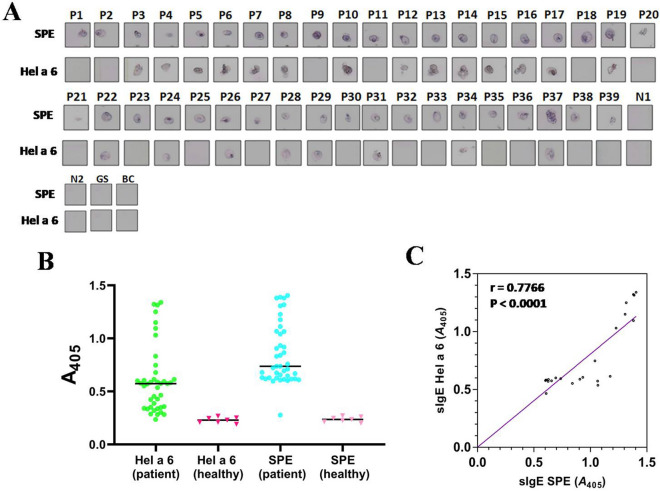
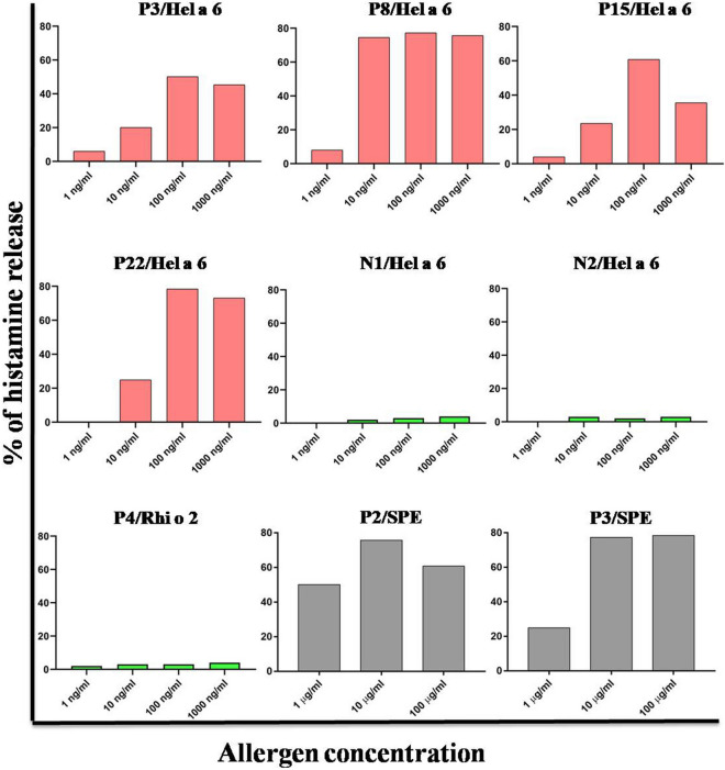
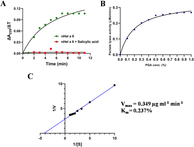
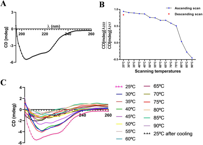
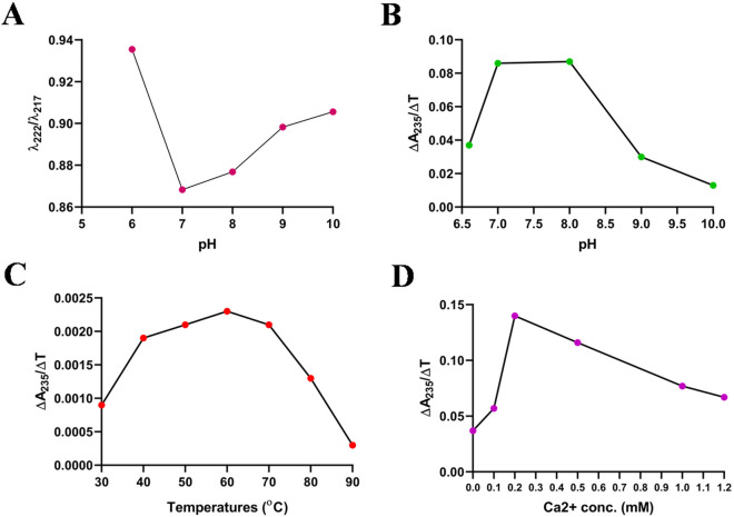
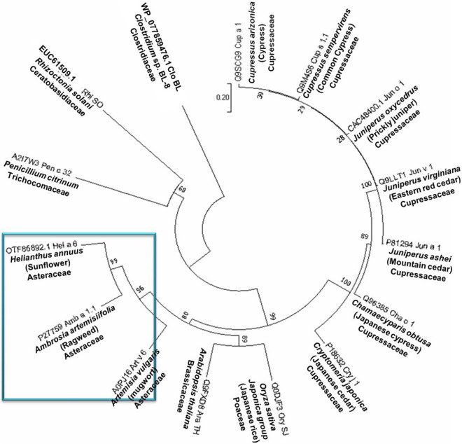
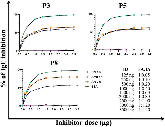
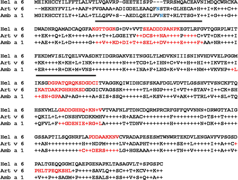
Sunflower pollen was reported to contain respiratory allergens responsible for occupational allergy and pollinosis. The present study describes the comprehensive characterization of a major sunflower allergen Hel a 6. Natural Hel a 6 was purified from sunflower pollen by anion exchange and gel filtration chromatography. Hel a 6 reacted with IgE-antibodies from 57% of 39 sunflower-sensitized patient sera suggesting it to be a major allergen. The patients were of Indian origin and suffering from pollinosis and allergic rhinitis. Hel a 6 exhibited allergenic activity by stimulating mediator release from basophils. Monomeric Hel a 6 displayed pectate lyase activity. The effect of various physicochemical parameters such as temperature, pH, and calcium ion on the functional activity of Hel a 6 revealed a stable nature of the protein. Hel a 6 was folded, and its melting curve showed reversible denaturation in which it refolded back to its native conformation from a denatured state. Hel a 6 displayed a high degree of sequence conservation with the pectate lyase allergens from related taxonomic families such as Amb a 1 (67%) and Art v 6 (57%). The IgE-cross reactivity was observed between Hel a 6 and its ragweed and mugwort homologs. The cross-reactivity was further substantiated by the mediator release when Hel a 6-sensitized effector cells were cross-stimulated with Art v 6 and Amb a 1. Several putative B cell epitopes were predicted and mapped on these 3 allergens. Two antigenic regions were found to be commonly shared by these 3 allergens, which could be crucial for cross-reactivity. In conclusion, Hel a 6 serves as a candidate molecule for diagnosis and immunotherapy for weed allergy.


Conflict of interest statement

The authors declare no competing interests.

Figures

Figure 1
Figure 1
Purification of natural Hel a 6 from sunflower pollen. (A) Chromatogram of the NaCl eluted fractions from anion exchanger Q column showing 3 peaks (Fr-1 to Fr-3). The elution volume (ml) in x-axis, the A280 (milli absorbance unit; mAu) of the elutes in left y-axis and NaCl concentration gradient (mM) in right y-axis. (B) 12% SDS-PAGE profile of the 3 eluted fractions (Fr-3a to 3c) of the peak Fr-3 (flanked by dashed lines) collected as 0.5 ml/fraction tube. Fr-3a was found to have Hel a 6 with highest purity. (C) Therefore, Fr-3a was immunoblotted with sera from 2 sunflower-sensitized patients (P2 and P3) to confirm the presence of Hel a 6. Non-atopic serum (N1) and buffer control (BC) were used as negative controls. (D) Chromatogram of the eluted fractions after separating Fr-3a in gel filtration column in which A280 values of the eluted fractions (y-axis) are plotted against elution volume (ml) in x-axis. 17 ml of void volume is marked with dashed lines. Fr-3a-IV was found to contain Hel a 6 with > 90% purity. (D) Monomeric status of Hel a 6 was checked in non-reducing (NR; without β-ME) SDS-PAGE as compared to reducing (R; with β-ME). (E) Presence of Hel a 6 in Fr-3a-IV was checked by immunoblotting with sera from 4 sunflower-sensitized patients (P1-P4).
Figure 2
Figure 2
Hel a 6 is a major sunflower pollen allergen. (A) Immuno-dot blot in which either 0.5 µg of purified Hel a 6 or 5 µg of sunflower pollen extract (SPE) was exposed to sera from 39 sunflower-sensitized patients (P1–P39). Negative controls are 2 non-atopic sera (N1–N2), one grass pollen sensitized serum (GS), and buffer control (BC) without serum. (B) Scattered dot plot of IgE-ELISA (in shades of green) showing quantitative estimation of specific IgE level (Absorbance at 405 nm in y-axis) against either Hel a 6 or SPE (x-axis) in sunflower-sensitized patients (n = 39). Horizontal lines represent mean and error bars as SD. For any patient, the IgE-titre ≥ twofold of the corresponding cut-off level was considered as positive for that antigen. 22 of 39 sunflower-allergic patients were found seropositive to Hel a 6 in ELISA as well as in immuno-dot blot. Two other scattered plots (in shades of red) showing distribution of IgE-titre values in 6 non-atopic healthy sera against either Hel a 6 or SPE for negative controls. (C) Association between the specific IgE titres (sIgE as A405) of these 22 patients against SPE (x-axis) and Hel a 6 (y-axis) by non-parametric Spearman correlation analysis showing positive correlation.
Figure 3
Figure 3
Hel a 6 displayed allergenic activity. Stimulation of effector cells in the blood of 4 sunflower-sensitized patients (red bars) with increasing concentrations of purified Hel a 6 (x-axis) resulted in mediator release (y-axis). For negative controls (green bars), either non-atopic bloods (N1 and N2) were stimulated with Hel a 6 or Hel a 6-specific atopic blood (P4) were stimulated with an unrelated allergen Rhi o 2. For positive control (gray bars), Hel a 6-specific atopic bloods (P2 and P3) were stimulated with sunflower pollen extract i.e. SPE. As it is a crude extract, hence, the protein concentration in extract was 10 times higher in for each of the 3 doses as compared to purified allergens.
Figure 4
Figure 4
Hel a 6 is a pectate lyase. Pectate lyase activity is monitored by recording the cleavage of polygalacturonic acid (PGA) substrate by Hel a 6 and a concomitant increase in A235 of the reaction mix. (A) Rate of pectate lyase activity by Hel a 6 was determined at a fixed PGA concentration and expressed as a ratio of change in A235 and change in incubation time (ΔA235:ΔT; y-axis) as a function of time (minutes; x-axis). Addition of pectate lyase inhibitor salicylic acid resulted in a sharp decrease in enzyme activity. Kinetics of pectate lyase activity of Hel a 6 (x-axis) as a function of increase PGA concentration (y-axis) shown as non-linear Michaelis–Menten curve (B) and linear double reciprocal Lineweaver Burk plot (C) to determine the Vmax and Km.
Figure 5
Figure 5
Folding pattern of Hel a 6. (A) CD spectra of 4 µM of Hel a 6 showing the CD milli degree (y-axis) at different wavelengths (nm; y-axis). (B) Melting curve of Hel a 6 showing the ratio of CD spectra at 222 nm and 217 nm taken at increasing temperatures (up scan) to a fully denatured state followed by decreasing the temperature up to 25 °C (down scan) to allow refolding of the protein. (C) Thermal denaturation of Hel a 6 showing the entire CD spectra (y-axis) as a function of various wavelengths (x-axis) taken at various temperatures.
Figure 6
Figure 6
Effects of various parameters on the folding and activity of Hel a 6. (A) Effect of pH (x-axis) on the folding pattern of Hel a 6 (λ222217) in y-axis. Effects of pH (B), temperature (C), and calcium concentration (D) plotted in y-axis, and the rate of enzyme activity plotted on x-axis.
Figure 7
Figure 7
Evolutionary relationship among the pectate lyases. The unrooted phylogenetic tree constructed with 10 allergenic pectate lyases of plant origin enlisted in IUIS allergen database along two non-allergenic pectate lyases from rice and Arabidopsis, and two pectate lyases of fungal origin using MEGA v7.0 software (https://www.megasoftware.net). A separate clad from by Hel a 6, Amb a 1, and Art v 6 are marked within a blue box.
Figure 8
Figure 8
Cross-reactivity by ELISA inhibition. ELISA inhibition in which the plate-bound 0.5 µg/ml Hel a 6 was exposed to sera from 3 sunflower-sensitized patients pre-incubated with increasing doses (x-axis) of either Art v 6 or Amb a 1 as fluid phase inhibitors. Sera were also pre-incubated with either Hel a 6 (auto-inhibition) or BSA (negative control). The percentage of IgE-binding inhibition was plotted on y-axis. Lower right panel shows the ratio between fluid phase allergen (FA) and plate-bound immobilized allergen (IA) against each of the inhibitor doses (ID).
Figure 9
Figure 9
Cross-reactivity by blot inhibition and cross-stimulated mediator release. Hel a 6 (A), Amb a 1 (B), and Art v 6 (C) transferred on PVDF membrane were exposed to a pool of sunflower-sensitized sera pre-incubated with 10 µg/ml of either the same allergen (auto-inhibition) or the other two pectate lyases. Immunoblot was also done with only sera (i.e., no pre-incubation) or negative control. (D) Histamine release (y-axis) was observed when the effector cells in the blood of 4 sunflower-sensitized patients (x-axis) were stimulated with optimal concentration (100 ng/ml) of either Hel a 6 or Amb a 1 or Art v 6. For negative controls, bloods from healthy donor (N2) and grass pollen allergic sera (GS) were used.
Figure 10
Figure 10
Sequence conservation within common pectate lyase allergens of weed origin. Multiple sequence alignment of Hel a 6 as reference sequence with 2 pectate lyases of Compositae family such as Art v 6 from mugwort and Amb a 1 from ragweed as aligned sequences. Identical residues and gaps in the aligned sequences are shown + and − respectively. Putative linear B cell epitopes are in red and N-linked glycosylation sites in blue. The regions containing B cell epitopes commonly present in all the 3 allergens are marked with a line that can be anticipated as the possible IgE-epitope responsible IgE-cross reactivity.
Figure 11
Figure 11
In silico Epitope mapping. The putative linear B cell epitopes (different shades of red) predicted in this study are mapped on the globular/surface representation of the corresponding 3D models (in cyan) of Amb a 1 (A), Art v 6 (B), and Hel a 6 (C) using PyMol v1.74 (https://pymol.org/academic).

References

    1. Wopfner, N. et al. The spectrum of allergens in ragweed and mugwort pollen. Int. Arch. Allergy Immunol.138, 337–346 (2005). - PubMed
    1. Pablos, I. et al. Proteomic profiling of the weed feverfew, a neglected pollen allergen source. Sci. Rep.7, 1–10 (2017). - PMC - PubMed
    1. Pichler, U. et al. Pectate lyase pollen allergens: Sensitization profiles and cross-reactivity pattern. PLoS ONE10, e0120038 (2015). - PMC - PubMed
    1. Shin, S. B., Golovkin, M. & Reddy, A. S. A pollen-specific calmodulin-binding protein, NPG1, interacts with putative pectate lyases. Sci. Rep.4, 1–8 (2014). - PMC - PubMed
    1. Jahn-Schmid, B. et al. Humoral and cellular cross-reactivity between Amb a 1, the major ragweed pollen allergen, and its mugwort homolog Art v 6. J. Immunol.188, 1559–1567 (2012). - PubMed

Publication types

MeSH terms