Review
. 2021 Feb;21(2):71-88.
doi: 10.1038/s41568-020-00312-2. Epub 2020 Nov 19.

Endoplasmic reticulum stress signals in the tumour and its microenvironment

Affiliations
Review

Endoplasmic reticulum stress signals in the tumour and its microenvironment

Xi Chen et al. Nat Rev Cancer. 2021 Feb.

Abstract

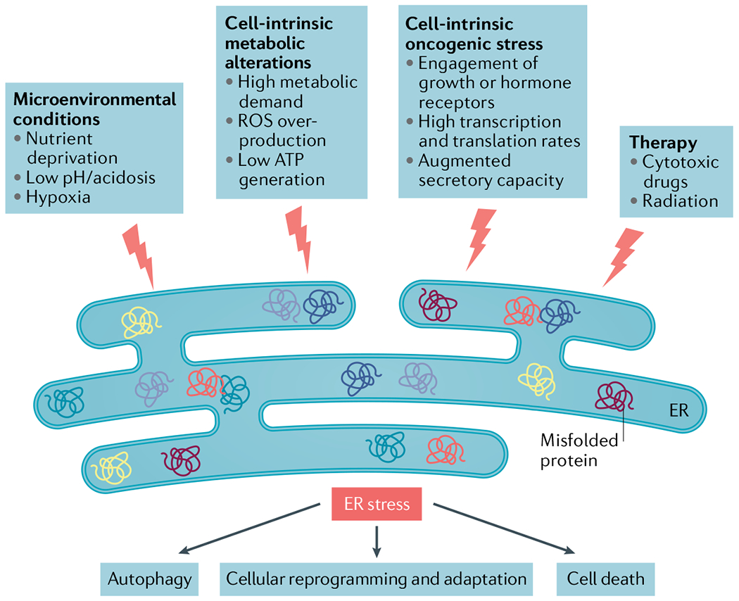
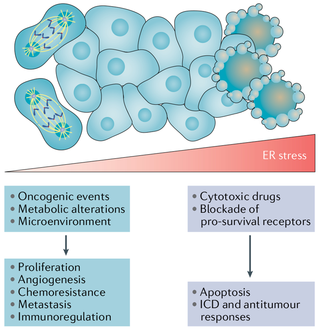
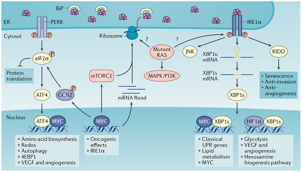
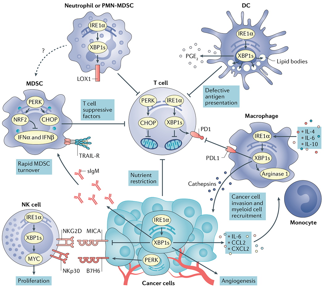
Protein handling, modification and folding in the endoplasmic reticulum (ER) are tightly regulated processes that determine cell function, fate and survival. In several tumour types, diverse oncogenic, transcriptional and metabolic abnormalities cooperate to generate hostile microenvironments that disrupt ER homeostasis in malignant and stromal cells, as well as infiltrating leukocytes. These changes provoke a state of persistent ER stress that has been demonstrated to govern multiple pro-tumoural attributes in the cancer cell while dynamically reprogramming the function of innate and adaptive immune cells. Aberrant activation of ER stress sensors and their downstream signalling pathways have therefore emerged as key regulators of tumour growth and metastasis as well as response to chemotherapy, targeted therapies and immunotherapy. In this Review, we discuss the physiological inducers of ER stress in the tumour milieu, the interplay between oncogenic signalling and ER stress response pathways in the cancer cell and the profound immunomodulatory effects of sustained ER stress responses in tumours.


Conflict of interest statement

Competing interests

X.C. and J.R.C.-R. hold patents on the use of ER stress and UPR modulators for cancer therapy. X.C. reports research funding from Fosun Pharma.

Figures

Fig. 1 |
Fig. 1 |. Inducers of endoplasmic reticulum stress in the tumour microenvironment.
The uncontrolled proliferative capacity of malignant cells in growing tumours engenders hostile microenvironments characterized by high metabolic demand, hypoxia, nutrient limitations and acidosis, which in turn provoke disruption of calcium and lipid homeostasis in multiple cell types inhabiting this milieu. Collectively, these harsh conditions alter the protein-folding capacity of the endoplasmic reticulum (ER) in both cancer cells and infiltrating immune cells, thereby promoting accumulation of misfolded or unfolded proteins within this organelle and, consequently, ER stress. Oncogenic events in cancer cells further contribute to this state by elevating their global transcription and translation rates. The unfolded protein response (UPR) is subsequently activated as an attempt to restore ER homeostasis and promote adaptation to diverse insults in the tumour. Certain therapeutic modalities can also trigger ER stress in the cancer cell to alter their normal behaviour in the tumour microenvironment (TME). Depending on the magnitude of ER stress, the cell type and the specific pathological context, ER stress responses can have multiple effects ranging from cellular reprogramming and adaptation to autophagy and apoptosis. Owing to the additive effects of various ER stressors concurrently enriched in the TME during cancer initiation, progression and therapy, robust and persistent UPR activation is mostly evidenced in cancer cells and tumour-infiltrating immune cells in vivo, which has been challenging to recapitulate under in vitro conditions. ROS, reactive oxygen species.
Fig. 2 |
Fig. 2 |. The magnitude of endoplasmic reticulum stress and its differential outcomes in malignant cells.
Persistent, yet moderate, endoplasmic reticulum (ER) stress responses fuelled by oncogenic pathways, metabolic changes and conditions of the tumour microenvironment stimulate several mechanisms that promote cancer cell proliferation, metastasis, chemoresistance, angiogenesis and immune evasion. By contrast, extreme ER stress caused by the uncontrolled accumulation of misfolded proteins in this organelle can lead to a terminal unfolded protein response (UPR) that induces cell death. For instance, proteasome inhibitors have been shown to trigger proapoptotic ER stress responses in multiple myeloma cells by hyperactivating the PRKR-like ER kinase (PERK)–eukaryotic translation initiation factor 2α (eIF2α)–activating transcription factor 4 (ATF4)–C/EBP homologous protein (CHOP) arm of the UPR. Of note, exposure to some cytotoxic agents, such as anthracyclines, can trigger ER stress responses that promote immunogenic cell death (ICD) capable of eliciting antitumour immunity (BOX 2). Hence, the consequences of UPR activation, either pro-survival or pro-apoptotic, are determined by the duration and intensity of the stress.
Fig. 3 |
Fig. 3 |. Integration of oncogenic programmes and endoplasmic reticulum stress responses in the cancer cell.
Oncogenic MYC activates the unfolded protein response (UPR) through multiple mechanisms. MYC-induced upregulation of global transcription (mRNA flood) and translation increases ribosome biogenesis and protein load in the endoplasmic reticulum (ER), thus activating all branches of the UPR. MYC further binds to promoter and enhancer regions in the gene encoding inositol-requiring protein 1α (IRE1α), positively regulating its transcription and augmenting IRE1α protein levels. MYC can also form a heterodimer with X-box binding protein 1s (XBP1s) in the nucleus to regulate classical UPR genes and lipid metabolism genes. Of note, XBP1s has been shown to promote MYC transcription in prostate cancer cells and natural killer cells. MYC engages PRKR-like ER kinase (PERK) and general control non-derepressible 2 (GCN2) kinase to induce eukaryotic translation initiation factor 2α (eIF2α) phosphorylation and the integrated stress response. MYC can also interact with activating transcription factor 4 (ATF4) to regulate amino acid transporters and biosynthesis, antioxidant pathways and autophagy. The MYC–ATF4 complex regulates eukaryotic translation initiation factor 4E-binding protein 1 (4EBP1) to reduce translation and proteotoxic stress. mTOR complex 1 (mTORC1) activation induces protein synthesis and ER overload that activates the UPR. In turn, the IRE1α–tumour necrosis factor receptor-associated factor 2 (TRAF2)–JUN N-terminal kinase (JNK)–insulin receptor substrate 1 (IRS1) axis has been shown to restrict mTORC1 activity. Mutant RAS is integrated within the UPR in a context-specific manner. Mutant HRAS preferentially induces IRE1α activity in keratinocytes through an unknown mechanism. In primary human melanocytes, HRAS-G12V–PI3K, but not BRAF-V600E, increases ER content and induces activation of all UPR branches. It is unclear whether mutant RAS enhances global protein translation and protein load in the ER, which promotes ER stress in all cancer types. BiP, binding-immunoglobulin protein; HIF1α, hypoxia-inducible factor 1α; P, phosphorylation; RIDD, regulated IRE1-dependent decay of RNA; VEGF, vascular endothelial growth factor.
Fig. 4 |
Fig. 4 |. Immunomodulatory effects of endoplasmic reticulum stress signals in the tumour microenvironment.
Cancer cells undergoing activation of inositol-requiring protein 1α (IRE1α) or PRKR-like ER kinase (PERK) modulate tumour recognition by natural killer (NK) cells while secreting mediators that promote angiogenesis and recruitment of myeloid cell types to tumour sites. Both IRE1α and PERK are well established to regulate angiogenesis. X-box binding protein 1s (XBP1s) and activating transcription factor 4 (ATF4) directly bind to the vascular endothelial growth factor (VEGF) promoter to regulate its expression. Nutrient restriction, reactive oxygen species (ROS) accumulation or the presence of soluble factors that blunt glucose uptake cause endoplasmic reticulum (ER) stress and chronic activation of the IRE1α–XBP1 and PERK–C/EBP homologous protein (CHOP) arms of the unfolded protein response (UPR) in intratumoural T cells, provoking mitochondrial dysfunction and inhibition of their optimal anticancer effector function. High levels of cholesterol in the tumour microenvironment (TME) can also activate IRE1α–XBP1 signalling in intratumoural T cells to induce programmed cell death protein 1 (PD1) expression and limit their protective activity. Myeloid-derived suppressor cells (MDSCs) exploit PERK to control antitumour immunity via CHOP-mediated expression of T cell suppressive factors and by inducing nuclear factor erythroid 2-related factor 2 (NRF2)-driven responses that inhibit production of protective type I interferon. ER stress in MDSCs has also been associated with their elevated expression of tumour necrosis factor-related apoptosis-inducing ligand-receptors (TRAIL-Rs) and rapid turnover in the TME. ER stress-related gene signatures and expression of lectin-type oxidized LDL receptor 1 (LOX1) distinguish normal neutrophils from polymorphonuclear (PMN)-MDSCs in patients with cancer. In addition, ER-stressed neutrophils acquire immunosuppressive attributes and overexpress LOX1 via IRE1α activation. ROS accumulation fuels ER stress and persistent IRE1α–XBP1 activation in tumour-associated dendritic cells (DCs), driving uncontrolled lipid droplet formation that inhibits their capacity to present local antigens to intratumoural T cells. ER-stressed DCs have also been shown to overproduce the immunosuppressive lipid mediator prostaglandin E2 (PGE2) via IRE1α–XBP1 activation, presumably contributing to immune escape in cancer. In macrophages, the IRE1α–XBP1 branch has been shown to promote expression of cathepsins, PD1 ligand 1 (PDL1) and Arginase 1, further promoting cancer cell invasion and immunosuppression in the TME. CCL2, CC-chemokine ligand 2; CXCL2, CXC-chemokine ligand 2; IFNα, interferon-α; IL-6, interleukin-6; MICA, MHC class I polypeptide-related sequence A; NKGD2, natural killer group 2D; sIgM, secretory immunoglobulin M.

References

    1. Hetz C, Zhang K & Kaufman RJ Mechanisms, regulation and functions of the unfolded protein response. Nat. Rev. Mol. Cell Biol 21, 421–438 (2020). - PMC - PubMed
    1. Wang M, Wey S, Zhang Y, Ye R & Lee AS Role of the unfolded protein response regulator GRP78/BiP in development, cancer, and neurological disorders. Antioxid. Redox Signal 11, 2307–2316 (2009). - PMC - PubMed
    1. Walter P & Ron D The unfolded protein response: from stress pathway to homeostatic regulation. Science 334, 1081–1086 (2011). - PubMed
    1. Lee AH, Iwakoshi NN, Anderson KC & Glimcher LH Proteasome inhibitors disrupt the unfolded protein response in myeloma cells. Proc. Natl Acad. Sci. USA 100, 9946–9951 (2003). - PMC - PubMed
    1. Iwawaki T, Akai R, Kohno K & Miura M A transgenic mouse model for monitoring endoplasmic reticulum stress. Nat. Med 10, 98–102 (2004). - PubMed

Publication types

MeSH terms