Review
. 2021 Jan;21(1):49-64.
doi: 10.1038/s41577-020-00470-2. Epub 2020 Nov 19.

Leukocyte trafficking to the lungs and beyond: lessons from influenza for COVID-19

Affiliations
Review

Leukocyte trafficking to the lungs and beyond: lessons from influenza for COVID-19

Ronen Alon et al. Nat Rev Immunol. 2021 Jan.

Abstract

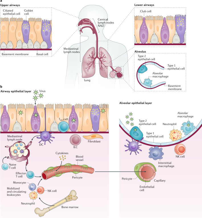
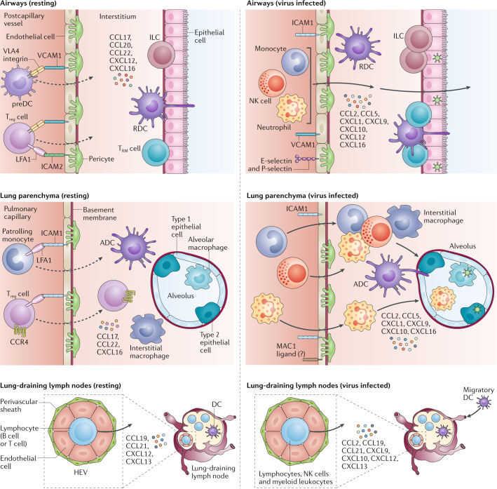
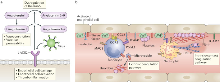
Severe acute respiratory syndrome coronavirus 2 (SARS-CoV-2) is the causative agent of coronavirus disease 2019 (COVID-19). Understanding of the fundamental processes underlying the versatile clinical manifestations of COVID-19 is incomplete without comprehension of how different immune cells are recruited to various compartments of virus-infected lungs, and how this recruitment differs among individuals with different levels of disease severity. As in other respiratory infections, leukocyte recruitment to the respiratory system in people with COVID-19 is orchestrated by specific leukocyte trafficking molecules, and when uncontrolled and excessive it results in various pathological complications, both in the lungs and in other organs. In the absence of experimental data from physiologically relevant animal models, our knowledge of the trafficking signals displayed by distinct vascular beds and epithelial cell layers in response to infection by SARS-CoV-2 is still incomplete. However, SARS-CoV-2 and influenza virus elicit partially conserved inflammatory responses in the different respiratory epithelial cells encountered early in infection and may trigger partially overlapping combinations of trafficking signals in nearby blood vessels. Here, we review the molecular signals orchestrating leukocyte trafficking to airway and lung compartments during primary pneumotropic influenza virus infections and discuss potential similarities to distinct courses of primary SARS-CoV-2 infections. We also discuss how an imbalance in vascular activation by leukocytes outside the airways and lungs may contribute to extrapulmonary inflammatory complications in subsets of patients with COVID-19. These multiple molecular pathways are potential targets for therapeutic interventions in patients with severe COVID-19.


Conflict of interest statement

The authors declare no competing interests.

Figures

Fig. 1
Fig. 1. The main molecular and cellular changes elicited by influenza virus infection of the respiratory system.
a | Shown are the major cell types (ciliated epithelial cells, goblet cells and basal cells) that form the epithelial layers of the nasal airway, lower airways and alveoli, which are targeted by respiratory viruses such as influenza virus and severe acute respiratory syndrome coronavirus 2 (SARS-CoV-2). An adult human lung contains more than 3 × 10⁸ alveoli, each covered by capillaries. The mediastinal lymph nodes are the major lung-draining lymph nodes. Some cervical lymph nodes and nasal-associated lymphoid tissue (NALT) play an additional role in priming antiviral adaptive immunity against viruses replicating in the upper airways. b | Events triggered by viral infection of the airway epithelial compartment (left) and of the alveolar epithelial compartment that constitute the lung parenchyma (right). The smooth muscle cell layers underneath the airway epithelial monolayers, neuroendocrine cells and the mucus layer on the apical aspects of these cells are omitted for clarity. Viral infection of both types of epithelial compartments triggers the release of inflammatory cytokines. These cytokines activate various epithelial-associated lymphocytes (such as innate lymphoid cells (ILCs) and resident memory T cells (TRM cells)), sentinel cells such as fibroblasts, interstitial macrophages and dendritic cells (DCs), and the pericytes associated with nearby blood vessels. In addition, some of the cytokines produced enter the circulation, reach the bone marrow and trigger generation and mobilization of innate leukocytes such as neutrophils, natural killer (NK) cells and monocytes, which are critical for viral clearance. The various blood vessels near the infected epithelial cells display arrays of leukocyte trafficking molecules (not shown), which are recognized by the circulating bone marrow-mobilized innate leukocytes. These various immune cells can then emigrate to the airway vessels and the capillaries of the virus-infected airway and alveolar compartments (Fig. 2). Virus-infected epithelial cells either die and release viral fragments or release live virus. Both fragments and particles are taken up and are processed by airway (respiratory) and alveolar DCs. On uptake and stimulation by viral particles, these different DCs leave their tissue, enter the lymphatic vessels and migrate into the draining lymph nodes (mediastinal lymph nodes). Naive T cells undergo priming by specific viral antigens in the T cell area of the lymph node, and antigen-activated CD4+ T cells then enter B cell follicles, where they provide critical signals to naive virus antigen-specific B cells, which subsequently become antibody-producing B cells (not shown). Within several days, T cells differentiate into effector T cells and egress from the lymph node, enter the blood vessels and migrate to the site of infection. These T cells are now equipped with specific receptors that allow them to emigrate through the inflamed airway blood vessels and/or the inflamed capillaries.
Fig. 2
Fig. 2. Trafficking of leukocytes to resting (left) and influenza virus-infected lungs (right).
Leukocyte trafficking is mediated by specific adhesion molecules, chemokines and their respective receptors expressed by leukocytes. Three major vascular beds (postcapillary vessels in the respiratory tract, alveolar capillaries in the lung parenchyma and high endothelial venules (HEVs) in the draining lymph nodes) and their adjacent epithelial cells are indicated. Dashed arrows depict the low-level homeostatic emigration of circulating immune cells through the different types of blood vessel. The vessels are surrounded by basement membrane and pericytes. Other sentinel cells depicted in Fig. 1 are omitted. Top panel: in the resting state, endothelial cells of postcapillary vessels near the lower airways constitutively express integrin ligands (VCAM1 and ICAM2) and present homeostatic chemokines (CCL17, CCL20, CCL22 and CXCL16) in gradients across the vessels and epithelial cell layers. These trafficking signals mainly promote the low-level entry of leukocyte precursors (for example, precursor dendritic cells (preDCs)) and regulatory T cells (Treg cells). On viral infection, the inducible expression of trafficking molecules such as endothelial selectins (E-selectin and P-selectin), pro-inflammatory chemokines such as CCL2, CCL5, CXCL1, CXCL9 and CXCL10, and de novo transcribed ICAM1, together with VCAM1 and ICAM2, triggers massive emigration (thick arrows) of various innate immune cells (such as neutrophils and inflammatory monocytes) and natural killer (NK) cells from blood towards the infected airways. During later stages of infection, virus-specific CD8+ and CD4+ effector T cells (not shown) enter these infected compartments. Homeostatic chemokines (such as CXCL16) allow recruited CD8+ effector T cells to differentiate into epithelial-associated resident memory T cells (TRM cells). Uncontrolled viral spread or dysregulated epithelial and endothelial cell activation results in destructive leukocyte recruitment. During infection, resident innate lymphoid cells (ILCs) and TRM cells proliferate, and DCs carrying viral antigens either enter lymphatic vessels or stay in the inflamed tissue, where they present viral antigens to recruited effector T cells. Unresolved infection gives rise to inducible bronchus-associated lymphoid tissue, which recruits naive and effector T and B cells through HEVs (not shown). Middle panel: in the resting state, distinct integrin ligands, such as ICAM1, are constitutively expressed by resting endothelial cells that line the pulmonary capillaries in the lung parenchyma. Various homeostatic chemokines (such as CCL17, CCL22 and CXCL16) are secreted by nearby alveolar epithelial cells and endothelial cells that line the capillaries, and these chemokines promote the low-level entry of Treg cells and leukocyte precursors such as preDCs (not shown). In addition, patrolling monocytes crawl on all these endothelial cells using endothelial ICAM1 and remove dying endothelial cells. Alveolar macrophages are attached to the epithelial cells that constitute individual alveoli and capture airborne particles without eliciting inflammation. Just like in the respiratory tract, on viral infection, the emigration of multiple types of immune cell towards the infected alveolus is guided by enhanced expression of chemokines. These are produced primarily by individual infected epithelial cells and by virus-infected alveolar macrophages. Myeloid leukocytes may use their integrin macrophage 1 antigen (MAC1) to attach to and crawl along inflamed capillaries by as yet unidentified ligands. Bottom panel: constitutive expression of integrin ligands (not shown) on the endothelial cells lining specialized lymph node blood vessels called HEVs and the chemokines displayed by these cells, such as CCL19, CCL21, CXCL12 and CXCL13, allows the constant entry of recirculating naive T and B lymphocytes into lung-draining lymph nodes. On viral infection, in addition to naive lymphocytes, NK cells and specific myeloid leukocytes can emigrate from the HEVs, responding to signals from inflammatory chemokines such as CCL2, CXCL9 and CXCL10 displayed on these venules in an inducible manner in addition to the constitutively displayed chemokines CCL19, CXCL12 and CXCL13. For more details on individual trafficking molecules used by these different leukocytes to cross each of these vascular beds, please refer to Table 2. ADCs, alveolar DCs; LFA1, lymphocyte function-associated antigen 1; RDCs, respiratory DCs; VLA4, very late antigen 4. Adapted from ref., Springer Nature Limited.
Fig. 3
Fig. 3. Involvement of endothelial and leukocyte trafficking molecules in the vascular pathophysiology of COVID-19 in the lungs and other organs.
a | Dysregulation of the renin–angiotensin–aldosterone system (RAAS) as a consequence of the downregulation of angiotensin-converting enzyme 2 (ACE2) by viral binding leads to decreased cleavage of angiotensin I and angiotensin II, resulting in elevated vasoconstriction and increased vascular permeability. Viral binding also results in endothelial cell damage, endothelial cell activation and thromboinflammation. b | Thromboinflammation in blood vessels is driven by the activation of endothelial cells and blood monocytes, which increase tissue factor membrane expression. Tissue factor is a key driver of thrombin generation. Severe acute respiratory syndrome coronavirus 2 (SARS-CoV-2)-infected or damaged endothelial cells, as well as endothelial cells stimulated by systemic or locally produced cytokines, upregulate the expression of adhesion molecules such as ICAM1 and of monocyte and neutrophil chemoattractants such as CXCL1 (not shown) and CCL2. The binding of thrombin to endothelial receptors (not shown) mobilizes vesicles containing prestored P-selectin and von Willebrand factor (vWF) (not shown), which, in turn, facilitate the recruitment of neutrophils and bind to and activate circulating platelets, respectively. The endothelial cell damage induced by the virus also exposes endothelial tissue factor, which further amplifies platelet deposition and thrombus formation. Moreover, monocytes and neutrophils are recruited to the damaged vessels by deposited platelets. Monocyte-derived tissue factor-rich microvesicles also activate the extrinsic coagulation pathway. Neutrophils recruited by the damaged endothelial cells and platelets can release neutrophil extracellular traps (NETs), which activate the intrinsic (contact activation) coagulation pathway, leading to massive fibrin deposition and blood clotting. Both neutrophils and monocytes express the integrin macrophage 1 antigen (MAC1), which allows them to bind to the damaged endothelial cells, activated platelets and deposited fibrin. COVID-19, coronavirus disease 2019. Adapted from ref. and ref., Springer Nature Limited.

Similar articles

Cited by

References

    1. Holt PG, Strickland DH, Wikstrom ME, Jahnsen FL. Regulation of immunological homeostasis in the respiratory tract. Nat. Rev. Immunol. 2008;8:142–152. - PubMed
    1. Tay MZ, Poh CM, Rénia L, MacAry PA, Ng LFP. The trinity of COVID-19: immunity, inflammation and intervention. Nat. Rev. Immunol. 2020 doi: 10.1038/s41577-020-0311-8. - DOI - PMC - PubMed
    1. Lazear HM, Schoggins JW, Diamond MS. Shared and distinct functions of type I and type III interferons. Immunity. 2019;50:907–923. - PMC - PubMed
    1. Thoms M, et al. Structural basis for translational shutdown and immune evasion by the Nsp1 protein of SARS-CoV-2. Science. 2020 doi: 10.1126/science.abc8665. - DOI - PMC - PubMed
    1. Blanco-Melo D, et al. Imbalanced host response to SARS-CoV-2 drives development of COVID-19. Cell. 2020;181:1036–1045.e9. - PMC - PubMed

Publication types