Case Reports
. 2020 Jun 15:9:604.
doi: 10.12688/f1000research.24353.2. eCollection 2020.

Case Report: Simultaneously diagnosed gastric adenocarcinoma and pernicious anemia - a classic association

Affiliations
Case Reports

Case Report: Simultaneously diagnosed gastric adenocarcinoma and pernicious anemia - a classic association

Syed Kamran et al. F1000Res. .

Abstract

Primary gastric cancer remains one of the most prevalent malignancies worldwide. Often patients remain asymptomatic until it is detected at an advanced stage with a poor prognosis. Thus, it's characteristically difficult to initially diagnose until it becomes late stage, at which point prognosis becomes poor. Pernicious anemia is a classic risk factor for the development of primary gastric cancer, but is uncommonly seen in clinical practice. Over time, patients who produce the autoantibodies to intrinsic factor that cause pernicious anemia typically will present initially with clinically significant megaloblastic anemia and peripheral neuropathy. However, patients can also present with more nonspecific signs and symptoms. Thus, clinicians should remain vigilant as circulating anti-intrinsic factor antibodies only worsen the disease over time and increase the risk of developing primary gastric cancer. This report not only presents the rare concurrent diagnosis of pernicious anemia and gastric cancer, but also aims to increase clinical awareness of these two conditions' classic association because early diagnosis and treatment significantly impacts morbidity and mortality.

Keywords: Autoimmune gastritis; Gastric adenocarcinoma; Parietal cells; Pernicious anemia; Stomach cancer.


Conflict of interest statement

No competing interests were disclosed.

Figures

Figure 1.
Figure 1.. Abdominopelvic CT scan with contrast is primarily nonrevealing for a malignant process.
Nonspecific gastric fold thickening in the fundus is observed ( arrow). An incidental finding in the liver was noted by a small focal hypoattenuation in the middle segment of the left lobe of the liver adjacent to the fissure for ligamentum teres ( arrowhead). This nodule was confirmed as PET-negative on later PET/CT studies.
Figure 2.
Figure 2.. EGD demonstrates esophagitis and an extensive ulcer involving the entire lesser curvature of the stomach.
(From proximal to distal.) ( A) Esophagitis. ( B) Gastro-esophageal junction. ( C) More esophagitis, and a tongue of columnar mucosa. ( D) Normal gastric cardia. ( E) Normal gastric fundus. ( F) Cavernous ulcer along the incisura (lesser curvature) with debris, food particles, and some central exudates ( G) Continued ulcer description. At the 12 o'clock position the scope is originating from the gastric cardia and fundus region. At 3 o'clock is the expected location of the pylorus. However, due to size and extent of the ulcer typical anatomy and landmarks were considerably distorted making visualization of the fundus from that particular EGD position not possible. At 6 o'clock the ulcer is shown to extend along the incisura. At 9 o'clock food debris is seen along the greater curvature.
Figure 3.
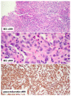
Figure 3.. Pathology demonstrates an invasive poorly differentiated adenocarcinoma.
Figure 4.
Figure 4.. Pre-chemotherapy staging PET scan shows a locally advanced gastric cancer.
PET from skull to mid-thigh reveals extensive, diffuse hypermetabolism throughout the gastric wall compatible with a PET-avid infiltrating gastric neoplasm. Imaging revealed involvement of at least one hepatogastric lymph node. Thus, the patient was determined to have stage III disease (T3N1). Scattered areas of contrast uptake within the bowel are likely physiologic and limit evaluation for lesions. Contrast uptake within the brain and genitourinary system are physiologic.
Figure 5.
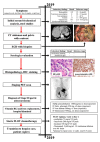
Figure 5.. Case report timeline.
Presented according to CARE guidelines.

References

    1. Rawla P, Barsouk A: Epidemiology of gastric cancer: global trends, risk factors and prevention. Prz Gastroenterol. 2019;14(1):26–38. 10.5114/pg.2018.80001 - DOI - PMC - PubMed
    1. Maconi G, Manes G, Porro GB: Role of symptoms in diagnosis and outcome of gastric cancer. World J Gastroenterol. 2008;14(8):1149–55. 10.3748/wjg.14.1149 - DOI - PMC - PubMed
    1. Layke JC, Lopez PP: Gastric cancer: diagnosis and treatment options. Am Fam Physician. 2004;69(5):1133–40. - PubMed
    1. Zali H, Rezaei-Tavirani M, Azodi M: Gastric cancer: prevention, risk factors and treatment. Gastroenterol Hepatol Bed Bench. 2011;4(4):175–85. - PMC - PubMed
    1. Pernicious Anemia.2019; Accessed: May 6, 2020. Reference Source

Publication types