

Linker histone H1.5 is an underestimated factor in differentiation and carcinogenesis
- PMID: 33214908
- PMCID: PMC7660118
- DOI: 10.1093/eep/dvaa013
Linker histone H1.5 is an underestimated factor in differentiation and carcinogenesis
Abstract
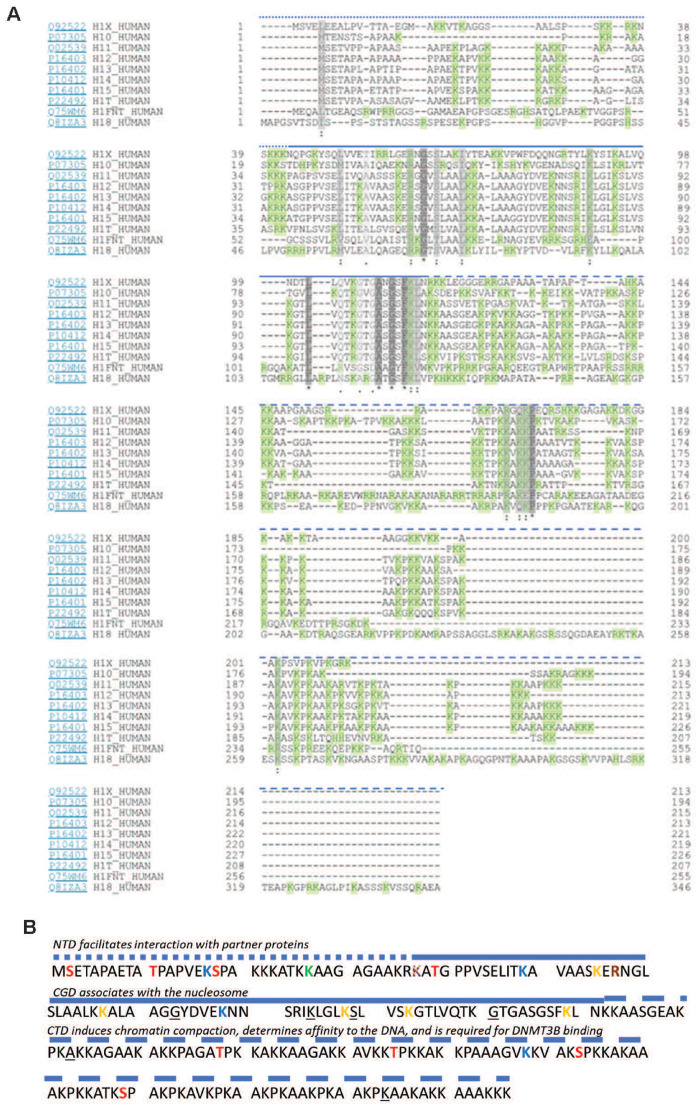
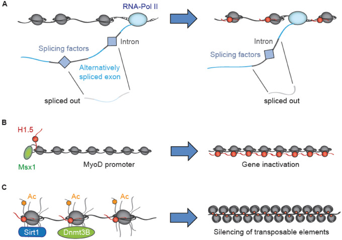
Human histone H1.5, in mice called H1b, belongs to the family of linker histones (H1), which are key players in chromatin organization. These proteins sit on top of nucleosomes, in part to stabilize them, and recruit core histone modifying enzymes. Through subtype-specific deposition patterns and numerous post-translational modifications, they fine-tune gene expression and chromatin architecture, and help to control cell fate and homeostasis. However, even though it is increasingly implicated in mammalian development, H1.5 has not received as much research attention as its relatives. Recent studies have focused on its prognostic value in cancer patients and its contribution to tumorigenesis through specific molecular mechanisms. However, many functions of H1.5 are still poorly understood. In this review, we will summarize what is currently known about H1.5 and its function in cell differentiation and carcinogenesis. We will suggest key experiments that are required to understand the molecular network, in which H1.5 is embedded. These experiments will advance our understanding of the epigenetic reprogramming occurring in developmental and carcinogenic processes.
Keywords: H1.5; cancer; development; histone isoforms; histones.
© The Author(s) 2020. Published by Oxford University Press.
Figures
Similar articles
-
The H1 linker histones: multifunctional proteins beyond the nucleosomal core particle.EMBO Rep. 2015 Nov;16(11):1439-53. doi: 10.15252/embr.201540749. Epub 2015 Oct 15. EMBO Rep. 2015. PMID: 26474902 Free PMC article. Review.
-
Linker histones in hormonal gene regulation.Biochim Biophys Acta. 2016 Mar;1859(3):520-5. doi: 10.1016/j.bbagrm.2015.10.016. Epub 2015 Oct 27. Biochim Biophys Acta. 2016. PMID: 26518266 Review.
-
Human linker histones: interplay between phosphorylation and O-β-GlcNAc to mediate chromatin structural modifications.Cell Div. 2011 Jul 12;6:15. doi: 10.1186/1747-1028-6-15. Cell Div. 2011. PMID: 21749719 Free PMC article.
-
Role of H1 linker histones in mammalian development and stem cell differentiation.Biochim Biophys Acta. 2016 Mar;1859(3):496-509. doi: 10.1016/j.bbagrm.2015.12.002. Epub 2015 Dec 13. Biochim Biophys Acta. 2016. PMID: 26689747 Free PMC article. Review.
-
Linker histone subtypes and their allelic variants.Cell Biol Int. 2012 Nov 1;36(11):981-96. doi: 10.1042/CBI20120133. Cell Biol Int. 2012. PMID: 23075301 Review.
Cited by
-
Linker histone H1 modulates defense priming and immunity in plants.Nucleic Acids Res. 2023 May 22;51(9):4252-4265. doi: 10.1093/nar/gkad106. Nucleic Acids Res. 2023. PMID: 36840717 Free PMC article.
-
Biochemical role of FOXM1-dependent histone linker H1B in human epidermal stem cells.Cell Death Dis. 2024 Jul 17;15(7):508. doi: 10.1038/s41419-024-06905-1. Cell Death Dis. 2024. PMID: 39019868 Free PMC article.
-
Post-Translation Modifications and Mutations of Human Linker Histone Subtypes: Their Manifestation in Disease.Int J Mol Sci. 2023 Jan 11;24(2):1463. doi: 10.3390/ijms24021463. Int J Mol Sci. 2023. PMID: 36674981 Free PMC article. Review.
-
Functional analysis of tumor-derived immunoglobulin lambda and its interacting proteins in cervical cancer.BMC Cancer. 2023 Oct 2;23(1):929. doi: 10.1186/s12885-023-11426-9. BMC Cancer. 2023. PMID: 37784026 Free PMC article.
-
HIST1H1B Promotes Basal-Like Breast Cancer Progression by Modulating CSF2 Expression.Front Oncol. 2021 Oct 22;11:780094. doi: 10.3389/fonc.2021.780094. eCollection 2021. Front Oncol. 2021. PMID: 34746019 Free PMC article.
References
-
- Rattray AMJ, Müller B. The control of histone gene expression. Biochem Soc Trans 2012;40:880–5. - PubMed
Publication types
LinkOut - more resources
Full Text Sources
