. 2021 Jan;23(1):64.
doi: 10.3892/mmr.2020.11702. Epub 2020 Nov 20.

S100a8 silencing attenuates inflammation, oxidative stress and apoptosis in BV2 cells induced by oxygen‑glucose deprivation and reoxygenation by upregulating GAB1 expression

Affiliations

S100a8 silencing attenuates inflammation, oxidative stress and apoptosis in BV2 cells induced by oxygen‑glucose deprivation and reoxygenation by upregulating GAB1 expression

Wenguang Hu et al. Mol Med Rep. 2021 Jan.

Abstract

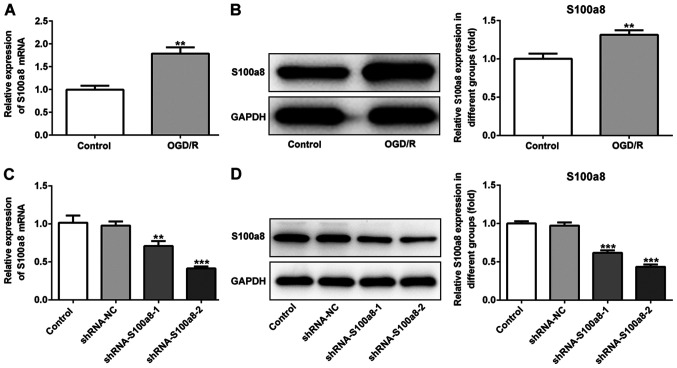
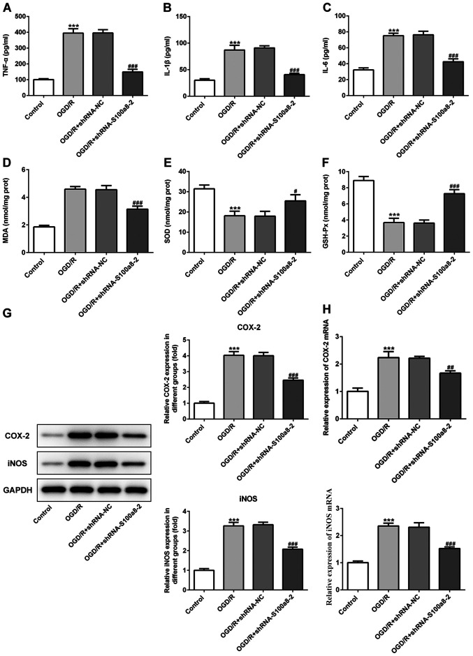
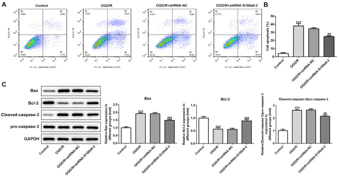
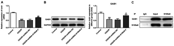
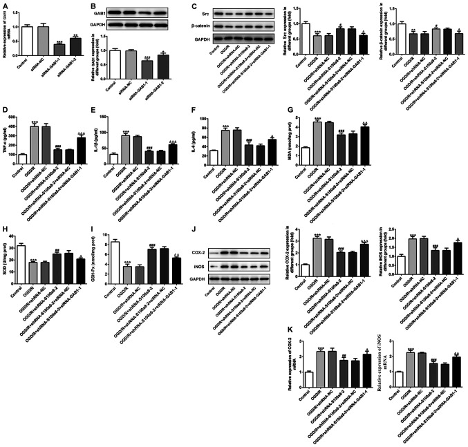
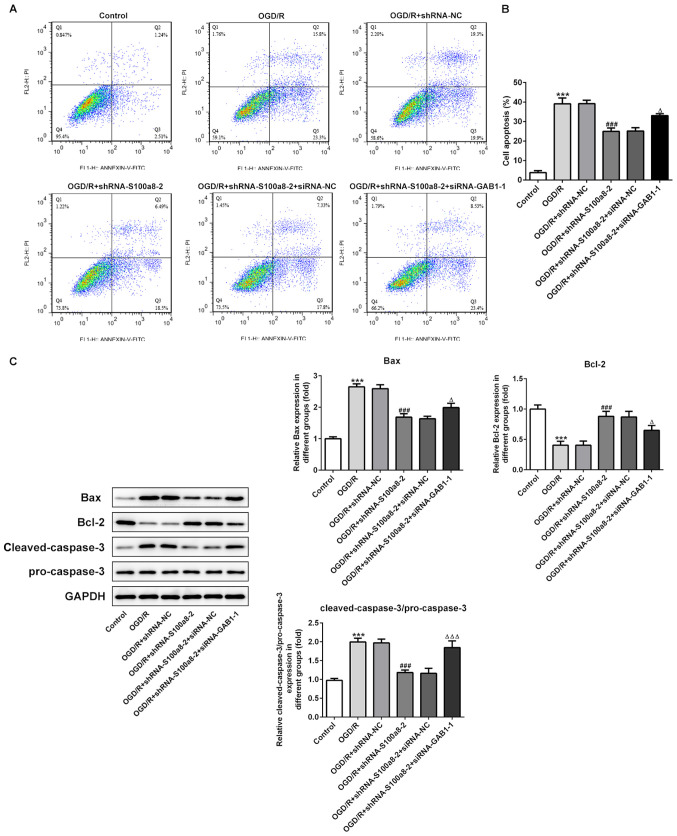
S100a8 serves an important role in cell differentiation and is abnormally expressed in common tumors, but there are few studies on the association between S100a8 and brain I/R injury. The present study aimed to investigate the role of S100a8 in oxygen‑glucose deprivation and reoxygenation (OGD/R)‑induced BV2 microglia cell injury, and to elucidate the potential underlying molecular mechanisms. BV2 cells were exposed to OGD/R to mimic ischemia/reperfusion (I/R) injury in vitro. S100a8 expression was detected via reverse transcription‑quantitative PCR and western blot analyses. Following transfection with short hairpin RNAs targeting S100a8, the levels of inflammatory cytokines and oxidative stress‑related factors were determined using commercial kits. Apoptosis was assessed using flow cytometric analysis and the expression levels of apoptosis‑related proteins were determined using western blot analysis. Subsequently, the mRNA and protein levels of Grb2‑associated binder 1 (GAB1) were assessed following S100a8 silencing. Immunoprecipitation (IP) was performed to verify the association between S100a8 and GAB1. The levels of inflammation, oxidative stress and apoptosis were assessed following GAB1 silencing, along with S100a8 silencing in BV2 cells subjected to OGD/R. The results indicated that exposure to OGD/R markedly upregulated S100a8 expression in BV2 cells. S100a8 silencing inhibited inflammation, oxidative stress and apoptosis, accompanied by changes in the expression of related proteins. The IP assay revealed a strong interaction between GAB1 and S100a8. In addition, GAB1 silencing reversed the inhibitory effects of S100a8 silencing on inflammation, oxidative stress and apoptosis in OGD/R‑stimulated BV2 cells. Taken together, the results of the present study demonstrated that S100a8 silencing alleviated inflammation, oxidative stress and the apoptosis of BV2 cells induced by OGD/R, partly by upregulating the expression of GAB1. Thus, these findings may potentially provide a novel direction to develop therapeutic strategies for cerebral I/R injury.

Keywords: inflammation; apoptosis; oxygen‑glucose deprivation; S100a8; Grb2‑associated binder 1.


Figures

Figure 1.
Figure 1.
S100a8 is highly expressed in OGD/R-exposed BV2 cells. (A) RT-qPCR and (B) western blot analyses were performed to determine S100a8 expression in BV2 cells following exposure to OGD/R. (C) RT-qPCR and (D) western blot analyses were performed to determine S100a8 expression in BV2 cells following transfection with shRNA-S100a8-1 or shRNA-S100a8-2. **P<0.01 and ***P<0.001 vs. the control. OGD/R, oxygen-glucose deprivation and reoxygenation; RT-qPCR, reverse transcription-quantitative PCR; sh, short hairpin; NC, negative control.
Figure 2.
Figure 2.
S100a8 silencing suppresses inflammation and oxidative stress in OGD/R-exposed BV2 cells. The expression levels of (A) TNF-α, (B) IL-1β and (C) IL-6 were assessed using ELISA kits. The concentration of (D) MDA, as well as the activities of (E) SOD and (F) GSH-Px were determined using commercial kits. (G) Western blot and (H) reverse transcription-quantitative PCR analyses were performed to determine the expression of COX-2 and iNOS following transfection with shRNA-S100a8-2. ***P<0.001 vs. control; #P<0.05, ##P<0.01, ###P<0.001 vs. OGD/R + shRNA-NC. OGD/R, oxygen-glucose deprivation and reoxygenation; TNF-α, tumor necrosis factor α; IL, interleukin; MDA, malondialdehyde; SOD, superoxide dismutase; GSH-Px, glutathione peroxidase; COX-2, cyclooxygenase-2; iNOS, induced nitric oxide synthase; sh, short hairpin; NC, negative control.
Figure 3.
Figure 3.
S100a8 silencing restrains apoptosis in OGD/R-exposed BV2 cells. (A) Apoptosis of BV2 cells was assessed via flow cytometric analysis. (B) Quantifications of apoptotic rate. (C) Expression of apoptosis-related proteins was assessed via western blot analysis. ***P<0.001 vs. control; ##P<0.01, ###P<0.001 vs. OGD/R + shRNA-NC. OGD/R, oxygen-glucose deprivation and reoxygenation; sh, short hairpin; NC, negative control.
Figure 4.
Figure 4.
GAB1 directly interacts with S100a8. (A) Reverse transcription-quantitative PCR and (B) western blot analyses were performed to determine GAB1 expression following transfection with shRNA-S100a8-2. (C) IP was performed in BV2 cells and the immunocomplexes were subjected to western blot analysis. **P<0.01, ***P<0.001 vs. control; #P<0.05, ##P<0.01 vs. OGD/R + shRNA-NC. OGD/R, oxygen-glucose deprivation and reoxygenation; GAB1, Grb2-associated binder 1; sh, short hairpin; NC, negative control; IP, immunoprecipitation.
Figure 5.
Figure 5.
GAB1 silencing alleviates the inhibitory effects of S100a8 silencing on inflammation and oxidative stress in OGD/R-exposed BV2 cells. (A) RT-qPCR and (B) western blot analyses were performed to determine GAB1 expression following transfection with siRNA-GAB1-1 or siRNA-GAB1-2. (C) Expression of downstream targets of GAB1, including Src and β-catenin was assessed via western blot analysis. Expression levels of (D) TNF-α, (E) IL-1β and (F) IL-6 were determined using ELISA kits. The concentration of (G) MDA, as well as the activities of (H) SOD and (I) GSH-Px were determined using commercial kits. (J) Western blot and (K) RT-qPCR analyses were performed to determine the expression levels of COX-2 and iNOS. *P<0.05, **P<0.01, ***P<0.001 vs. control; #P<0.05, ##P<0.01, ###P<0.001 vs. OGD/R + shRNA-NC; ΔP<0.05, ΔΔP<0.01, ΔΔΔP<0.001 vs. OGD/R + shRNA-S100a8-2 + siRNA-NC. GAB1, Grb2-associated binder 1; OGD/R, oxygen-glucose deprivation and reoxygenation; RT-qPCR, reverse transcription-quantitative PCR; si, small interfering; TNF-α, tumor necrosis factor α; IL, interleukin; MDA, malondialdehyde; SOD, superoxide dismutase; GSH-Px, glutathione peroxidase; COX-2, cyclooxygenase-2; iNOS, induced nitric oxide synthase; sh, short hairpin; NC, negative control.
Figure 6.
Figure 6.
GAB1 silencing abrogates the inhibitory effects of S100a8 silencing on the apoptosis of OGD/R-exposed BV2 cells. (A) Apoptosis of BV2 cells was assessed via flow cytometric analysis. (B) Quantifications of apoptotic rate. (C) The expression of apoptosis-related proteins was assessed via western blot analysis. ***P<0.001 vs. the control; ###P<0.001 vs. OGD/R + shRNA-NC; ΔP<0.05, ΔΔΔP<0.001 vs. OGD/R + shRNA-S100a8-2 + siRNA-NC. GAB1, Grb2-associated binder 1; OGD/R, oxygen-glucose deprivation and reoxygenation; sh, short hairpin; NC, negative control; si, small interfering.

Similar articles

Cited by

References

    1. Hayakawa K, Tanda K, Koshino S, Nishimura A, Kizaki Z, Ohno K. Pontine and cerebellar injury in neonatal hypoxic-ischemic encephalopathy: MRI features and clinical outcomes. Acta Radiol. 2020 Jan 24; doi: 10.1177/0284185119900442. (Epub ahead of print). doi: 10.1177/0284185119900442. - DOI - PubMed
    1. Douglas-Escobar M, Weiss MD. Hypoxic-ischemic encephalopathy: A review for the clinician. JAMA Pediatr. 2015;169:397–403. doi: 10.1001/jamapediatrics.2014.3269. - DOI - PubMed
    1. Dixon BJ, Reis C, Ho WM, Tang J, Zhang JH. Neuroprotective strategies after neonatal hypoxic ischemic encephalopathy. Int J Mol Sci. 2015;16:22368–22401. doi: 10.3390/ijms160922368. - DOI - PMC - PubMed
    1. Howlett JA, Northington FJ, Gilmore MM, Tekes A, Huisman TA, Parkinson C, Chung SE, Jennings JM, Jamrogowicz JJ, Larson AC, et al. Cerebrovascular autoregulation and neurologic injury in neonatal hypoxic-ischemic encephalopathy. Pediatr Res. 2013;74:525–535. doi: 10.1038/pr.2013.132. - DOI - PMC - PubMed
    1. Busl KM, Greer DM. Hypoxic-ischemic brain injury: Pathophysiology, neuropathology and mechanisms. NeuroRehabilitation. 2010;26:5–13. doi: 10.3233/NRE-2010-0531. - DOI - PubMed

MeSH terms