Review
. 2020 Dec;18(6):716-726.
doi: 10.1007/s11914-020-00640-0. Epub 2020 Nov 20.

Trabecular Architecture and Mechanical Heterogeneity Effects on Vertebral Body Strength

Affiliations
Review

Trabecular Architecture and Mechanical Heterogeneity Effects on Vertebral Body Strength

Joshua D Auger et al. Curr Osteoporos Rep. 2020 Dec.

Abstract

Purpose of review: We aimed to synthesize the recent work on the intra-vertebral heterogeneity in density, trabecular architecture and mechanical properties, its implications for fracture risk, its association with degeneration of the intervertebral discs, and its implications for implant design.

Recent findings: As compared to the peripheral regions of the centrum, the central region of the vertebral body exhibits lower density and more sparse microstructure. As compared to the anterior region, the posterior region shows higher density. These variations are more pronounced in vertebrae from older persons and in those adjacent to degenerated discs. Mixed results have been reported in regard to variation along the superior-inferior axis and to relationships between the heterogeneity in density and vertebral strength and fracture risk. These discrepancies highlight that, first, despite the large amount of study of the intra-vertebral heterogeneity in microstructure, direct study of that in mechanical properties has lagged, and second, more measurements of vertebral loading are needed to understand how the heterogeneity affects distributions of stress and strain in the vertebra. These future areas of study are relevant not only to the question of spine fractures but also to the design and selection of implants for spine fusion and disc replacement. The intra-vertebral heterogeneity in microstructure and mechanical properties may be a product of mechanical adaptation as well as a key determinant of the ability of the vertebral body to withstand a given type of loading.

Keywords: Density; Intervertebral disc; Loading; Microstructure; Vertebra.


Conflict of interest statement

Conflict of Interest

The authors declare no conflict of interest.

Figures

Figure 1.
Figure 1.
Components of the human vertebral body and adjacent tissues. The individual vertebra is split into the vertebral arch and vertebral body, with the arch to the posterior side. The vertebral body is made up of the trabecular centrum (containing trabecular bone) surrounded by a cortical shell. At the superior and inferior ends of this body is the endplate region. This area contains the bony vertebral endplate adjacent to the cartilage endplate. The intervertebral disc is located between the vertebrae, and consists of the gel-like nucleus pulposus surrounded by the comparatively more fibrous annulus fibrosus.
Figure 2.
Figure 2.
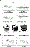
(A) Four different ratios of BMD between two different regions of the L3 vertebral body (defined in the schematic shown at the bottom), plotted against age for n=377 men and women; (B) BMD of the entire vertebral body (“integral BMD”) and of just the trabecular centrum (“Trabecular BMD”) for the same data set [11].
Figure 3.
Figure 3.
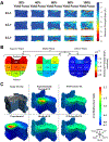
(A) Strains incurred on the surface of the L1 vertebral body during compression to yield, as measured by digital image correlation [42]. Each row shows a different vertebra. (Used with permission from Wolters Kluwer.) (B) Strains incurred in 27 different regions of the L1 trabecular centrum during compression to yield, as measured by digital volume correlation [16]. The color of each region corresponds to the median value over n=26 vertebrae, while the number that labels each region is the interquartile range over all vertebrae with the same units and on the same scale as the median values. *Difference between transverse planes. (C) Displacements incurred throughout the T8 vertebral body (representative specimen) during compression to just past the ultimate point, as measured by digital volume correlation (left column: top panel shows the microCT rendering before (grey) and upon (blue) loading to just past the ultimate point; bottom panel shows displacements) and as predicted by QCT-based FE simulations for four different types of boundary conditions (middle and right columns) [44]: using displacements measured by digital volume correlation across the endplates (“Experimentally Matched FE”); using uniform displacement boundary conditions (“Idealized FE”); using force boundary conditions calculated from distributions of intradiscal pressure averaged over intervertebral discs (IVDs) at different stages of degeneration (“IVD-Generic FE”); and using force boundary conditions calculated from distributions of intradiscal pressure averaged over intervertebral discs at the stage of degeneration exhibited by the given specimen (“IVD-Specific FE”). Positive values are downward displacements.

Similar articles

Cited by

References

    1. Mosekilde L (1998) The effect of modelling and remodelling on human vertebral body architecture. Technol Heal Care 6:287–297 - PubMed
    1. Ulrich D, Van Rietbergen B, Laib A, Rueegsegger P (1999) The ability of three-dimensional structural indices to reflect mechanical aspects of trabecular bone. Bone 25:55–60 - PubMed
    1. Carter DR, Hayes WC (1976) Bone compressive strength: the influence of density and strain rate. Science (80- ) 194:1174–1176 - PubMed
    1. Morgan EF, Bayraktar HH, Keaveny TM (2003) Trabecular bone modulus-density relationships depend on anatomic site. J Biomech 36:. 10.1016/S0021-9290(03)00071-X - DOI - PubMed
    1. Morgan EF, Keaveny TM (2001) Dependence of yield strain of human trabecular bone on anatomic site. J Biomech 34:. 10.1016/S0021-9290(01)00011-2 - DOI - PubMed

Publication types