. 2020 Nov;25(11):116503.
doi: 10.1117/1.JBO.25.11.116503.

Combined transmission, dark field and fluorescence microscopy for intact, 3D tissue analysis of biopsies

Affiliations

Combined transmission, dark field and fluorescence microscopy for intact, 3D tissue analysis of biopsies

Marius I Boamfa et al. J Biomed Opt. 2020 Nov.

Abstract

Significance: Currently, tissue biopsies are sectioned into 3- to 5-μm-thick slices that are used for conventional pathology analysis. Previous work by confocal microscopy and light-sheet microscopy has shown that analyzing biopsies intact in three-dimensions (3D) is possible and may lead to a better understanding of cancer growth patterns. Although accurate, these methods require fluorescent staining of the tissue, in addition to tissue clearing. If the 3D biopsy analysis could be done sufficiently swiftly, this approach may be used for on-site assessment of the adequacy of a biopsy taken.

Aim: We aim to show that, by transmission microscopy of optically cleared tissue punches, the tissue architecture can be determined without the need for fluorescent staining.

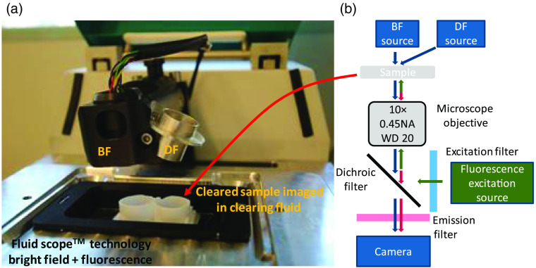
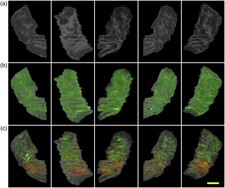
Approach: Transmission microscopy is used by combining bright field microscopy with dark field and epifluorescent microscopy to compare samples that have also been analyzed by fluorescent confocal microscopy.

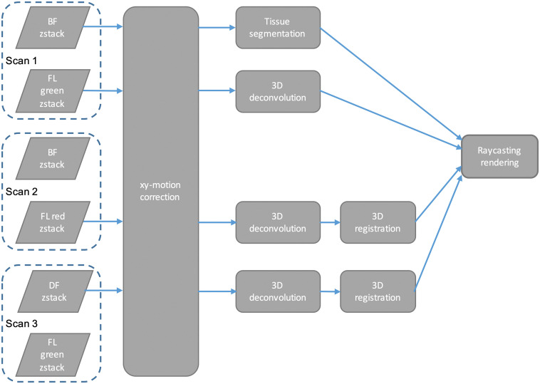
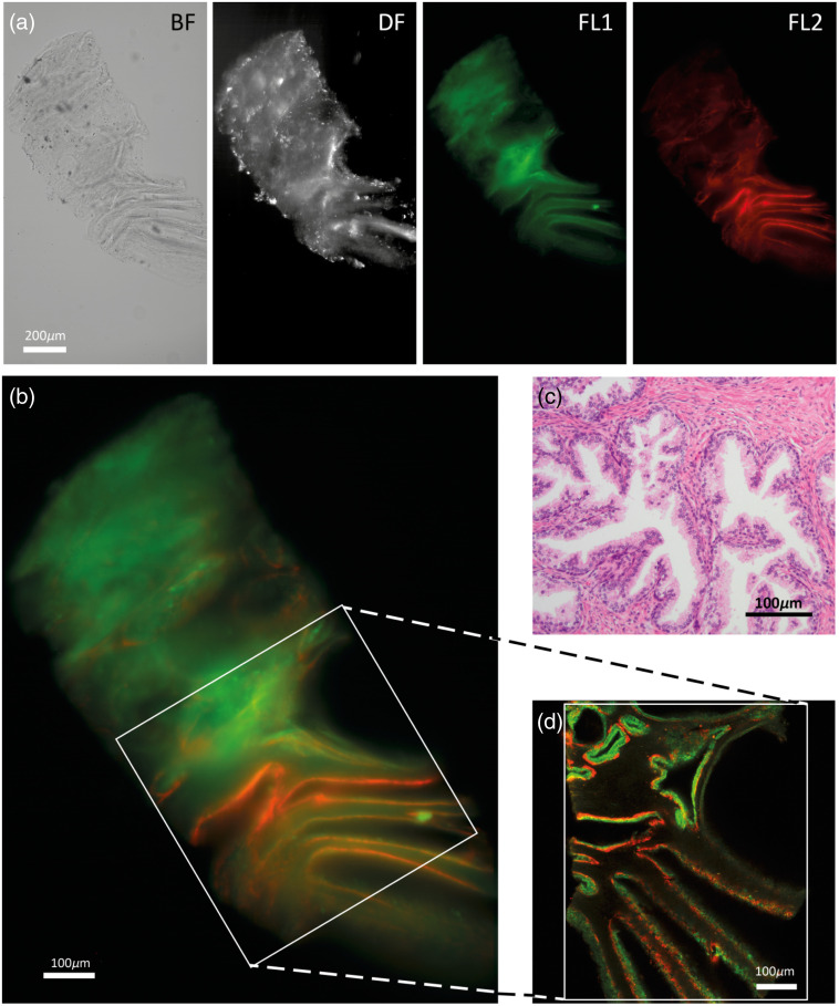
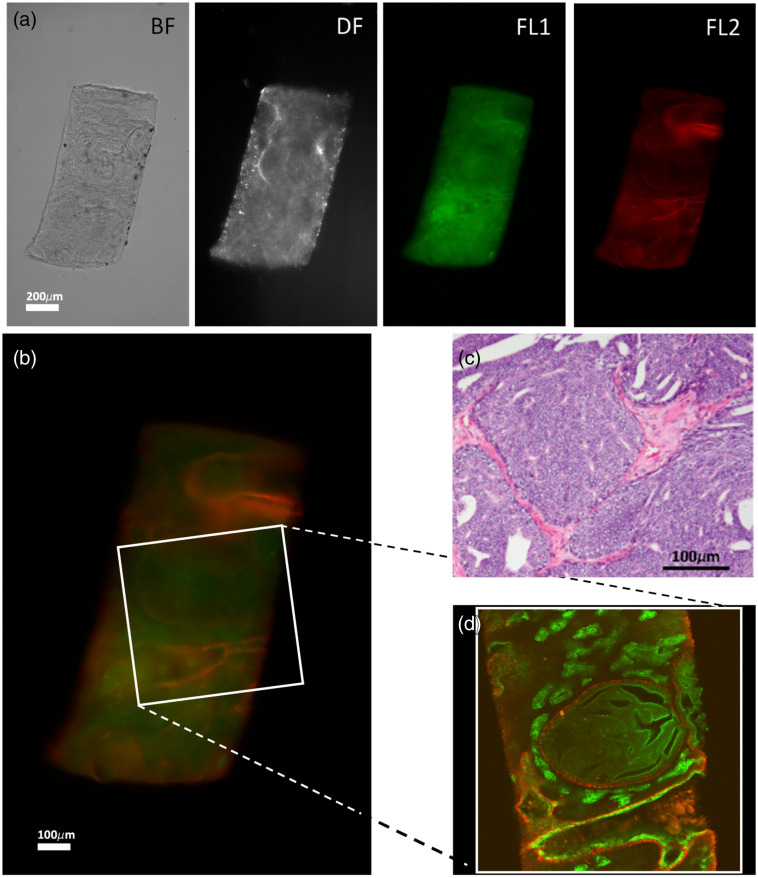
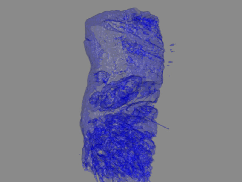
Results: With increasing distance to the focal plane, the higher-frequency part of the spatial frequency spectrum of transmitted light is attenuated increasingly. This property is exploited for tissue segmentation, detecting whether tissue is present at a certain position in the focal plane image. Using this approach, we show that a 3D rendering of the internal cavity or tubules structure of punch biopsies, which are up to 1-mm thick, can be acquired in ≈1 min scan time per imaging modality. The images of the overall tissue architecture that are obtained are similar to those from the confocal microscopy benchmark, without requiring fluorescent staining.

Conclusions: Images of the overall tissue architecture can be obtained from transmission microcopy; they are similar to those from the confocal microscopy benchmark without requiring fluorescent staining. Tissue clearing is still needed. The total scan time of the present method is significantly shorter at a fraction of the device costs.

Keywords: 3D biopsy imaging;; bright field imaging;; image processing;; optical imaging;; tissue clearing; transmission microscopy;.


Figures

Fig. 1
Fig. 1
Overview of the setup, showing the BF light source and the DF option. (a) DF imaging is be achieved by simply relocating the light source into the DF option holder. (b) Schematic representation of the setup and light path.
Fig. 2
Fig. 2
Image processing algorithm.
Fig. 3
Fig. 3
(a) and (b) BF, DF and Alexa488 and Alexa647 Fluorescence (FL1 and FL2, respectively) typical images of the z-stack obtained with the oCelloscope. The scale bar depicted in the right panel represents 200  μm. (c) H&E-stained tissue section, and (d) cross section of the confocal microscope showing the same structures with CK8-18 positive luminal epithelial cells (green) and CK5 positive basal epithelial cells (red) in benign transition zone prostate glands.
Fig. 4
Fig. 4
(a) and (b) BF, DF and Alexa488 and Alexa647 Fluorescence (FL1 and FL2, respectively) typical images of the z-stack obtained with the oCelloscope. The scale bar depicted in the right panel represents 200  μm. (c) H&E-stained tissue section, and (d) cross section of the confocal microscope showing the same structures with CK8-18 positive luminal epithelial cells (green) and CK5 positive basal epithelial cells (red) in a Gleason 4 prostate sample with ill-formed growth pattern with intraductal carcinoma.
Fig. 5
Fig. 5
Three-dimensional rendering of the ducts in a prostate sample using BF data and the tissue segmentation algorithm based on the spatial frequency spectrum as discusssed in Sec. 2.4. The ducts are detected in this 900-μm-thick optically cleared prostate biopsy sample. (Video 1, gives the 360° rotation movie of this sample. The clipping of the signal on the sample’s side is due to the biopsy resting on the bottom of the sample container.) (Video 1, mp4, 5324 KB [URL: https://doi.org/10.1117/1.JBO.25.11.116503.1]).
Fig. 6
Fig. 6
Projection of the ray-casting rendering with the sample in different orientations. (a) Tissue segmentation, (b) tissue segmentation (white) and DF signal (green), and (c) tissue segmentation (white) and two fluorescence channels, CK5 (red), CK 8-18 (green). The scale bar represents 250  μm Movies of the ray-casting renderings are available: Video 2 (top row), Video 3 (middle row) and Video 4 (bottom row). (Video 2, mp4, 1335 KB [URL: https://doi.org/10.1117/1.JBO.25.11.116503.2]; Video 3, mp4, 1339 KB [URL: https://doi.org/10.1117/1.JBO.25.11.116503.3]; Video 4, mp4, 1473 KB [URL: https://doi.org/10.1117/1.JBO.25.11.116503.4]).

References

    1. Weeber F., et al. , “Tumor organoids as a pre-clinical cancer model for drug discovery,” Cell Chem. Biol. 24(9), 1092–1100 (2017).10.1016/j.chembiol.2017.06.012 - DOI - PubMed
    1. Fong E. L. S., et al. , “3D culture as a clinically relevant model for personalized medicine,” SLAS Technol. 22(3), 245–253 (2017).10.1177/2472630317697251 - DOI - PubMed
    1. Breslin S., O’Driscoll L., “Three-dimensional cell culture: the missing link in drug discovery,” Drug Discov. Today 18(5–6), 240–249 (2013).DDTOFS10.1016/j.drudis.2012.10.003 - DOI - PubMed
    1. Helmchen F., Denk W., “Deep tissue two-photon microscopy,” Nat. Methods 2(12), 932–940 (2005).10.1038/nmeth818 - DOI - PubMed
    1. Richardson D. S., Lichtman J. W., “Clarifying tissue clearing,” Cell 162(2), 246–257 (2015).CELLB510.1016/j.cell.2015.06.067 - DOI - PMC - PubMed