

Utility of Plasma Neurofilament Light in the 1Florida Alzheimer's Disease Research Center (ADRC)
- PMID: 33216030
- PMCID: PMC7902971
- DOI: 10.3233/JAD-200901
Utility of Plasma Neurofilament Light in the 1Florida Alzheimer's Disease Research Center (ADRC)
Abstract
Background: Plasma NfL (pNfL) levels are elevated in many neurological disorders. However, the utility of pNfL in a clinical setting has not been established.
Objective: In a cohort of diverse older participants, we examined: 1) the association of pNfL to age, sex, Hispanic ethnicity, diagnosis, and structural and amyloid imaging biomarkers; and 2) its association to baseline and longitudinal cognitive and functional performance.
Methods: 309 subjects were classified at baseline as cognitively normal (CN) or with cognitive impairment. Most subjects had structural MRI and amyloid PET scans. The most frequent etiological diagnosis was Alzheimer's disease (AD), but other neurological and neuropsychiatric disorders were also represented. We assessed the relationship of pNfL to cognitive and functional status, primary etiology, imaging biomarkers, and to cognitive and functional decline.
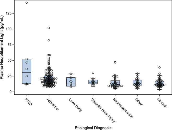
Results: pNfL increased with age, degree of hippocampal atrophy, and amyloid load, and was higher in females among CN subjects, but was not associated with Hispanic ethnicity. Compared to CN subjects, pNfL was elevated among those with AD or FTLD, but not those with neuropsychiatric or other disorders. Hippocampal atrophy, amyloid positivity and higher pNfL levels each added unique variance in predicting greater functional impairment on the CDR-SB at baseline. Higher baseline pNfL levels also predicted greater cognitive and functional decline after accounting for hippocampal atrophy and memory scores at baseline.
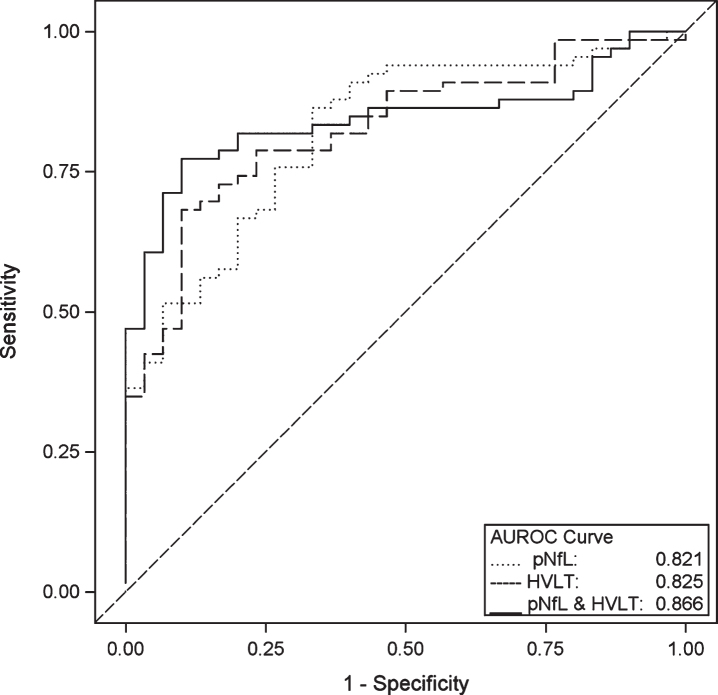
Conclusion: pNfL may have a complementary and supportive role to brain imaging and cognitive testing in a memory disorder evaluation, although its diagnostic sensitivity and specificity as a stand-alone measure is modest. In the absence of expensive neuroimaging tests, pNfL could be used for differentiating neurodegenerative disease from neuropsychiatric disorders.
Keywords: Alzheimer’s disease; amyloid; diagnosis; hippocampal atrophy; magnetic resonance imaging; plasma neurofilament light; positron emission tomography.
Conflict of interest statement
Authors’ disclosures available online (
Figures
Similar articles
-
Association Between Longitudinal Plasma Neurofilament Light and Neurodegeneration in Patients With Alzheimer Disease.JAMA Neurol. 2019 Jul 1;76(7):791-799. doi: 10.1001/jamaneurol.2019.0765. JAMA Neurol. 2019. PMID: 31009028 Free PMC article.
-
Performance of Plasma Biomarkers Combined with Structural MRI to Identify Candidate Participants for Alzheimer's Disease-Modifying Therapy.J Prev Alzheimers Dis. 2024;11(5):1198-1205. doi: 10.14283/jpad.2024.110. J Prev Alzheimers Dis. 2024. PMID: 39350364 Free PMC article.
-
Potential role of blood pressure variability and plasma neurofilament light in the mechanism of comorbidity between Alzheimer's disease and cerebral small vessel disease.Alzheimers Dement. 2024 Jul;20(7):4891-4902. doi: 10.1002/alz.14056. Epub 2024 Jun 19. Alzheimers Dement. 2024. PMID: 38895921 Free PMC article.
-
Molecular imaging of dementia.Psychogeriatrics. 2012 Jun;12(2):106-14. doi: 10.1111/j.1479-8301.2012.00409.x. Psychogeriatrics. 2012. PMID: 22712644 Review.
-
Molecular imaging of Alzheimer disease pathology.AJNR Am J Neuroradiol. 2014 Jun;35(6 Suppl):S12-7. doi: 10.3174/ajnr.A3847. Epub 2014 Feb 6. AJNR Am J Neuroradiol. 2014. PMID: 24503555 Free PMC article. Review.
Cited by
-
Association of Cognitive Impairment With Free Water in the Nucleus Basalis of Meynert and Locus Coeruleus to Transentorhinal Cortex Tract.Neurology. 2022 Feb 15;98(7):e700-e710. doi: 10.1212/WNL.0000000000013206. Epub 2021 Dec 14. Neurology. 2022. PMID: 34906980 Free PMC article.
-
Serum neurofilament light concentrations are associated with cortical thinning in anorexia nervosa.Psychol Med. 2023 Nov;53(15):7053-7061. doi: 10.1017/S0033291723000387. Epub 2023 Mar 27. Psychol Med. 2023. PMID: 36967674 Free PMC article.
-
Associations Between Vascular Risk Factors and Perivascular Spaces in Adults with Intact Cognition, Mild Cognitive Impairment, and Dementia.J Alzheimers Dis. 2022;89(2):437-448. doi: 10.3233/JAD-215585. J Alzheimers Dis. 2022. PMID: 35871327 Free PMC article.
-
Neuropathology, Neuroimaging, and Fluid Biomarkers in Alzheimer's Disease.Diagnostics (Basel). 2024 Mar 27;14(7):704. doi: 10.3390/diagnostics14070704. Diagnostics (Basel). 2024. PMID: 38611617 Free PMC article. Review.
-
Multi-Omic Blood Biomarkers as Dynamic Risk Predictors in Late-Onset Alzheimer's Disease.Int J Mol Sci. 2024 Jan 19;25(2):1231. doi: 10.3390/ijms25021231. Int J Mol Sci. 2024. PMID: 38279230 Free PMC article. Review.
References
-
- Sjogren M, Blomberg M, Jonsson M, Wahlund LO, Edman A, Lind K, Rosengren L, Blennow K, Wallin A (2001) Neurofilament protein in cerebrospinal fluid: A marker of white matter changes. J Neurosci Res 66, 510–516. - PubMed
-
- Idland AV, Sala-Llonch R, Borza T, Watne LO, Wyller TB, Brækhus A, Zetterberg H, Blennow K, Walhovd KB, Fjell AM (2017) CSF neurofilament light levels predict hippocampal atrophy in cognitively healthy older adults. Neurobiol Aging 49, 138–144. - PubMed
Publication types
MeSH terms
Substances
Grants and funding
LinkOut - more resources
Full Text Sources
Other Literature Sources
Medical
