Review
. 2021;8(2):169-183.
doi: 10.3233/JND-200568.

Molecular Mechanisms of Skeletal Muscle Hypertrophy

Affiliations
Review

Molecular Mechanisms of Skeletal Muscle Hypertrophy

Stefano Schiaffino et al. J Neuromuscul Dis. 2021.

Abstract

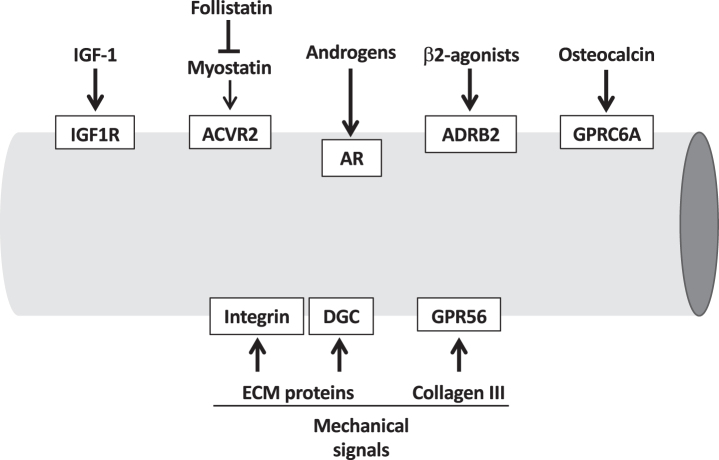
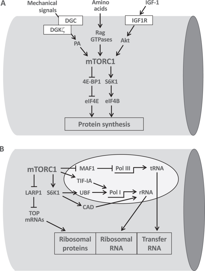
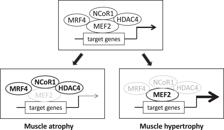
Skeletal muscle hypertrophy can be induced by hormones and growth factors acting directly as positive regulators of muscle growth or indirectly by neutralizing negative regulators, and by mechanical signals mediating the effect of resistance exercise. Muscle growth during hypertrophy is controlled at the translational level, through the stimulation of protein synthesis, and at the transcriptional level, through the activation of ribosomal RNAs and muscle-specific genes. mTORC1 has a central role in the regulation of both protein synthesis and ribosomal biogenesis. Several transcription factors and co-activators, including MEF2, SRF, PGC-1α4, and YAP promote the growth of the myofibers. Satellite cell proliferation and fusion is involved in some but not all muscle hypertrophy models.

Keywords: MEF2; SRF; Skeletal muscle; mTOR; muscle hypertrophy; ribosomal biogenesis; transcriptional control; translational control.


Conflict of interest statement

The authors have no conflict of interest to report.

Figures

Fig. 1
Fig. 1
Extracellular pro-hypertrophic signals, including hormo-nes and growth factors, and mechanical signals acting on the muscle cell membrane. The receptors mediating these signals are indicated in the white boxes. ACVR2, activin receptor type II; AR, androgen receptor; ADRB2, adrenergic receptor b2; DGC, dystrophin glycoprotein complex; GPRC6A, G Protein-Coupled ReceptorC6A; GPR56, G protein-coupled receptor 56; IGF1R, IGF-1 receptor.
Fig. 2
Fig. 2
The central role of mTORC1 in the translational and transcriptional control of protein synthesis. A. mTORC1 integrates the pro-hypertrophic input from growth factors, amino acids and mechanical signals. mTORC1 stimulates protein synthesis by acting at the translational level through phosphorylation of 4E-BP1 and S6K1 which in turn activate initiation factors eIF4E and eIF4B. B. mTORC1 controls ribosomal biogenesis at the transcriptional level by stimulating PolI-mediated synthesis of ribosomal RNA (rRNA) via TIF-1A and Pol III-mediated synthesis of transfer RNA (tRNA) via MAF1. S6K1 also activates PolI through UBF and stimulates pyrimidine biosynthesis required for rRNA synthesis by CAD phosphorylation. The translational activation of TOP mRNAs, controlled by mTORC1 via LARP1, leads to the formation of ribosomal proteins. See text for further details.
Fig. 3
Fig. 3
Wide interindividual variability in the hypertrophic response. Percent change in type II myofiber cross-sectional area (CSA) from pre- to post-resistance exercise training in older adults (age 60–75 yr) subjected for 4 wk to the same resistance exercise protocol. Based on the hypertrophic response, 3 groups were identified: nonresponders, moderate responders and extreme responders. Variations in resistance training-induced myofiber hypertrophy were found to correlate with parallel changes in markers of ribosomal biogenesis (Modified from [63]).
Fig. 4
Fig. 4
The transcriptional activity of myocyte enhancer factor-2 (MEF2) factors is controlled by different repressors. MEF2 factors promote muscle growth during development and in the adult by regulating the expression of muscle-specific genes. MEF2 transcriptional activity is controlled by different repressors, including muscle-specific repressors like myogenic regulatory factor 4 (MRF4, coded by MYF6) and ubiquitous repressor as nuclear receptor co-repressor 1 (NCoR1) and class II histone deacetylases (HDACs), like HDAC4. Under normal conditions (upper panel) muscle size is maintained in the adult by a balance between these inhibitory factors and different stimulatory influences, including MEF2 post-translational changes, not depicted in the scheme. Loss of repressor activity (lower right panel), such as muscle-specific knockout of NCoR1 or muscle-specific knockdown of MRF4, lead to upregulation of MEF2 transcriptional activity and muscle hypertrophy. Increased repressor function (lower left panel), e.g. denervation-induced up-regulation and nuclear translocation of MRF4 and HDAC4, reduce MEF2 transcriptional activity and contribute to muscle atrophy.
Fig. 5
Fig. 5
Signaling pathways regulating the activity of the transcription factor SRF (serum response factor). SRF is activated by high intensity resistance exercise via nuclear translocation of myocardin related transcription factor B (MRTF-B), which is induced by ERK-dependent phopshorylation on serine 66, and by actin polymerization induced by STARS and RhoA, thus relieving the G-actin inhibitory effect on MRTF. SRF activation also requires chromatin remodeling which is induced by histone 3 phosphorylation on serine 10 (H3S10). H3S10 phosphorylation is mediated by mitogen- and stress-activated kinases (MSK1/2), which are in turn activated by p38 MAPK. (Modified from [94]).

References

    1. Haun CT, Vann CG, Roberts BM, Vigotsky AD, Schoenfeld BJ, Roberts MD. A critical evaluation of the biological construct skeletal muscle hypertrophy: size matters but so does the measurement. Front Physiol. 2019;10:247. - PMC - PubMed
    1. Blaauw B, Schiaffino S, Reggiani C. Mechanisms modulating skeletal muscle phenotype. Compr Physiol. 2013;3(4):1645–87. - PubMed
    1. Schiaffino S, Dyar KA, Ciciliot S, Blaauw B, Sandri M. Mechanisms regulating skeletal muscle growth and atrophy. FEBS J. 2013;280(17):4294–314. - PubMed
    1. Spangenburg EE, Le Roith D, Ward CW, Bodine SC. A functional insulin-like growth factor receptor is not necessary for load-induced skeletal muscle hypertrophy. J Physiol. 2008;586(1):283–91. - PMC - PubMed
    1. Lee SJ, McPherron AC. Regulation of myostatin activity and muscle growth. Proc Natl Acad Sci U S A. 2001;98(16):9306–11. - PMC - PubMed

LinkOut - more resources