. 2022 Feb 15;225(4):661-674.
doi: 10.1093/infdis/jiaa714.

Human Immunodeficiency Virus-Infected Immunological Nonresponders Have Colon-Restricted Gut Mucosal Immune Dysfunction

Affiliations

Human Immunodeficiency Virus-Infected Immunological Nonresponders Have Colon-Restricted Gut Mucosal Immune Dysfunction

Malin Holm Meyer-Myklestad et al. J Infect Dis. .

Abstract

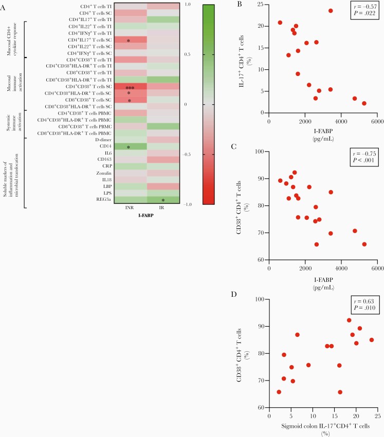
Background: Human immunodeficiency virus (HIV)-infected immunological nonresponders (INRs) fail to reconstitute their CD4+ T-cell pool after initiation of antiretroviral therapy, and their prognosis is inferior to that of immunological responders (IRs). A prevailing hypothesis is that the INR phenotype is caused by a persistently disrupted mucosal barrier, but assessments of gut mucosal immunology in different anatomical compartments are scarce.

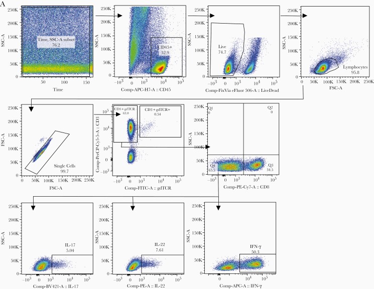
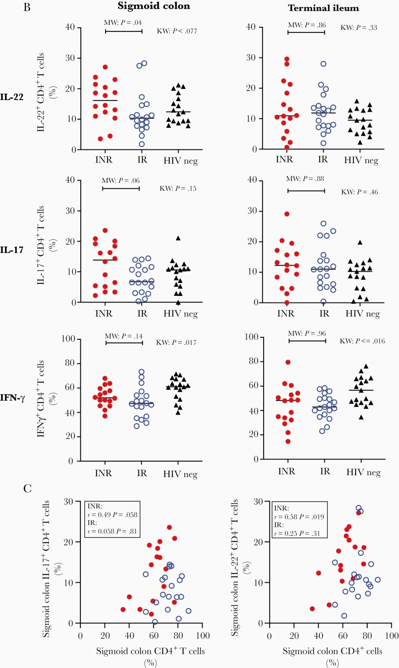
Methods: We investigated circulating markers of mucosal dysfunction, immune activation, mucosal Th17 and Th22 cells, and mucosa-adherent microbiota signatures in gut mucosal specimens from sigmoid colon and terminal ileum of 19 INRs and 20 IRs in addition to 20 HIV-negative individuals.

Results: INRs had higher blood levels of the enterocyte damage marker intestinal fatty acid-binding protein than IRs. In gut mucosal biopsies, INRs had lower fractions of CD4+ T cells, higher fractions of interleukin 22, and a tendency to higher fractions of interleukin 17-producing CD4+ T cells. These findings were all restricted to the colon and correlated to circulating markers of enterocyte damage. There were no observed differences in gut microbial composition between INRs and IRs.

Conclusions: Restricted to the colon, enterocyte damage and mucosal immune dysfunction play a role for insufficient immune reconstitution in HIV infection independent of the gut microbiota.

Keywords: HIV; gut microbiota; immunological nonresponders; mucosal immunology.


Figures

Figure 1.
Figure 1.
Higher level of enterocyte damage in immunological nonresponders (INR). A, Serum concentration of intestinal fatty acid–binding protein (I-FABP). B, Plasma concentration of regenerating islet-derived protein 3 alpha (REG3α) analyzed by enzyme-linked immunosorbent assay in INR compared to immunological responders (IR); human immunodeficiency virus (HIV)–negative individuals (HIV neg) served as controls. Horizontal bars represent median values. Kruskal–Wallis (KW) and Mann–Whitney (MW) test values. Spearman correlation between I-FABP and REG3α in people living with HIV (INR: dots, IR: open circles) (C) and HIV-negative individuals (triangles) (D).
Figure 2.
Figure 2.
Immunologic nonresponders (INR) have lower sigmoid colon CD4+ T-cell fractions that correlate with enterocyte damage. Fractions of CD4+ T cells of total T cells analyzed by flow cytometry on lamina propria mononuclear cells in sigmoid colon (A) and terminal ileum (B) of INR compared to immunological responders (IR); human immunodeficiency virus (HIV)–negative individuals (HIV neg) served as controls. Horizontal bars represent median values. Kruskal–Wallis (KW) and Mann–Whitney (MW) test values. Spearman correlation between CD4+ T-cell fractions of total T cells in sigmoid colon and terminal ileum in people living with HIV (INR: dots, IR: open circles) (C) and HIV-negative individuals (triangles) (D).
Figure 3.
Figure 3.
Higher interleukin (IL) 22 production in sigmoid colon CD4+ T cells in immunological nonresponders (INR). A, Representative flow cytometry analysis of mitogen-stimulated lamina propria mononuclear cells. B, Sigmoid colon fractions of IL-22+, IL-17+, and interferon (IFN)–γ + CD4+ T cells in INR compared to immunological responders (IR); human immunodeficiency virus (HIV)–negative individuals (HIV neg) served as controls. Terminal ileum fractions of IL-22+, IL-17+, and IFN-γ + CD4+ T cells in INR compared to IR, with HIV-negative individuals as controls. All analyses by flow cytometry on lamina propria mononuclear cells. Horizontal bars represent median values. Kruskal–Wallis (KW) and Mann–Whitney (MW) test values. C, Spearman correlation between sigmoid colon CD4+ T-cell fractions and sigmoid colon IL-17+ CD4+ T cells and sigmoid colon IL-22+ CD4+ T cells in people living with HIV (INR: dots, IR: open circles).
Figure 3.
Figure 3.
Higher interleukin (IL) 22 production in sigmoid colon CD4+ T cells in immunological nonresponders (INR). A, Representative flow cytometry analysis of mitogen-stimulated lamina propria mononuclear cells. B, Sigmoid colon fractions of IL-22+, IL-17+, and interferon (IFN)–γ + CD4+ T cells in INR compared to immunological responders (IR); human immunodeficiency virus (HIV)–negative individuals (HIV neg) served as controls. Terminal ileum fractions of IL-22+, IL-17+, and IFN-γ + CD4+ T cells in INR compared to IR, with HIV-negative individuals as controls. All analyses by flow cytometry on lamina propria mononuclear cells. Horizontal bars represent median values. Kruskal–Wallis (KW) and Mann–Whitney (MW) test values. C, Spearman correlation between sigmoid colon CD4+ T-cell fractions and sigmoid colon IL-17+ CD4+ T cells and sigmoid colon IL-22+ CD4+ T cells in people living with HIV (INR: dots, IR: open circles).
Figure 4.
Figure 4.
No differences in gut microbiota between immunological nonresponders (INR) and immunological responders (IR). A, Alpha diversity analyzed by Shannon index and observed operational taxonomic units (OTUs) for differences between INR, IR, and human immunodeficiency virus (HIV)–negative individuals (HIV neg) in sigmoid colon, terminal ileum, and feces. Horizontal bars represent median values. Kruskal–Wallis (KW) and Mann–Whitney (MW) test values. B, β-diversity analyzed by weighted UniFrac analyses of differences between INR, IR, and HIV-negative controls in sigmoid colon, terminal ileum, and feces. Permutational multivariate analysis of variance test values (INR: dots, IR: open circles, HIV negative: triangles). C, Relative abundance of taxonomic phylum (top 3 rows) and family (lower 3 rows) in sigmoid colon, terminal ileum, and feces of INR, IR, and HIV-negative controls.
Figure 4.
Figure 4.
No differences in gut microbiota between immunological nonresponders (INR) and immunological responders (IR). A, Alpha diversity analyzed by Shannon index and observed operational taxonomic units (OTUs) for differences between INR, IR, and human immunodeficiency virus (HIV)–negative individuals (HIV neg) in sigmoid colon, terminal ileum, and feces. Horizontal bars represent median values. Kruskal–Wallis (KW) and Mann–Whitney (MW) test values. B, β-diversity analyzed by weighted UniFrac analyses of differences between INR, IR, and HIV-negative controls in sigmoid colon, terminal ileum, and feces. Permutational multivariate analysis of variance test values (INR: dots, IR: open circles, HIV negative: triangles). C, Relative abundance of taxonomic phylum (top 3 rows) and family (lower 3 rows) in sigmoid colon, terminal ileum, and feces of INR, IR, and HIV-negative controls.
Figure 5.
Figure 5.
Intestinal fatty acid–binding protein (I-FABP) correlates inversely with sigmoid colon T-cell cytokine response and activation in immunological nonresponders (INR). ​A, Heat map illustrating Spearman correlations between I-FABP and listed parameters in INR and immunological responders (IR) in terminal ileum (TI), sigmoid colon (SC), and peripheral blood mononuclear cells (PBMCs). R values are shown on the scale. P values lower than the defined level of significance are marked as *P < .05 and ***P < .001. Spearman correlations between I-FABP and SC interleukin (IL) 17+ CD4+ T cells (B) and SC CD38+CD+4 T cells (C), and between SC IL-17+ CD4+ T cells and CD38+CD4+ T cells (D) within INR (dots).

References

    1. Klatt NR, Estes JD, Sun X, et al. Loss of mucosal CD103+ DCs and IL-17+ and IL-22+ lymphocytes is associated with mucosal damage in SIV infection. Mucosal Immunol 2012; 5:646–57. - PMC - PubMed
    1. Kim CJ, Nazli A, Rojas OL, et al. A role for mucosal IL-22 production and Th22 cells in HIV-associated mucosal immunopathogenesis. Mucosal Immunol 2012; 5:670–80. - PubMed
    1. Hensley-McBain T, Berard AR, Manuzak JA, et al. Intestinal damage precedes mucosal immune dysfunction in SIV infection. Mucosal Immunol 2018; 11:1429–40. - PMC - PubMed
    1. Mudd JC, Brenchley JM. Gut mucosal barrier dysfunction, microbial dysbiosis, and their role in HIV-1 disease progression. J Infect Dis 2016; 214(Suppl 2):S58–66. - PMC - PubMed
    1. Brenchley JM, Price DA, Schacker TW, et al. Microbial translocation is a cause of systemic immune activation in chronic HIV infection. Nat Med 2006; 12:1365–71. - PubMed

Publication types