. 2020 Nov 3;11(44):3984-3997.
doi: 10.18632/oncotarget.27775.

Anti-tumor activities of the new oral pan-RAF inhibitor, TAK-580, used as monotherapy or in combination with novel agents in multiple myeloma

Affiliations

Anti-tumor activities of the new oral pan-RAF inhibitor, TAK-580, used as monotherapy or in combination with novel agents in multiple myeloma

Rikio Suzuki et al. Oncotarget. .

Abstract

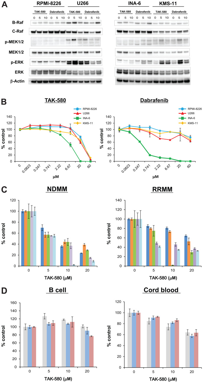
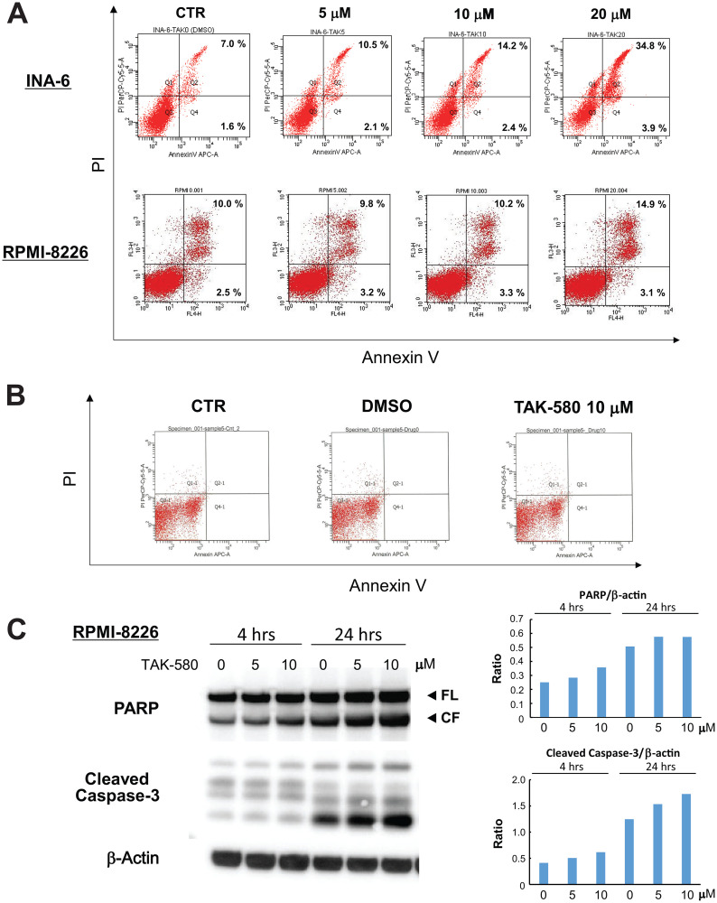
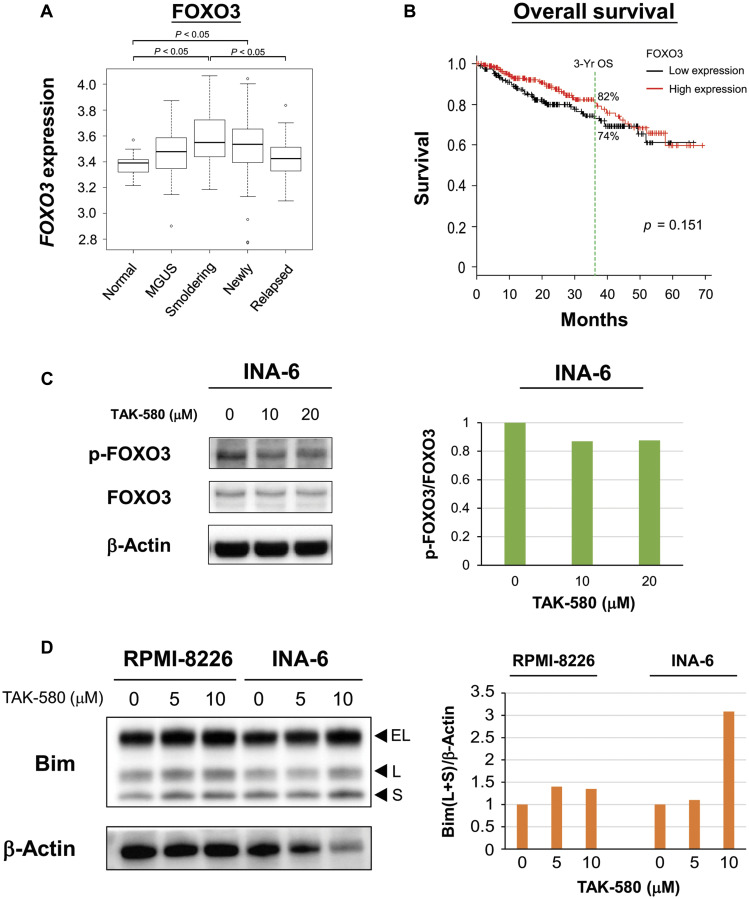
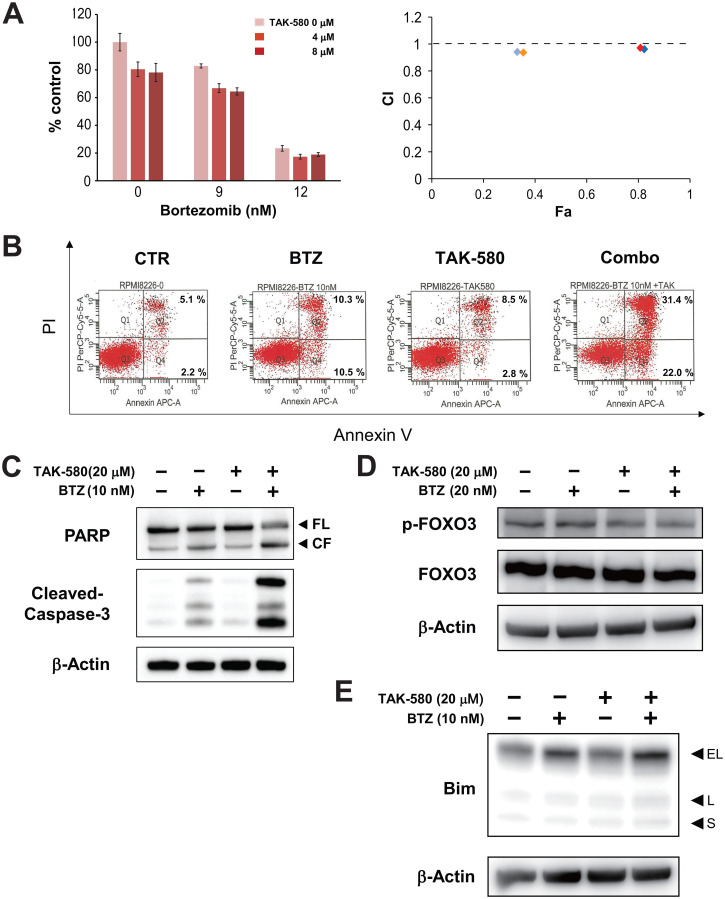
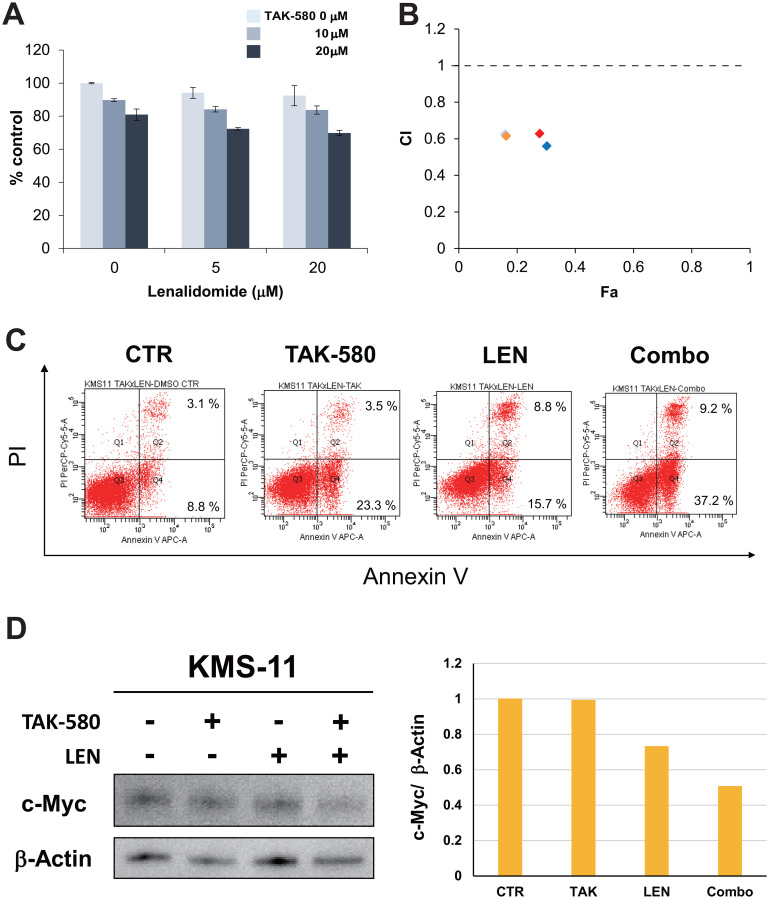
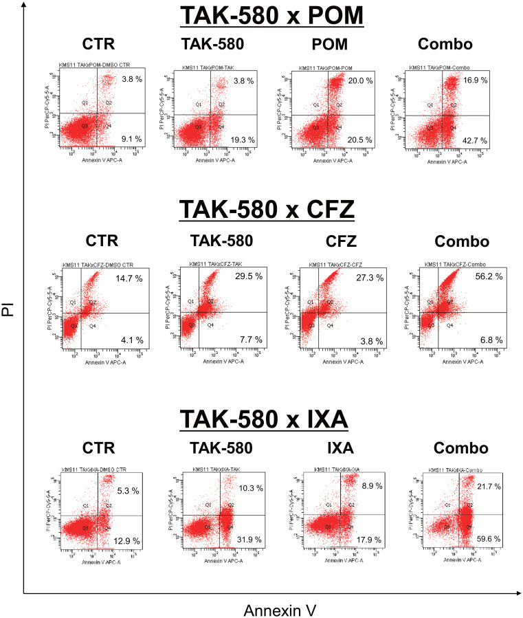
Many RAS pathway inhibitors, including pan-RAF inhibitors, have shown significant anti-tumor activities in both solid and hematological tumors. The pan-RAF inhibitor, TAK-580, is a representative of the novel RAF inhibitors that act by disrupting RAF homo- or heterodimerization. In this study, we examined the anti-tumor effects of TAK-580 used as monotherapy or in combination with bortezomib, lenalidomide, or other novel agents in multiple myeloma (MM) cells in vitro. TAK-580 monotherapy potently targeted proteins in the RAS-RAF-MEK-ERK signaling pathway and induced potent cytotoxicity and apoptosis in MM cell lines and myeloma cells from patients with newly diagnosed and relapsed and/or refractory MM, compared with a representative RAF inhibitor, dabrafenib. Normal donor peripheral blood B lymphocytes and cord blood CD34-positive cells were not affected. Importantly, TAK-580 significantly inhibited phospho-FOXO3 and induced upregulation of BimL and BimS in a dose-dependent manner, finally leading to apoptosis in MM cells. Moreover, TAK-580 enhanced bortezomib-induced cytotoxicity and apoptosis in MM cells via the FOXO3-Bim axis and the terminal unfolded protein response. Importantly, TAK-580 also enhanced lenalidomide-induced cytotoxicity and apoptosis in MM cells. Taken together, our results provide the rationale for TAK-580 monotherapy and/or treatment in combination with novel agents to improve outcomes in patients with MM.

Keywords: Bim; FOXO3a; bortezomib; lenalidomide; pan-RAF inhibitor.


Conflict of interest statement

CONFLICTS OF INTEREST Authors have no conflicts of interest to declare.

Figures

Figure 1
Figure 1. TAK-580 specifically inhibits the RAS-RAF-MEK-ERK pathway and induces anti-myeloma effects in MM cells.
(A) RPMI-8226, U266, INA-6, and KMS-11 MM cell lines were treated with the indicated doses of TAK-580 (0–10 μM) or dabrafenib (0–10 μM) for 24 h. Whole-cell lysates were subjected to western blotting using B-Raf, C-Raf, p-MEK1/2, MEK1/2, p-ERK, ERK, and β-Actin Abs. (B) RPMI-8226, U266, INA-6, and KMS-11 MM cell lines were cultured with TAK-580 (0–60 μM) or dabrafenib (0–60 μM) for 48 h. In each case, the cell viability of triplicate cultures was assessed with the Cell Titer-Glo® Cell Viability Assay and expressed as the percentage of the untreated control. Data are the mean ± standard deviation (SD). (C) Cells from five patients with newly diagnosed multiple myeloma (NDMM) and five patients with relapsed/refractory multiple myeloma (RRMM) were cultured with TAK-580 (0–20 μM) for 48 h. In each case, the cell viability of triplicate cultures was assessed with the Cell Titer-Glo® Cell Viability Assay and expressed as the percentage of the untreated control. Data are the mean ± SD. (D) Peripheral blood B lymphocytes from three normal donors and CD34-positive cells from three donors of cord blood were cultured with TAK-580 (0–20 μM) for 48 h. In each case, the cell viability of triplicate cultures was assessed with the Cell Titer-Glo® Cell Viability Assay and expressed as the percentage of the untreated control. Data are the mean ± SD.
Figure 2
Figure 2. TAK-580 induces apoptosis in MM cells.
(A) INA-6 cells were treated with TAK-580 (0–20 μM) for 24 h; RPMI-8226 cells were treated with TAK-580 (0–20 μM) for 48 h. Apoptotic cells were analyzed with flow cytometry using annexin V/PI staining. Apoptosis was assessed as the percentage of annexin V-positive cells. (B) Normal donor peripheral blood B lymphocytes were treated with dimethylsulfoxide or TAK-580 (10 μM) for 48 h. Apoptotic cells were analyzed with flow cytometry using annexin V/PI staining. Apoptosis was assessed as the percentage of annexin V-positive cells. (C) RPMI-8226 cells were treated with TAK-580 (0–10 μM) for 4 or 24 h. Whole-cell lysates were subjected to western blotting using PARP, cleaved caspase-3, and β-Actin Abs. FL, full-length; CF, cleaved form. (Upper right panel): The graph represents ratios of PARP CF density relative to β-Actin in Figure 2C. (Lower right panel): The graph represents ratios of cleaved caspase-3 density relative to β-Actin in Figure 2C.
Figure 3
Figure 3. TAK-580 induces apoptosis via the FOXO3-Bim axis in MM cells.
(A) Publicly available microarray GSE6477 data sets were analyzed for mRNA expression of FOXO3 in normal plasma cells (Normal), monoclonal gammopathy of undetermined significance (MGUS), smoldering multiple myeloma (Smoldering), newly diagnosed multiple myeloma (Newly), and relapsed multiple myeloma (Relapsed) using the Kruskal-Wallis test. (B) Kaplan-Meier overall survival curves of MM patients according to FOXO3 expression above or below the value of 4000, based on gene expression omnibus dataset GSE4581. The black line indicates the patient group with lower FOXO3 expression, whereas the red line represents the group of patients with higher FOXO3 expression. (C) (Left panel): INA-6 cells were treated with TAK-580 (0–20 μM) for 3 h. Whole-cell lysates were subjected to western blotting using phospho-FOXO3 (p-FOXO3), FOXO3, and β-Actin Abs. (Right panel): The graph represents ratios of p-FOXO3 density relative to FOXO3 in Figure 3C. (D) (Left panel): RPMI-8226 and INA-6 cells were treated with TAK-580 (0–10 μM) for 48 h. Whole-cell lysates were subjected to western blotting using Bim and β-Actin Abs. Bim has three main isoforms generated by alternative splicing: BimEL, BimL, and the most pro-apoptotic variant, BimS. (Right panel): The graph represents ratios of Bim(L+S) density relative to β-Actin in Figure 3D.
Figure 4
Figure 4. The combination of TAK-580 and BTZ triggers synergistic anti-MM activity.
(A) (Left panel): KMS-11 cells were treated with TAK-580 (0–8 μM) in combination with BTZ (0–12 nM) for 48 h. In each case, the cell viability of triplicate cultures was assessed with the Cell Titer-Glo® Cell Viability Assay and expressed as the percentage of the untreated control. Data are the mean ± SD. (Right panel): Isobologram analysis shows the synergistic or additive cytotoxic effect of TAK-580 and BTZ. Light blue, blue, orange, and red rhombuses indicate CI values of the combination of TAK-580 and BTZ; 0.94 (TAK-580 4 μM and BTZ 9 nM), 0.96 (TAK-580 4 μM and BTZ 12 nM), 0.94 (TAK-580 8 μM and BTZ 9 nM), and 0.97 (TAK-580 4 μM and BTZ 12 nM), respectively. CI < 1.0 indicates synergism; CI = 1.0 indicates an additive effect; and CI > 1.0 indicates antagonism. CI, combination index; Fa, fraction affected. (B) RPMI-8226 cells were cultured with TAK-580 (20 μM), BTZ (10 nM), or TAK-580 plus BTZ for 24 h. Apoptotic cells were analyzed with flow cytometry using annexin V/PI staining. Apoptosis was assessed as the percentage of annexin V-positive cells. (C) RPMI-8226 cells were treated with TAK-580 (20 μM) alone or in combination with BTZ (10 nM) for 24 h. Whole-cell lysates were subjected to western blotting using PARP, cleaved caspase-3, and β-Actin Abs. FL, full-length; CF, cleaved form. (D) KMS-11 cells were treated with TAK-580 (20 μM) alone or in combination with BTZ (20 nM) for 5 h. Whole-cell lysates were subjected to western blotting using phospho-FOXO3, FOXO3, and β-Actin Abs. (E) RPMI-8226 cells were treated with TAK-580 (20 μM) alone or in combination with BTZ (10 nM) for 5 h. Whole-cell lysates were subjected to western blotting using Bim and β-Actin Abs. Bim has three main isoforms generated by alternative splicing: BimEL, BimL, and the most pro-apoptotic variant, BimS.
Figure 5
Figure 5. TAK-580 induces synergistic cytotoxicity with lenalidomide (LEN) in MM cells.
(A) (Left panel): KMS-11 cells were treated with TAK-580 (0–20 μM) in combination with LEN (0–20 μM) for 48 h. In each case, the cell viability of triplicate cultures was assessed with the Cell Titer-Glo® Cell Viability Assay and expressed as the percentage of the untreated control. Data are the mean ± SD. (B): Isobologram analysis shows the synergistic or additive cytotoxic effect of TAK-580 and LEN in Figure 5A. Light blue, orange, red, and blue rhombuses indicate CI values of the combination of TAK-580 and LEN; 0.63 (TAK-580 10 μM and LEN 5 μM), 0.62 (TAK-580 10 μM and LEN 20 μM), 0.63 (TAK-580 20 μM and LEN 5 μM), and 0.56 (TAK-580 20 μM and LEN 20 μM), respectively. CI < 1.0 indicates synergism; CI = 1.0 indicates an additive effect; and CI > 1.0 indicates antagonism. CI, combination index; Fa, fraction affected. (C) KMS-11 cells were cultured with TAK-580 (20 μM), LEN (40 μM), or TAK-580 plus LEN for 72 h. Apoptotic cells were analyzed with flow cytometry using annexin V/PI staining. Apoptosis was assessed as the percentage of annexin V-positive cells. (D) (Left panel): KMS-11 cells were treated with TAK-580 (20 μM) alone or in combination with LEN (20 μM) for 48 h. Whole-cell lysates were subjected to western blotting using c-Myc and β-Actin Abs. (Right panel): The graph represents ratios of c-Myc density relative to β-Actin in Figure 5D.
Figure 6
Figure 6. TAK-580 induces synergistic cytotoxicity with other novel agents in MM cells.
KMS-11 cells were cultured with TAK-580 (20 μM), POM (40 μM), or TAK-580 plus POM for 72 h; TAK-580 (20 μM), CFZ (1 nM), or TAK-580 plus CFZ for 24 h; TAK-580 (20 μM), IXA (30 nM), or TAK-580 plus IXA for 48 h. Apoptotic cells were analyzed with flow cytometry using annexin V/PI staining. Apoptosis was assessed as the percentage of annexin V-positive cells.

Similar articles

Cited by

References

    1. Palumbo A, Anderson K. Multiple myeloma. N Engl J Med. 2011; 364:1046–1060. 10.1056/NEJMra1011442. - DOI - PubMed
    1. Raab MS, Podar K, Breitkreutz I, Richardson PG, Anderson KC. Multiple myeloma. Lancet. 2009; 374:324–339. 10.1016/S0140-6736(09)60221-X. - DOI - PubMed
    1. Kumar SK, Rajkumar SV, Dispenzieri A, Lacy MQ, Hayman SR, Buadi FK, Zeldenrust SR, Dingli D, Russell SJ, Lust JA, Greipp PR, Kyle RA, Gertz MA. Improved survival in multiple myeloma and the impact of novel therapies. Blood. 2008; 111:2516–2520. 10.1182/blood-2007-10-116129. - DOI - PMC - PubMed
    1. Giuliani N, Accardi F, Marchica V, Dalla Palma B, Storti P, Toscani D, Vicario E, Malavasi F. Novel targets for the treatment of relapsing multiple myeloma. Expert Rev Hematol. 2019; 12:481–496. 10.1080/17474086.2019.1624158. - DOI - PubMed
    1. Montagut C, Settleman J. Targeting the RAF-MEK-ERK pathway in cancer therapy. Cancer Lett. 2009; 283:125–134. 10.1016/j.canlet.2009.01.022. - DOI - PubMed