. 2021 Jan 15;203(2):192-201.
doi: 10.1164/rccm.202008-3265OC.

Tissue-Specific Immunopathology in Fatal COVID-19

Affiliations

Tissue-Specific Immunopathology in Fatal COVID-19

David A Dorward et al. Am J Respir Crit Care Med. .

Abstract

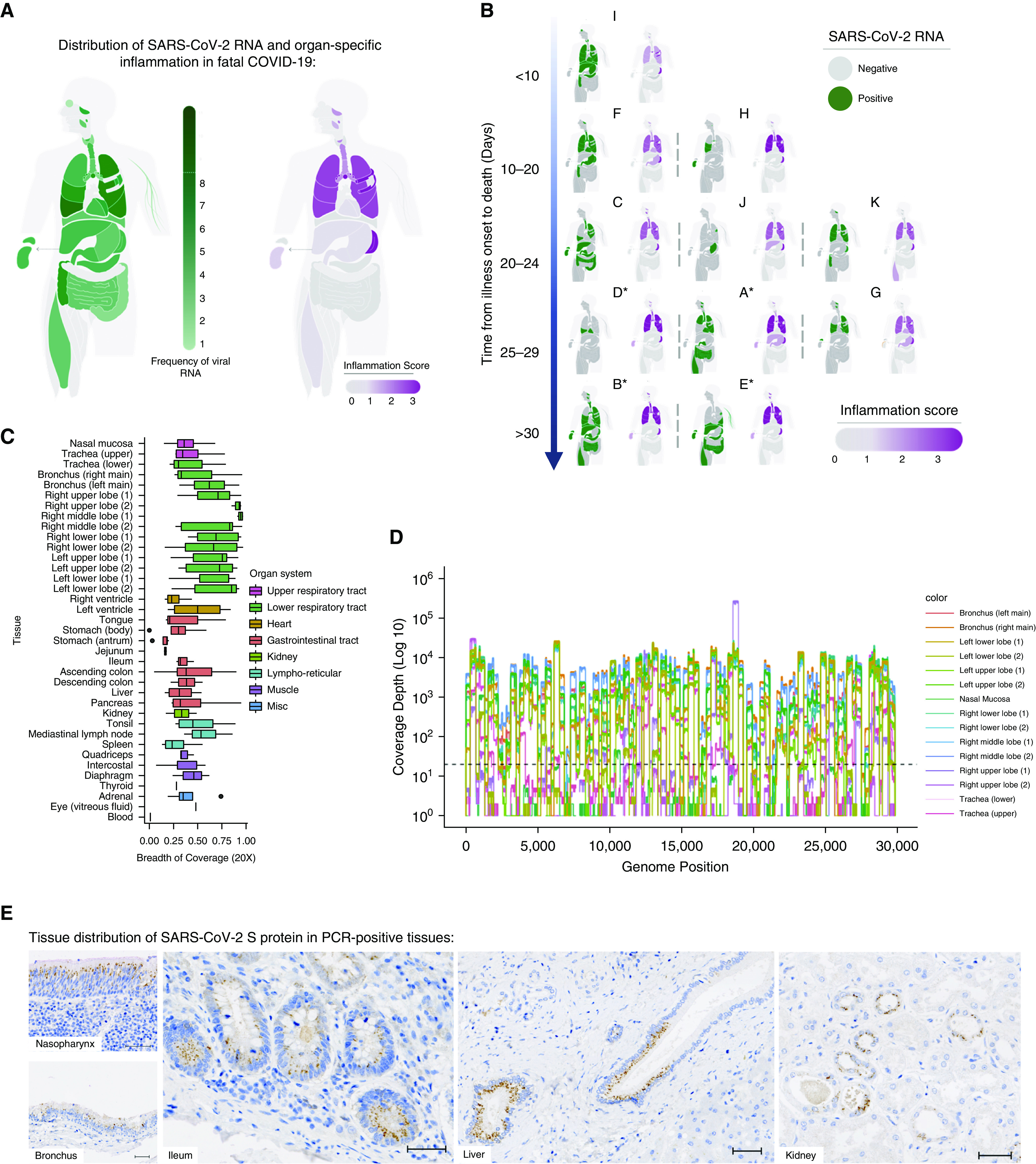
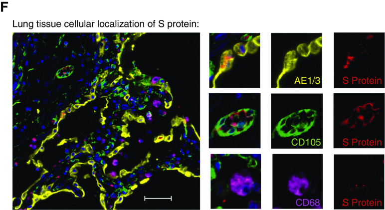
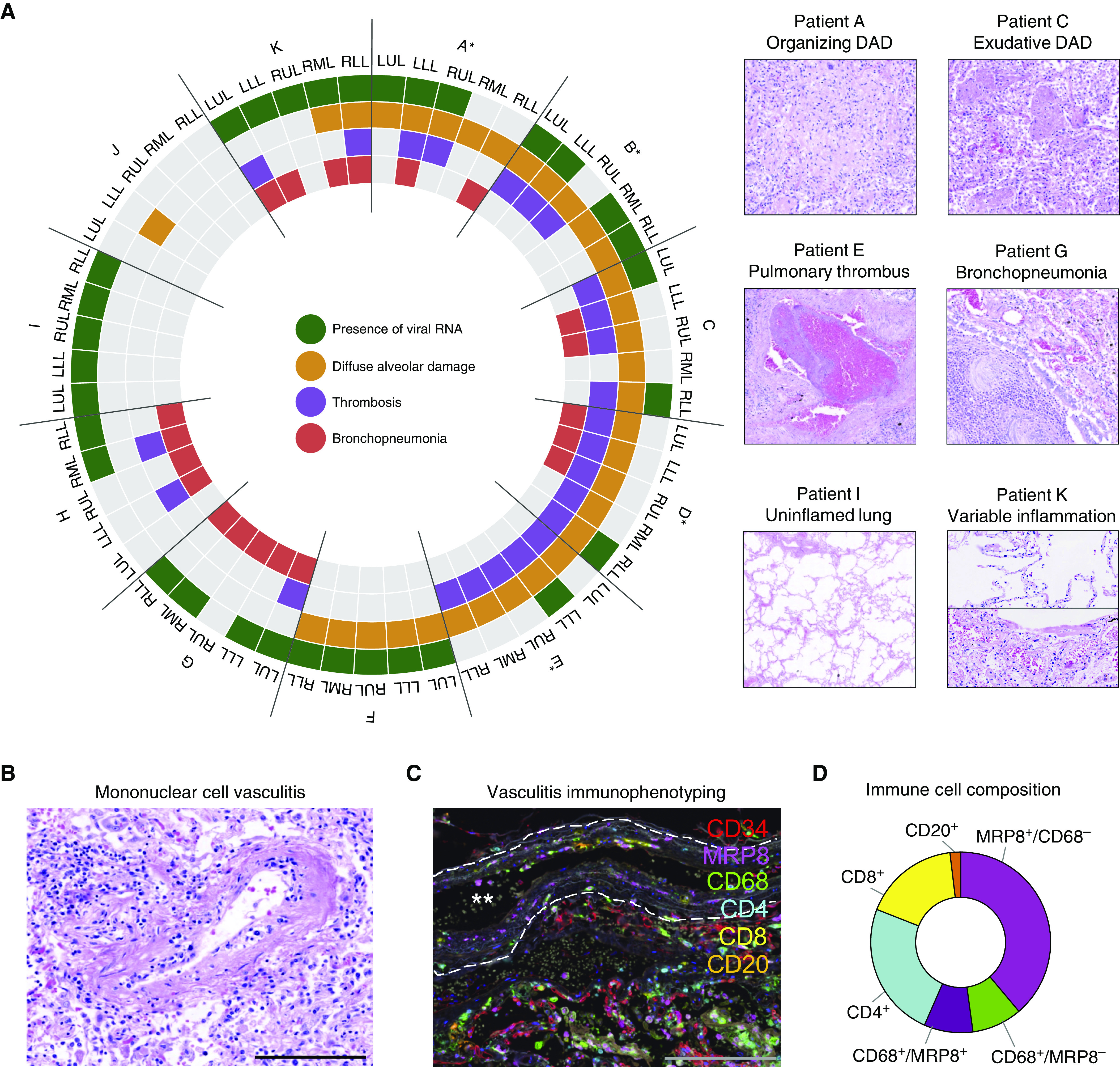
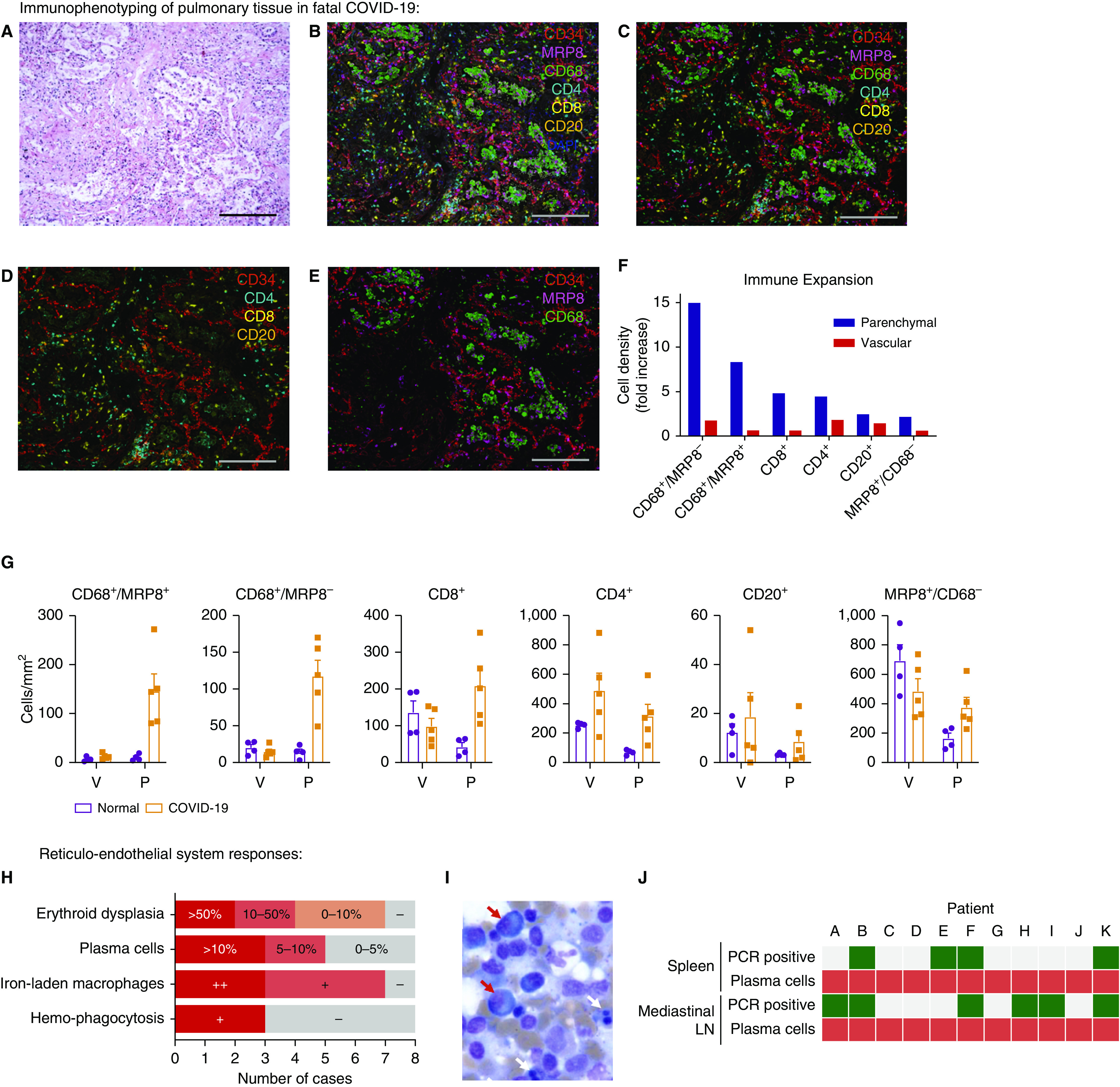
Rationale: In life-threatening coronavirus disease (COVID-19), corticosteroids reduce mortality, suggesting that immune responses have a causal role in death. Whether this deleterious inflammation is primarily a direct reaction to the presence of severe acute respiratory syndrome coronavirus 2 (SARS-CoV-2) or an independent immunopathologic process is unknown.Objectives: To determine SARS-CoV-2 organotropism and organ-specific inflammatory responses and the relationships among viral presence, inflammation, and organ injury.Methods: Tissue was acquired from 11 detailed postmortem examinations. SARS-CoV-2 organotropism was mapped by using multiplex PCR and sequencing, with cellular resolution achieved by in situ viral S (spike) protein detection. Histologic evidence of inflammation was quantified from 37 anatomic sites, and the pulmonary immune response was characterized by using multiplex immunofluorescence.Measurements and Main Results: Multiple aberrant immune responses in fatal COVID-19 were found, principally involving the lung and reticuloendothelial system, and these were not clearly topologically associated with the virus. Inflammation and organ dysfunction did not map to the tissue and cellular distribution of SARS-CoV-2 RNA and protein between or within tissues. An arteritis was identified in the lung, which was further characterized as a monocyte/myeloid-rich vasculitis, and occurred together with an influx of macrophage/monocyte-lineage cells into the pulmonary parenchyma. In addition, stereotyped abnormal reticuloendothelial responses, including excessive reactive plasmacytosis and iron-laden macrophages, were present and dissociated from viral presence in lymphoid tissues.Conclusions: Tissue-specific immunopathology occurs in COVID-19, implicating a significant component of the immune-mediated, virus-independent immunopathologic process as a primary mechanism in severe disease. Our data highlight novel immunopathologic mechanisms and validate ongoing and future efforts to therapeutically target aberrant macrophage and plasma-cell responses as well as promote pathogen tolerance in COVID-19.

Keywords: COVID-19; autopsy; inflammation; lung; macrophages.


Figures

Figure 1.
Figure 1.
Mapping severe acute respiratory syndrome coronavirus 2 (SARS-CoV-2) organotropism and cellular distribution in fatal coronavirus disease (COVID-19) in relation to tissue inflammation. (A) Distribution of SARS-CoV-2 RNA for all patients was determined by using multiplex PCR (color intensity denotes frequency of detectable RNA; dotted line on legend denotes maximal frequency within the patient cohort) (n = 11). The extent of organ-specific inflammation was assessed semiquantitatively (0–3; no inflammation [0] to severe inflammatory changes [3]), with aggregate scores visualized (n = 11). (B) Distribution of individual-patient (patients A–K) viral RNA presence within organs plotted against the time interval between illness onset and death compared with organ-specific inflammation scores for each patient. *Denotes invasive mechanical ventilation. (C and D) Multiplex PCR–confirmed SARS-CoV-2–positive samples were confirmed by sequencing, with the proportion of the SARS-CoV-2 genome mapped calculated (C) and a representative sequence-coverage map of the respiratory tract of one patient shown (D). (E and F) Tissue and cellular distribution of SARS-CoV-2 S (spike) protein was evaluated by using immunohistochemistry and multiplex immunofluorescence on randomly selected PCR-confirmed SARS-CoV-2–positive formalin-fixed, paraffin-embedded tissue (n = 4 patients). (E) Representative images demonstrate the tissue distribution of S protein within the nasal mucosa, bronchial epithelium, small-bowel enterocytes, distal biliary epithelium within the liver, and distal renal tubular epithelium. (F) Within the lung, cellular localization of S protein is demonstrated within the alveolar epithelium (AE1/3) and is rarely demonstrated in macrophages (CD68) or in the endothelium (CD105) within the lung parenchyma. Scale bars, 50 μm. Misc = miscellaneous.
Figure 1.
Figure 1.
Mapping severe acute respiratory syndrome coronavirus 2 (SARS-CoV-2) organotropism and cellular distribution in fatal coronavirus disease (COVID-19) in relation to tissue inflammation. (A) Distribution of SARS-CoV-2 RNA for all patients was determined by using multiplex PCR (color intensity denotes frequency of detectable RNA; dotted line on legend denotes maximal frequency within the patient cohort) (n = 11). The extent of organ-specific inflammation was assessed semiquantitatively (0–3; no inflammation [0] to severe inflammatory changes [3]), with aggregate scores visualized (n = 11). (B) Distribution of individual-patient (patients A–K) viral RNA presence within organs plotted against the time interval between illness onset and death compared with organ-specific inflammation scores for each patient. *Denotes invasive mechanical ventilation. (C and D) Multiplex PCR–confirmed SARS-CoV-2–positive samples were confirmed by sequencing, with the proportion of the SARS-CoV-2 genome mapped calculated (C) and a representative sequence-coverage map of the respiratory tract of one patient shown (D). (E and F) Tissue and cellular distribution of SARS-CoV-2 S (spike) protein was evaluated by using immunohistochemistry and multiplex immunofluorescence on randomly selected PCR-confirmed SARS-CoV-2–positive formalin-fixed, paraffin-embedded tissue (n = 4 patients). (E) Representative images demonstrate the tissue distribution of S protein within the nasal mucosa, bronchial epithelium, small-bowel enterocytes, distal biliary epithelium within the liver, and distal renal tubular epithelium. (F) Within the lung, cellular localization of S protein is demonstrated within the alveolar epithelium (AE1/3) and is rarely demonstrated in macrophages (CD68) or in the endothelium (CD105) within the lung parenchyma. Scale bars, 50 μm. Misc = miscellaneous.
Figure 2.
Figure 2.
Delineating pulmonary injury and vascular involvement in fatal coronavirus disease (COVID-19). (A) Detailed spatial evaluation of lung injury and key pathologic abnormalities were determined within each lobe of lung for each patient (patients A–K) and compared with the presence or absence of severe acute respiratory syndrome coronavirus 2 (SARS-CoV-2) viral RNA by using multiplex PCR (*denotes invasive mechanical ventilation; n = 11). Representative images of organizing and exudative diffuse alveolar damage, pulmonary thrombus, bronchopneumonia, uninflamed lung, and variable inflammation within the same lung. (B) In four individuals, frequent pulmonary vasculature immune infiltration was seen, with (C) multiplex immunofluorescence defining these immune-cell populations (CD4, CD8 [T cells]; CD20 [B cells]; CD68 [macrophages]; MRP8 [neutrophils and myeloid lineage cells]) demonstrating MRP8 immunopositive mononuclear cells to be the predominant cell type (representative image, white stars denote vessel lumen and white dashed line denotes elastic lamina). (D) Analysis of 50 arteries/arterioles from two selected patients quantifying cell types involved in vasculitis. Scale bars, 200 μm. DAD = diffuse alveolar damage; LLL = left lower lobe; LUL = left upper lobe; RLL = right lower lobe; RML = right middle lobe; RUL = right upper lobe.
Figure 3.
Figure 3.
Pulmonary tissue and reticuloendothelial immune responses to fatal coronavirus disease (COVID-19). Regions of interest were defined by histologic examination of hematoxylin and eosin–stained lung tissue to identify areas of diffuse alveolar damage in tissue from five patients. (A) Representative image. Corresponding multiplex immunofluorescence was used to define vascular-endothelium populations (CD34) relative to immune-cell populations: CD4, CD8 (T cells); CD20 (B cells); CD68 (macrophages); and MRP8 (neutrophils and myeloid lineage cells) (B) with and (C) without autofluorescence. (D and E) Separate cell populations are highlighted. Immune-cell populations were quantified, with the (F) relative abundance of cell types compared between COVID-19 (n = 5) and normal, uninflamed lung from patients undergoing lung-cancer resection (n = 4) and being (G) spatially stratified into vascular/perivascular and parenchymal regions. (H) Key pathologic abnormalities within bone marrow included erythroid dysplasia, iron-laden macrophages, and hemophagocytosis; plasma cells were confirmed by immunohistochemical staining and quantified in bone-marrow aspirates. + indicates present; ++ indicates frequent. (I) Representative image of bone marrow aspirate analysis demonstrating erythroid dysplasia (white arrows) and frequent plasma cells (red arrows). (J) Mismatch between stereotyped plasma-cell abnormalities in the spleen and mediastinal LN (red) and detection of severe acute respiratory syndrome coronavirus 2 (SARS-CoV-2) by using multiplex PCR (green, positive; gray, negative). Scale bars, 200 μm. LN = lymph node; P = parenchymal; V = vascular.

Comment in

Similar articles

  • Tissue Proteomic Analysis Identifies Mechanisms and Stages of Immunopathology in Fatal COVID-19.
    Russell CD, Valanciute A, Gachanja NN, Stephen J, Penrice-Randal R, Armstrong SD, Clohisey S, Wang B, Al Qsous W, Wallace WA, Oniscu GC, Stevens J, Harrison DJ, Dhaliwal K, Hiscox JA, Baillie JK, Akram AR, Dorward DA, Lucas CD. Russell CD, et al. Am J Respir Cell Mol Biol. 2022 Feb;66(2):196-205. doi: 10.1165/rcmb.2021-0358OC. Am J Respir Cell Mol Biol. 2022. PMID: 34710339 Free PMC article.
  • Tissue-based SARS-CoV-2 detection in fatal COVID-19 infections: Sustained direct viral-induced damage is not necessary to drive disease progression.
    El Jamal SM, Pujadas E, Ramos I, Bryce C, Grimes ZM, Amanat F, Tsankova NM, Mussa Z, Olson S, Salem F, Miorin L, Aydillo T, Schotsaert M, Albrecht RA, Liu WC, Marjanovic N, Francoeur N, Sebra R, Sealfon SC, García-Sastre A, Fowkes M, Cordon-Cardo C, Westra WH. El Jamal SM, et al. Hum Pathol. 2021 Aug;114:110-119. doi: 10.1016/j.humpath.2021.04.012. Epub 2021 May 4. Hum Pathol. 2021. PMID: 33961839 Free PMC article.
  • COVID-19 tissue atlases reveal SARS-CoV-2 pathology and cellular targets.
    Delorey TM, Ziegler CGK, Heimberg G, Normand R, Yang Y, Segerstolpe Å, Abbondanza D, Fleming SJ, Subramanian A, Montoro DT, Jagadeesh KA, Dey KK, Sen P, Slyper M, Pita-Juárez YH, Phillips D, Biermann J, Bloom-Ackermann Z, Barkas N, Ganna A, Gomez J, Melms JC, Katsyv I, Normandin E, Naderi P, Popov YV, Raju SS, Niezen S, Tsai LT, Siddle KJ, Sud M, Tran VM, Vellarikkal SK, Wang Y, Amir-Zilberstein L, Atri DS, Beechem J, Brook OR, Chen J, Divakar P, Dorceus P, Engreitz JM, Essene A, Fitzgerald DM, Fropf R, Gazal S, Gould J, Grzyb J, Harvey T, Hecht J, Hether T, Jané-Valbuena J, Leney-Greene M, Ma H, McCabe C, McLoughlin DE, Miller EM, Muus C, Niemi M, Padera R, Pan L, Pant D, Pe'er C, Pfiffner-Borges J, Pinto CJ, Plaisted J, Reeves J, Ross M, Rudy M, Rueckert EH, Siciliano M, Sturm A, Todres E, Waghray A, Warren S, Zhang S, Zollinger DR, Cosimi L, Gupta RM, Hacohen N, Hibshoosh H, Hide W, Price AL, Rajagopal J, Tata PR, Riedel S, Szabo G, Tickle TL, Ellinor PT, Hung D, Sabeti PC, Novak R, Rogers R, Ingber DE, Jiang ZG, Juric D, Babadi M, Farhi SL, Izar B, Stone JR, Vlachos IS, Solomon IH, Ashenberg O, Porter CBM, Li B, Shalek AK, Villani AC, Rozenblatt-Rosen O, Regev A. Delorey TM, et al. Nature. 2021 Jul;595(7865):107-113. doi: 10.1038/s41586-021-03570-8. Epub 2021 Apr 29. Nature. 2021. PMID: 33915569 Free PMC article.
  • The immune response to SARS-CoV-2 and COVID-19 immunopathology - Current perspectives.
    Boechat JL, Chora I, Morais A, Delgado L. Boechat JL, et al. Pulmonology. 2021 Sep-Oct;27(5):423-437. doi: 10.1016/j.pulmoe.2021.03.008. Epub 2021 Apr 9. Pulmonology. 2021. PMID: 33867315 Free PMC article. Review.
  • Inflammation Triggered by SARS-CoV-2 and ACE2 Augment Drives Multiple Organ Failure of Severe COVID-19: Molecular Mechanisms and Implications.
    Iwasaki M, Saito J, Zhao H, Sakamoto A, Hirota K, Ma D. Iwasaki M, et al. Inflammation. 2021 Feb;44(1):13-34. doi: 10.1007/s10753-020-01337-3. Epub 2020 Oct 8. Inflammation. 2021. PMID: 33029758 Free PMC article. Review.

Cited by

References

    1. Smatti MK, Cyprian FS, Nasrallah GK, Al Thani AA, Almishal RO, Yassine HM. Viruses and autoimmunity: a review on the potential interaction and molecular mechanisms. Viruses. 2019;11:762. - PMC - PubMed
    1. Medzhitov R, Schneider DS, Soares MP. Disease tolerance as a defense strategy. Science. 2012;335:936–941. - PMC - PubMed
    1. Schneider DS, Ayres JS. Two ways to survive infection: what resistance and tolerance can teach us about treating infectious diseases. Nat Rev Immunol. 2008;8:889–895. - PMC - PubMed
    1. Glass EJ. The molecular pathways underlying host resistance and tolerance to pathogens. Front Genet. 2012;3:263. - PMC - PubMed
    1. Mehta P, McAuley DF, Brown M, Sanchez E, Tattersall RS, Manson JJ HLH Across Speciality Collaboration, UK. COVID-19: consider cytokine storm syndromes and immunosuppression. Lancet. 2020;395:1033–1034. - PMC - PubMed

MeSH terms