. 2020 Dec 3;107(6):1078-1095.
doi: 10.1016/j.ajhg.2020.11.002. Epub 2020 Nov 19.

Pathogenic Variants in the Myosin Chaperone UNC-45B Cause Progressive Myopathy with Eccentric Cores

Affiliations

Pathogenic Variants in the Myosin Chaperone UNC-45B Cause Progressive Myopathy with Eccentric Cores

Sandra Donkervoort et al. Am J Hum Genet. .

Abstract

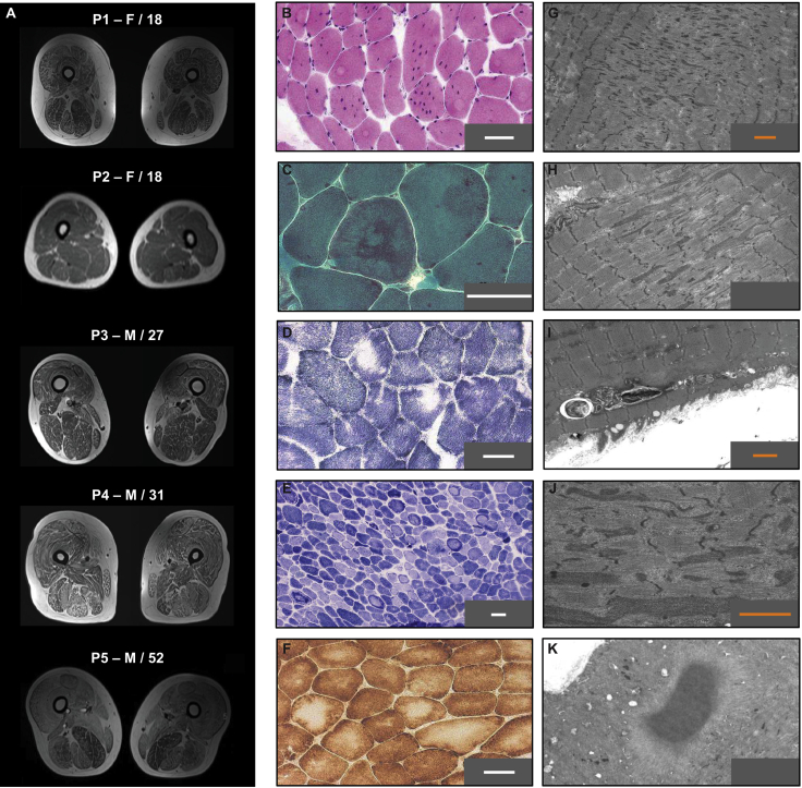
The myosin-directed chaperone UNC-45B is essential for sarcomeric organization and muscle function from Caenorhabditis elegans to humans. The pathological impact of UNC-45B in muscle disease remained elusive. We report ten individuals with bi-allelic variants in UNC45B who exhibit childhood-onset progressive muscle weakness. We identified a common UNC45B variant that acts as a complex hypomorph splice variant. Purified UNC-45B mutants showed changes in folding and solubility. In situ localization studies further demonstrated reduced expression of mutant UNC-45B in muscle combined with abnormal localization away from the A-band towards the Z-disk of the sarcomere. The physiological relevance of these observations was investigated in C. elegans by transgenic expression of conserved UNC-45 missense variants, which showed impaired myosin binding for one and defective muscle function for three. Together, our results demonstrate that UNC-45B impairment manifests as a chaperonopathy with progressive muscle pathology, which discovers the previously unknown conserved role of UNC-45B in myofibrillar organization.

Keywords: C. elegans; UNC-45; UNC45B; chaperone; core myopathy; muscle; myofibrillar; myosin; sarcomere.


Conflict of interest statement

The authors declare no competing interests.

Figures

Figure 1
Figure 1
Bi-allelic Variants in the Myosin-Directed Chaperone UNC-45B (A) UNC-45 contains four domains: an N-terminal TPR domain (light green), a conserved central domain (dark gray), a neck domain (light gray), and a C-terminal UCS domain (orange). The N-terminal TPR domain is important for the interaction with Hsp90 and Hsp70 chaperones, and the C-terminal UCS domain binds directly to the myosin head domain. The recurring UNC45B variant p.Arg754Gln (dark blue) and the p.Arg778Trp (light blue) variant impact conserved arginines in the myosin-binding UCS domain. Variants p.Ser403Pro (pink) and p.Cys514Arg (dark green) are located in the UNC-45B neck domain. (B) Overview of the muscle sarcomere. UNC-45 is involved in myofibrillogenesis, cooperating with Hsp90 to fold and to incorporate myosin into the thick filament. In adulthood, UNC-45 is stored at the Z-disk. Upon injury to the muscle fiber, UNC-45 shuttles to the A-band to help refold damaged myosin.
Figure 2
Figure 2
Muscle Magnetic Resonance Imaging and Histological Findings in Individuals with UNC45B-Related Myopathy (A) Lower extremity muscle MR imaging was available for five (P1–P5) individuals and showed mild fat infiltration evident in all lower extremity muscles, resulting in a marbled-like appearance. In individuals P1, P4, and P5, there is abnormal (increased) T1 signal without an apparent pattern of muscle involvement except for relative sparing of the semimembranosus muscle in individuals P1 and P5. P1 and P3 had evidence of mild, generalized muscle atrophy, whereas in P2, P4 and P5, muscle bulk appeared normal. (B–K) UNC45B-related myopathy manifests histologically with unstructured cores. On histological analyses there are findings of the following: increased internalized nuclei with numerous multinucleated fibers seen on H&E staining (P1) (B); uneven deposits of fuchsinophilic material is seen on GT staining (P4) (C); core-like regions often located along the periphery of fibers and consistent with eccentric cores are seen on SDH staining (P3) (D); areas of increased staining along the periphery of fibers with decreased staining centrally is seen on NADH staining (P6) (E); and large irregular areas of decreased staining seen on COX staining (P4) (F); on EM, there are findings of myofibrillar disarray (P3) (G); diffusion of Z-line material (P4) (H); autophagy lesions (P3) (I); wide bands of diffusion of the Z-line material that have spread in the disorganized areas (P3) (J); and a cytoplasmic body (P4) (K); white scale bar represents 50 μm; orange scale bar represents 5 μm.
Figure 3
Figure 3
Mislocalized UNC-45B Protein in Affected Individuals’ Muscle (A) Longitudinal sections of muscle biopsies of control and P1, P9, and P10 stained for UNC-45B (red) and M-line protein myomesin (green) (top row). UNC-45B is reduced in the M-line and mislocalized from the A-band, around the M-line, to the Z-line in these three individuals compared to control. Overlay of intensity profile of UNC-45B and myomesin (bottom row) from the cropped area (middle row) shows mislocalization of UNC-45B away from the A-band to the Z-disk. (B) Longitudinal sections of muscle biopsies of control and P1 stained for UNC-45B (red) and Z-disk protein α-actinin (green), revealing colocalization of UNC-45B at the Z-line. Overlay of intensity profile of UNC-45B and α-actinin in P1 from the cropped area (middle row) shows loss of co-location of UNC-45B with the M-line.
Figure 4
Figure 4
UNC45B Variant c.2261G>A Creates a Complex Hypomorph Splice Variant (A) The last base pair of exon 17 in UNC45B c.2261G (orange) is adjacent to the splice donor site of intron 17 (bold black). The c.2261G>A transition, located in the exonic part of the 5′ recognition sequence (G-G-U-G-A-G-U), leads to the activation of a nearby cryptic splice donor site (bold black). The resulting spliced mRNA transcript is elongated by 9 additional bases including an in-frame STOP codon (bold red). (B) Schematics of (1) normal UNC45B exon 17–18 splicing and of the two splice products seen in UNC45B c.2261G>A muscle: (2) full length product including the p.Arg754Gln substitution and (3) activation of the nearby cryptic splice donor site generating an elongated splice product that includes an in-frame STOP codon. (C) Immunoblot analysis of UNC-45B in muscle extracts from P1, P2, P9, and P10 compared to control. Quantification shows a significant reduction of UNC-45B in P1, P2, P9, while levels in P10 were slightly reduced. Mouse monoclonal anti-desmin was used as a loading control.
Figure 5
Figure 5
UNC-45B Mutant Proteins Are Prone to Aggregation (A) Structure of human UNC-45B based on the C. elegans UNC-45 3D structure (PDB: 4i2z) modelled with Swiss-Model, and corresponding residues mutated in PyMOL to display the reported variants. The recurring p.Arg754Gln (dark blue) and the p.Arg778Trp (P2) UNC45B variant (light blue) impact conserved arginine residues in the myosin-binding UCS domain (orange) and can be precisely mapped to helices 1 and 3, respectively, in the armadillo (ARM) repeat 14. ARM 14 is located in the hinge region, which connects the C-terminal half of the UCS domain (ARM repeats 14–17) with the N-terminal half (ARM repeats 10–13) and forms part of the myosin-binding canyon. The p.Ser403Pro (pink, P9) and p.Cys514Arg (dark green, P10) variants impact the UNC-45B neck domain. Van der Waals overlaps of the newly incorporated amino acid residues with the unmodified protein structure are depicted as red disks. (B) Purified UNC-45BWT, UNC-45Bp.Arg754Gln, UNC-45Bp.Arg778Trp, UNC-45Bp.Ser403Pro, and UNC-45Bp.Cys514Arg buffered solutions were subjected to partial proteolysis with trypsin at 37°C, and samples were loaded on SDS-PAGE gels for separation. Coomassie-stained gels of one of two repetitions are shown. (C) Quantification of full-length protein (∼109 kDa) compared to time point 0 at 37°C of two repetitions. Mean and SD can be found in Table S4. (D) Purified UNC-45BWT, UNC-45Bp.Arg754Gln, UNC-45Bp.Arg778Trp, UNC-45Bp.Ser403Pro, and UNC-45B p.Cys514Arg buffered solutions were slowly heated from 10°C to 95°C in the presence of SYPRO Orange protein stain. Boltzmann sigmoidal curves were fit to normalized combined melt curves of three experiments. Half maximal temperatures indicate melting temperatures in the Tris-based buffer. Mean and SD can be found in Table S5. (E) Slot blot of purified UNC-45BWT, UNC-45Bp.Arg754Gln, UNC-45Bp.Arg778Trp, UNC-45Bp.Ser403Pro, and UNC-45Bp.Cys514Arg Tris-buffered solutions incubated for 1 h at room temperature filtered through a 0.2 μm acetyl-cellulose membrane. Three decreasing amounts of protein solution were blotted for comparison. (F) Baseline-subtracted SYPRO fluorescence values at 22°C obtained in two experiments in (D) are plotted for each protein solution. No significant differences between WT and mutant proteins were found in non-parametric Kruskal-Wallis test.
Figure 6
Figure 6
Various Missense UNC-45 Mutant Proteins Cannot Rescue a Conditional Loss-of-Function Allele Transgenic UNC-45p.Arg792Trp, UNC-45p.Ser422Pro, and UNC-45p.Cys532Arg mutant proteins are unable to rescue the motility defect of unc-45(m94) worms grown at the restrictive temperature of 25°C, whereas UNC-45p.Arg767Gln rescues the m94 motility defect. (A) Body bends of 30 young adult unc-45(m94) worms expressing the indicated UNC-45 variants were counted in at least three different experiments. Values are mean ± SEM; p < 0.0001 compared to control in non-parametric Kruskal-Wallis test. (B) Expression of UNC-45-FLAG transgenes in young adult unc-45(m94) worms. (C) Population motility of 60 young adult unc-45(m94) worms expressing the indicated UNC-45 variants on a 24-well plate filled with NGM and seeded with OP50 was measured with the ARENA WMicrotracker system (NemaMetrix) at 25°C for 24 h after reaching adulthood. Motility data was binned in 15 min time buckets before plotting for easier interpretability. (D) Phalloidin-staining of F-actin-containing I-bands in unc-45(m94) mutant worms expressing the indicated UNC-45 variants. (E) The number of I-bands is given per body wall muscle cell (indicated by dashed white line in [D]), with all 12 analyzed cells located in the same area between pharynx and vulva. p < 0.005 compared to control in non-parametric Kruskal-Wallis test. (F) Co-immunoprecipitation of myosin heavy chain B (MHC B) and Hsp90 (DAF-21) from cell lysates of unc-45(m94) mutant worms expressing the indicated FLAG-tagged UNC-45 variants grown at 25°C. Representative result of one of three experiments is shown.

References

    1. Kim J., Löwe T., Hoppe T. Protein quality control gets muscle into shape. Trends Cell Biol. 2008;18:264–272. - PubMed
    1. Vicart P., Caron A., Guicheney P., Li Z., Prévost M.C., Faure A., Chateau D., Chapon F., Tomé F., Dupret J.M. A missense mutation in the alphaB-crystallin chaperone gene causes a desmin-related myopathy. Nat. Genet. 1998;20:92–95. - PubMed
    1. Selcen D., Muntoni F., Burton B.K., Pegoraro E., Sewry C., Bite A.V., Engel A.G. Mutation in BAG3 causes severe dominant childhood muscular dystrophy. Ann. Neurol. 2009;65:83–89. - PMC - PubMed
    1. Smith D.A., Carland C.R., Guo Y., Bernstein S.I. Getting folded: chaperone proteins in muscle development, maintenance and disease. Anat. Rec. (Hoboken) 2014;297:1637–1649. - PMC - PubMed
    1. Harms M.B., Sommerville R.B., Allred P., Bell S., Ma D., Cooper P., Lopate G., Pestronk A., Weihl C.C., Baloh R.H. Exome sequencing reveals DNAJB6 mutations in dominantly-inherited myopathy. Ann. Neurol. 2012;71:407–416. - PMC - PubMed

MeSH terms