. 2021 Jan 7;81(1):139-152.e10.
doi: 10.1016/j.molcel.2020.10.035. Epub 2020 Nov 19.

Structural Basis for Virulence Activation of Francisella tularensis

Affiliations

Structural Basis for Virulence Activation of Francisella tularensis

Brady A Travis et al. Mol Cell. .

Abstract

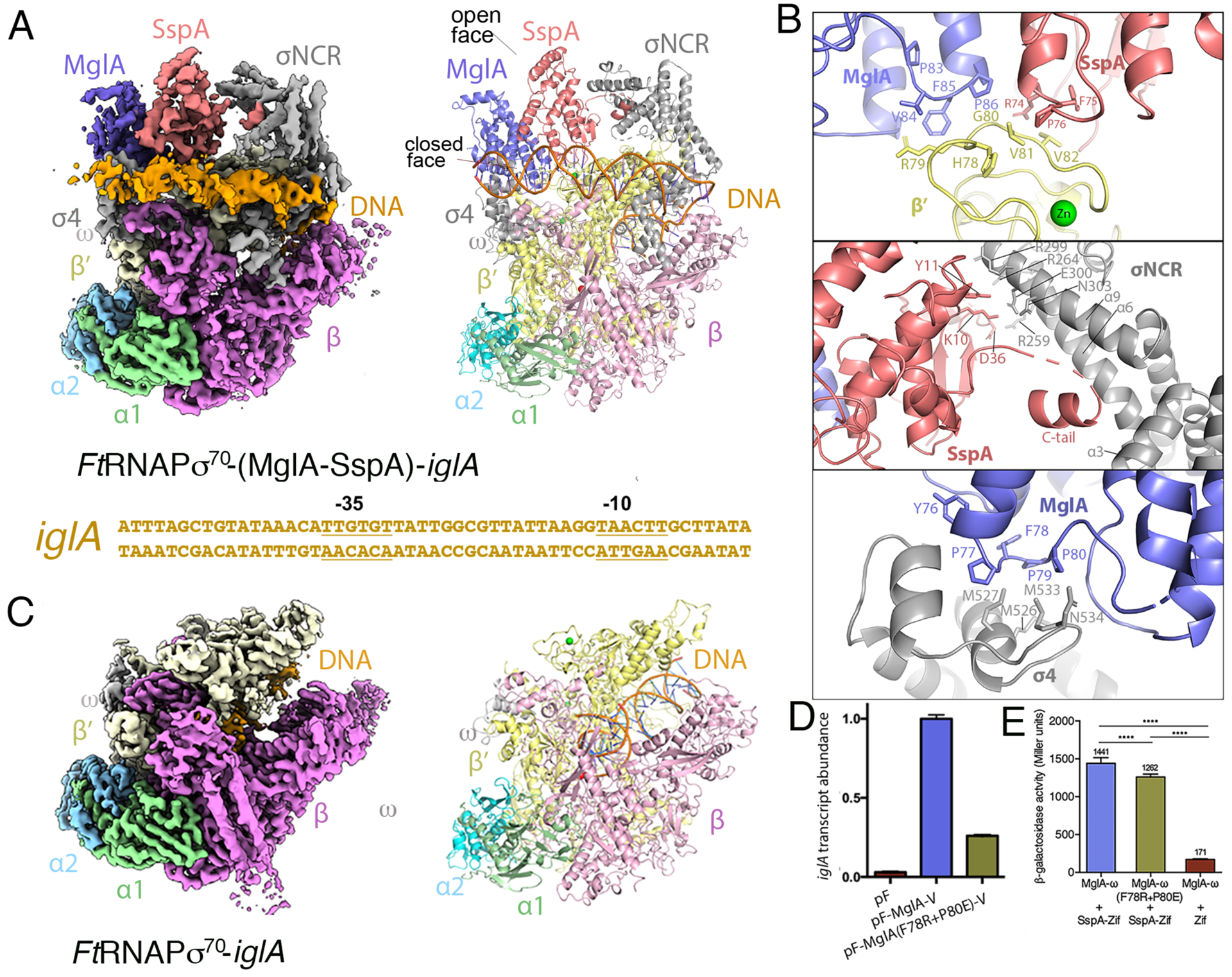
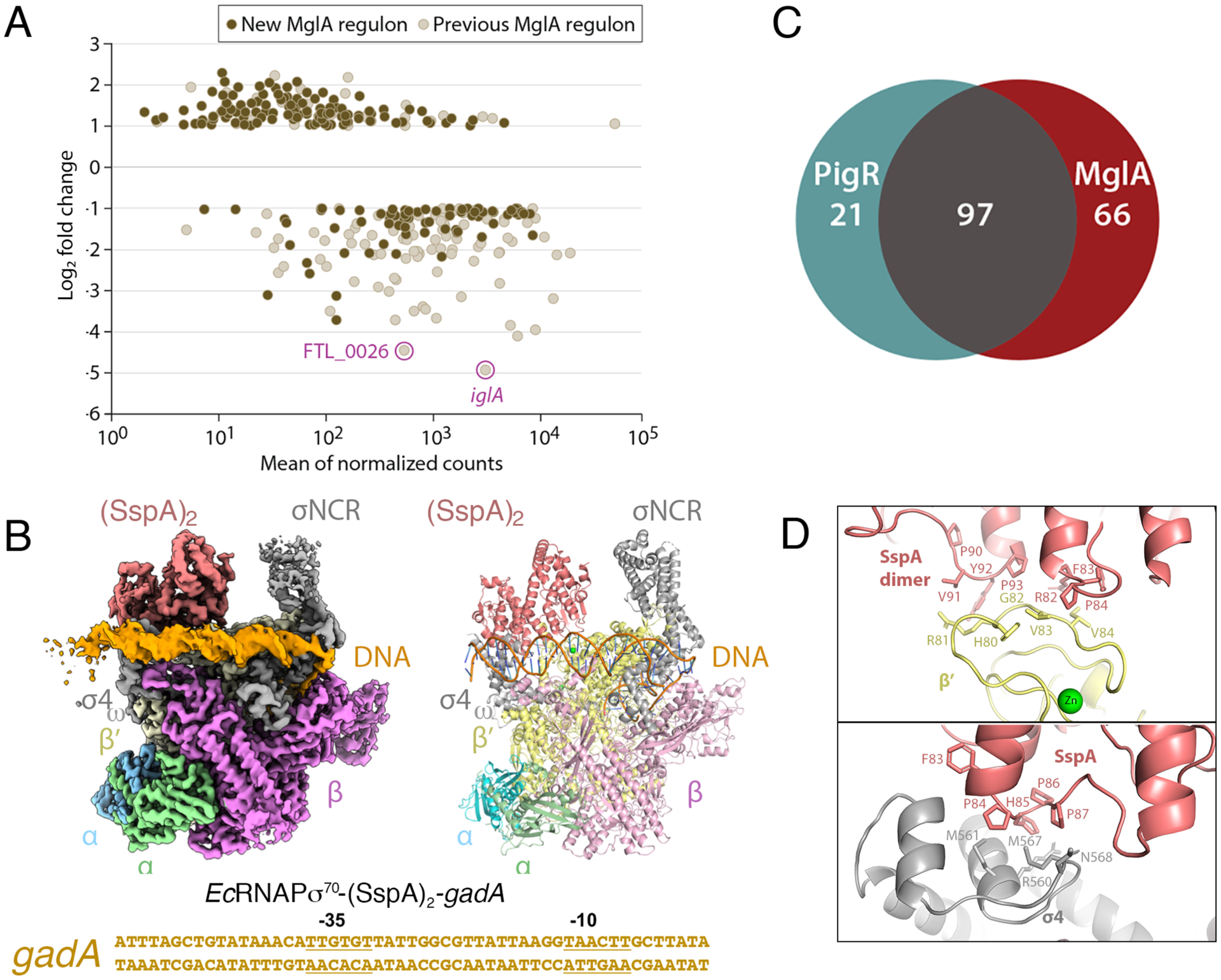
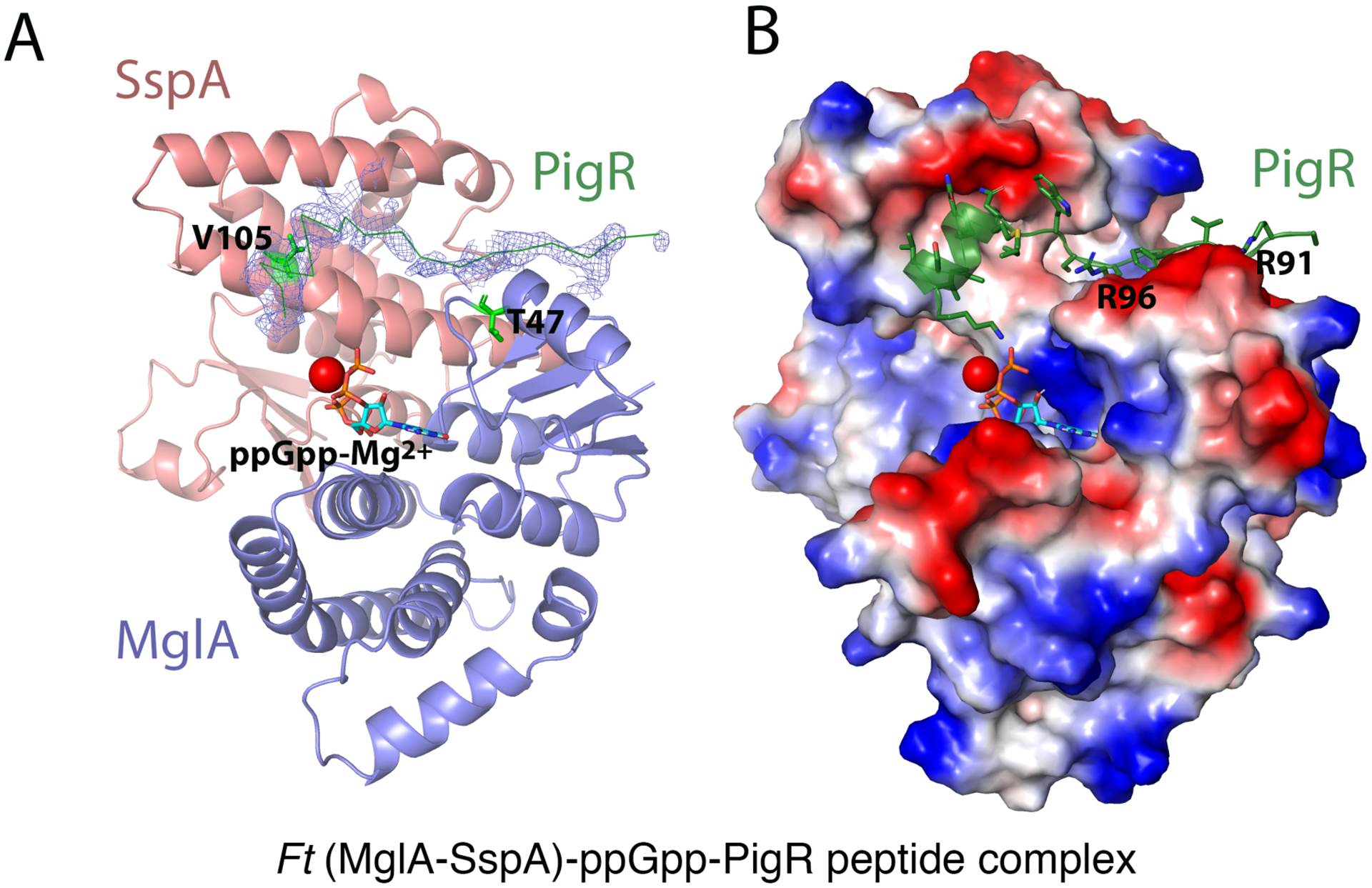
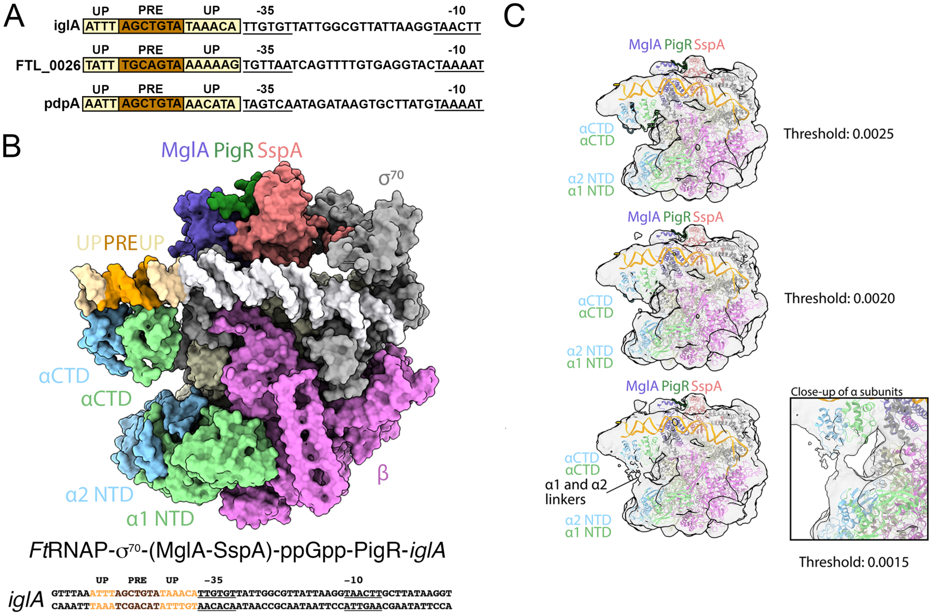
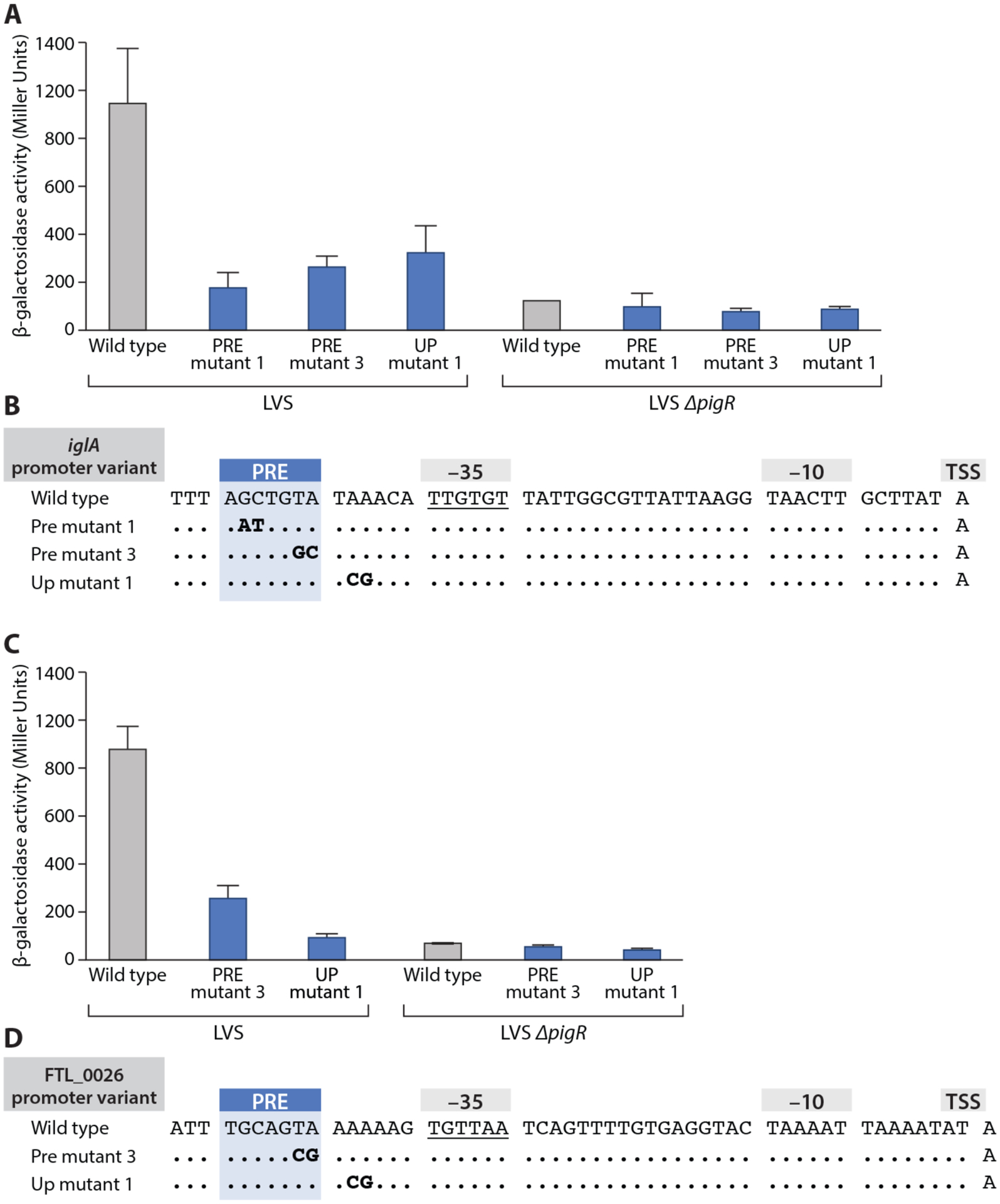
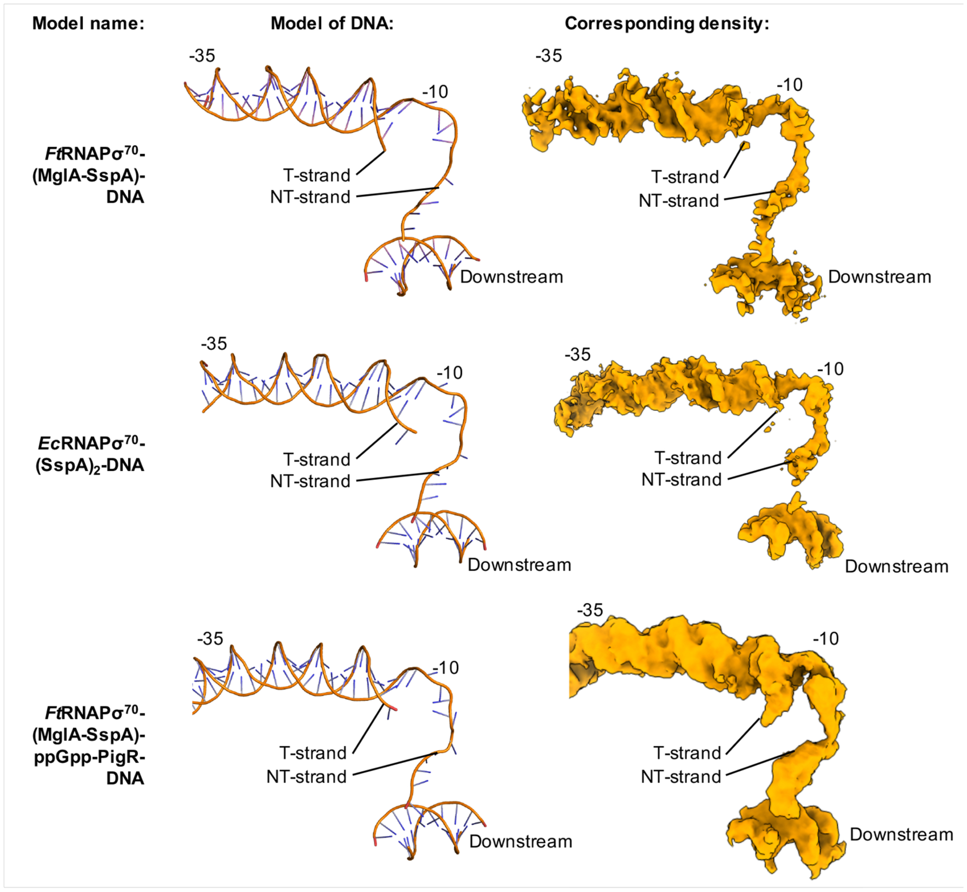
The bacterium Francisella tularensis (Ft) is one of the most infectious agents known. Ft virulence is controlled by a unique combination of transcription regulators: the MglA-SspA heterodimer, PigR, and the stress signal, ppGpp. MglA-SspA assembles with the σ70-associated RNAP holoenzyme (RNAPσ70), forming a virulence-specialized polymerase. These factors activate Francisella pathogenicity island (FPI) gene expression, which is required for virulence, but the mechanism is unknown. Here we report FtRNAPσ70-promoter-DNA, FtRNAPσ70-(MglA-SspA)-promoter DNA, and FtRNAPσ70-(MglA-SspA)-ppGpp-PigR-promoter DNA cryo-EM structures. Structural and genetic analyses show MglA-SspA facilitates σ70 binding to DNA to regulate virulence and virulence-enhancing genes. Our Escherichia coli RNAPσ70-homodimeric EcSspA structure suggests this is a general SspA-transcription regulation mechanism. Strikingly, our FtRNAPσ70-(MglA-SspA)-ppGpp-PigR-DNA structure reveals ppGpp binding to MglA-SspA tethers PigR to promoters. PigR in turn recruits FtRNAP αCTDs to DNA UP elements. Thus, these studies unveil a unique mechanism for Ft pathogenesis involving a virulence-specialized RNAP that employs two (MglA-SspA)-based strategies to activate virulence genes.

Keywords: Cryo-EM; Francisella tularensis; MglA-SspA; PigR; RNA polymerase; pathogenicity island; ppGpp; transcription; αCTD; σ70.


Conflict of interest statement

Declaration of Interests The authors declare no competing interests.

Figures

Figure 1.
Figure 1.. Structures of FtRNAPσ70-(MglA-SspA)-DNA and FtRNAPσ70-DNA complexes.
A FtRNAPσ70-(MglA-SspA)-iglA promoter DNA cryo-EM structure. Left, density map with protein subunits and DNA labeled. Right, ribbon diagram of the complex (obtained in the absence of ppGpp). Below the structure is the iglA promoter sequence used in the structure with −35 and −10 elements labeled. B Close up views of the three main MglA-SspA and FtRNAPσ70 contact points. C FtRNAPσ70-iglA promoter DNA complex. Left, density map and right is the corresponding ribbon diagram; density is absent for promoter DNA and σ70 in this structure. D qRT-PCR data showing the relative abilities of VSV-G tagged WT and mutant MglA, supplied by vectors pF-MglA-V and pF-MglA(F78R-P80E)-V respectively, to promote iglA expression in ΔmglA mutant cells. Transcripts were normalized to tul4, whose expression is independent of MglA. pF is an empty vector control. Error bars represent standard deviations of the mean. Statistical significance was assessed in Prism using a two-tailed t-test assuming equal variance; ***P<0.001, ****P<0.0001. E Bacterial two-hybrid assay of the ability of MglA(F78R-P80E)-ω to interact with the SspA-Zif fusion protein. Assays were performed with cells of the E. coli reporter strain KDZif1ΔZ containing compatible plasmids directing the IPTG-controlled synthesis of the indicated proteins. Cells were grown in LB supplemented with IPTG (50 μM) and then assayed for β-galactosidase activity. Statistical significance was assessed in Prism using one-way ANOVA with Tukey’s multiple comparisons; **** P<0.0001.
Figure 2.
Figure 2.. MglA-SspA mediated-PigR independent transcription and general binding mode of SspA proteins for RNAPσ70.
A DESeq2 was used to conduct differential gene expression analysis between WT and ΔmglA mutant RNA-Seq libraries. The graph shows log2 fold change in transcript abundance in ΔmglA compared to WT cells. Previously described MglA-controlled transcripts (such as iglA and FTL_0026) are in beige (with the iglA and FTL_0026 transcripts highlighted). Newly identified MglA-transcripts are in brown. B Venn diagram indicating MglA-regulated genes that are controlled by PigR and those that are not. Most genes regulated by PigR are also regulated by MglA-SspA but MglA-SspA regulates multiple genes independently of PigR. C Cryo-EM EcRNAPσ70-(SspA)2-gadA DNA structure. Left is cryo-EM map, right is ribbon diagram and below is the sequence of the gadA promoter used in the structure, with −35 and −10 elements labeled. The complex is shown in the same orientation as Figure 1A. D Close up views of contact points between EcSspA and EcRNAPσ70.
Figure 3.
Figure 3.. X-ray structure of (MglA-SspA)-ppGpp-PigR peptide complex.
A Ribbon diagram of MglA-SspA with 2Fo-Fc electron density (0.65 σ) shown before the PigR peptide was added. Shown also are the locations of SspA V105 and MglA T47 (green sticks). The PigR peptide is shown as a green ribbon, ppGpp as sticks and Mg2+ as a red sphere. B Electrostatic surface of MglA-SspA with the PigR peptide depicted as a green ribbon. Positive and negative regions are blue and red, respectively.
Figure 4.
Figure 4.. Cryo-EM structure of the FtRNAPs70-(MglA-SspA)-ppGpp-PigR-DNA complex.
A Cryo-EM structure of the FtRNAPσ70-(MglA-SspA)-ppGpp-PigR-DNA complex. The PigR C-tail is modeled from the crystal structure. Above the structure is the full promoter sequence used in the structure, with promoter elements labeled. B DNA sequences of PigR controlled genes showing just the promoter regions. −35 and −10 elements are indicated, PRE elements are colored brown and AT-rich UP elements are light yellow. C Images of the FtRNAPσ70-(MglA-SspA)-ppGpp-PigR-DNA density map at various contour levels. Right shows a close up of the linker region, which leaves ambiguous which NTD is linked to which CTD.
Figure 5.
Figure 5.. Test of importance of UP elements in activity of PigR-regulated promoters.
A Mutations were made in the PRE (PRE mutant 1 and PRE mutant 3 in bold) as well as in the UP element downstream of the PRE (UP mutant 1, in bold). Wild type is also labeled. In PRE mutant 3, the TA bps of the PRE at positions 6 and 7 are mutated to CG. In UP mutant 1, bps 2 and 3 in the UP element downstream of the PRE were changed from AA to GC. All mutations impaired PigR-dependent regulation. B Quantification of iglA-lacZ expression in LVS wild-type (LVS) and ΔpigR mutant (LVS ΔpigR) cells containing the indicated iglA promoter variants (X-axis) by β-galactosidase assay (Miller units). Promoter variants linked to a lacZ reporter gene were integrated into the FTL_0111 locus. Statistical significance was assessed in Prism using one-way ANOVA with Tukey’s multiple comparisons; **** P<0.0001. C Double mutations were made in the PRE (PRE mutant 3, as indicated in bold) and in the UP element downstream of the PRE (UP mutant 1, as indicated in bold). Wild type is labeled. In PRE mutant 3, the TA bps of the PRE at positions 6 and 7 are mutated to CG. In UP mutant 1, bps 2 and 3 in the FTL_0026 promoter downstream UP element were changed from AA to GC. All mutations impaired PigR-dependent regulation. D Quantification of FTL_0026-lacZ expression in LVS wild-type (LVS) and ΔpigR mutant (LVS ΔpigR) cells containing the indicated FTL_0026 promoter variants (X-axis) by β-galactosidase assay (Miller units). Promoter variants linked to a lacZ reporter gene were integrated into the FTL_0026 locus. Statistical significance was assessed in Prism using one-way ANOVA with Tukey’s multiple comparisons; **** P<0.0001.
Figure 6.
Figure 6.. Open-promoter complexes are formed in Ft(MglA-SspA)- and Ec(SspA)2-containing transcription complexes.
Protein subunits were removed from each model for clarity. Left shows ribbon diagrams of the DNA and right shows the corresponding DNA density.
Figure 7.
Figure 7.. Two roles of MglA-SspA in Ft virulence activation.
Schematic model for Ft virulence gene activation. Upper, PigR-independent genes are activated by MglA-SspA stabilizing σ70 and DNA interactions with FtRNAP. Lower, PigR-dependent genes involve PigR binding to (MglA-SspA)-ppGpp to recruit αCTDs to UP elements.

Comment in

References

    1. Afonine PW, Grosse-Kunstleve RW, Echols N, Headd JJ, Moriarty NW, Mustyakimov M, Terwilliger TC, Urzhumtsev A, Zwart PH, and Adams PD (2012). Towards automated crystallographic structure refinement with phenix.refine. Acta Crystallogr. D Biol. Crystallogr 68, 352–367. - PMC - PubMed
    1. Anders S, Pyl PT, and Huber W (2014). HTSeq--a Python framework to work with high throughput sequencing data. Bioinformatics 31,166–169. - PMC - PubMed
    1. Anthony LC, Foley KM, Thompson NE, and Burgess RR (2003). Expression, purification of, and monoclonal antibodies to sigma factors from Escherichia coli. Methods Enzymol. 370, 181–192. - PubMed
    1. Bae B, Feklistov A, Lass-Napiorkowska A, Landick R, and Darst SA (2015). Structure of a bacterial RNA polymerase open promoter complex. eLife 4, e08504. - PMC - PubMed
    1. Badger JL, and Miller VL (1998). Expression of invasion and motility are coordinately regulated in Yersnia enterocolitica. J. Bacteriol 180, 793–800. - PMC - PubMed

Publication types

MeSH terms