Review
. 2021 Jan;9(1):107-116.
doi: 10.1016/S2213-2600(20)30407-0. Epub 2020 Nov 17.

Beyond the clot: perfusion imaging of the pulmonary vasculature after COVID-19

Affiliations
Review

Beyond the clot: perfusion imaging of the pulmonary vasculature after COVID-19

Ranju T Dhawan et al. Lancet Respir Med. 2021 Jan.

Abstract

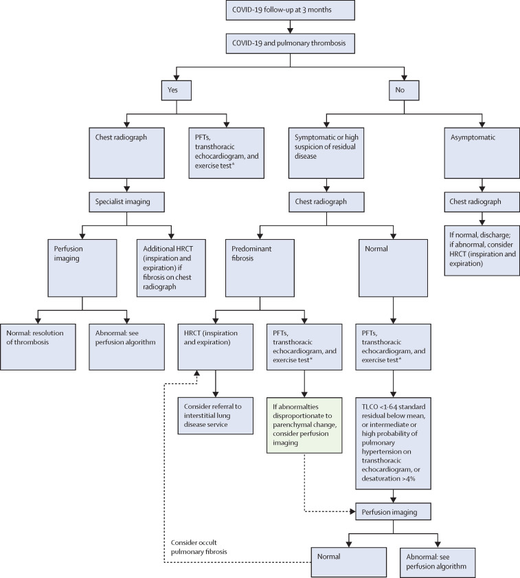
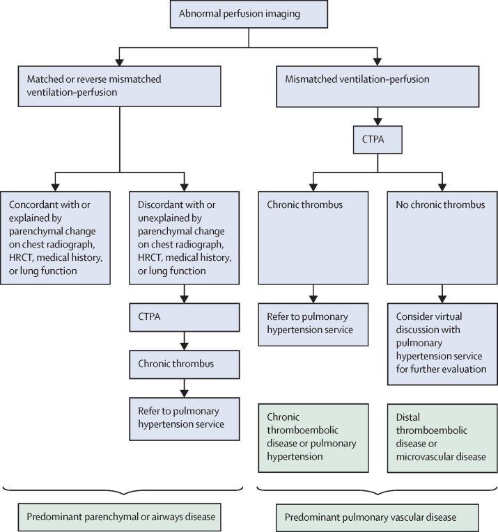
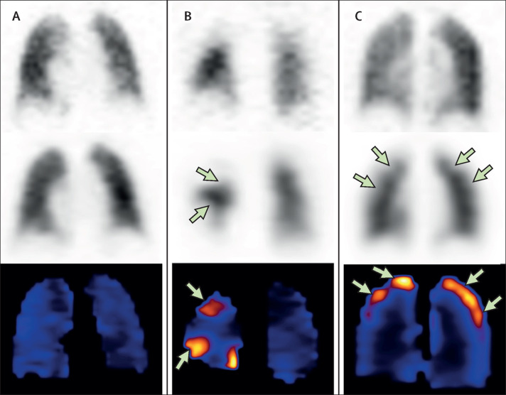
A compelling body of evidence points to pulmonary thrombosis and thromboembolism as a key feature of COVID-19. As the pandemic spread across the globe over the past few months, a timely call to arms was issued by a team of clinicians to consider the prospect of long-lasting pulmonary fibrotic damage and plan for structured follow-up. However, the component of post-thrombotic sequelae has been less widely considered. Although the long-term outcomes of COVID-19 are not known, should pulmonary vascular sequelae prove to be clinically significant, these have the potential to become a public health problem. In this Personal View, we propose a proactive follow-up strategy to evaluate residual clot burden, small vessel injury, and potential haemodynamic sequelae. A nuanced and physiological approach to follow-up imaging that looks beyond the clot, at the state of perfusion of lung tissue, is proposed as a key triage tool, with the potential to inform therapeutic strategies.


Figures

Figure 1
Figure 1
Follow-up algorithm at approximately 3 months after COVID-19 HRCT=high-resolution CT. PFT=pulmonary function test. TLCO=transfer factor of the lung for carbon monoxide. *The exercise test is an incremental shuttle walk test, a 6-min walk test, or a sit-to-stand test (a cardiopulmonary exercise test might be available in some centres to evaluate breathlessness following COVID-19).
Figure 2
Figure 2
Follow-up algorithm in the event of abnormal perfusion imaging CTPA=CT pulmonary angiogram. HRCT=high-resolution CT.
Figure 3
Figure 3
Coronal VQ SPECT, lung CT, and DECT in patient X Imaging in patient X, a 29-year-old woman, during the acute phase of COVID-19 and 6 weeks after the acute illness. (A, B) Coronal VQ SPECT 6 weeks after acute illness. (A) 81mKr ventilation coronal SPECT showing relatively preserved ventilation. (B) 99mTc MAA coronal perfusion SPECT showing subsegmental perfusion deficits (green arrows) in upper and middle lung zones. (C, D) Coronal lung CT and DECT lung perfusion 6 weeks earlier, during the acute phase of illness, showing no air-space changes in the lung. (D) Small perfusion defect on DECT lung perfusion (green arrow) identified retrospectively with the benefit of findings on the 6 week VQ SPECT scan. A CT pulmonary angiogram (not depicted) done in the acute phase of illness showed no macrothrombus. DECT=dual-energy CT. MAA=macroaggregated albumin. SPECT=single-photon emission computed tomography. VQ=ventilation perfusion.
Figure 4
Figure 4
VQ SPECT and coronal lung CT in patient Y VQ SPECT-CT in a 22-year-old woman, 6 weeks after the acute COVID-19 illness. (A, B) 99mTc MAA perfusion coronal MIP (B), showing subtle, tiny peripheral subpleural perfusion deficits (green arrows) in the upper zones with relatively preserved 99mTc DTPA aerosol ventilation (A). (C) Coronal lung CT (from VQ SPECT-CT template) showing no abnormal lung morphology. (D) VQ parametric images showing VQ quotient abnormalities highlighted in pink-orange colour tones (green arrows) at sites of ventilation–perfusion discordance, representing ventilation–perfusion mismatch. This pattern of tiny subpleural upper zone ventilation–perfusion mismatches is atypical for classical pulmonary thromboembolism and taken in the COVID-19 context to represent residual small vessel insult. MAA=macroaggregated albumin. MIP=maximum intensity projection. DTPA=diethylenetriamine pentacetic acid. SPECT-CT=single-photon emission computed tomography with CT fusion. VQ=ventilation perfusion.
Figure 5
Figure 5
Predicted COVID-19 VQ imaging phenotypes with normal, classical, and small vessel patterns These images of classical vascular patterns are drawn from pre-COVID experience of VQ imaging. The proposed phenotypes are deduced from a combination of our CTPA and VQ experience in post-COVID-19 patients and the DECT COVID-19 literature. The top row shows 99mTc DTPA aerosol ventilation coronal SPECT slices, the middle row 99mTc MAA perfusion coronal SPECT slices, and the bottom row VQ quotient parametric images. (A) Uniform ventilation and perfusion pattern. (B) Classical segmental geometry of perfusion defects typical of venous thromboembolism, with corresponding ventilation–perfusion mismatch on VQ quotient images. (C) Non-geometric small, peripheral subpleural perfusion defects, with corresponding peripheral mismatch pattern of a small vessel angiopathy. CTPA=CT pulmonary angiogram. DECT=dual-energy CT. DTPA=diethylenetriamine pentacetic acid. MAA=macroaggregated albumin. SPECT=single-photon emission computed tomography. VQ=ventilation perfusion.

References

    1. Spagnolo P, Balestro E, Aliberti S. Pulmonary fibrosis secondary to COVID-19: a call to arms? Lancet Respir Med. 2020;8:750–752. - PMC - PubMed
    1. Lee L, Iyer S, Jose RJ, Manuel A. COVID-19 follow-up planning: what will we be missing? ERJ Open Res. 2020;6:00198–02020. - PMC - PubMed
    1. Mo X, Jian W, Su Z. Abnormal pulmonary function in COVID-19 patients at time of hospital discharge. Eur Respir J. 2020;55 - PMC - PubMed
    1. Wichmann D, Sperhake J-P, Lütgehetmann M. Autopsy findings and venous thromboembolism in patients with COVID-19: a prospective cohort study. Ann Intern Med. 2020;173:268–277. - PMC - PubMed
    1. Klok FA, Kruip MJHA, van der Meer NJM. Incidence of thrombotic complications in critically ill ICU patients with COVID-19. Thromb Res. 2020;191:145–147. - PMC - PubMed

MeSH terms