. 2021 Jan:185:104974.
doi: 10.1016/j.antiviral.2020.104974. Epub 2020 Nov 17.

A bacterial artificial chromosome (BAC)-vectored noninfectious replicon of SARS-CoV-2

Affiliations

A bacterial artificial chromosome (BAC)-vectored noninfectious replicon of SARS-CoV-2

Yang Zhang et al. Antiviral Res. 2021 Jan.

Abstract

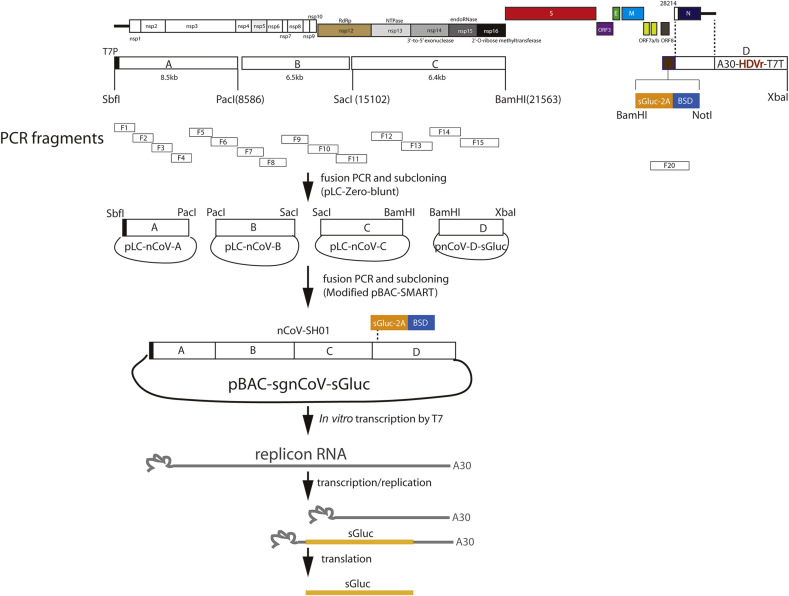
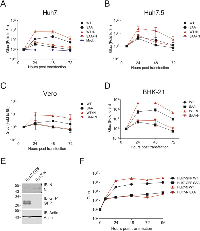
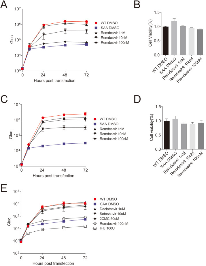
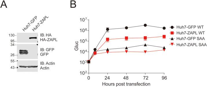
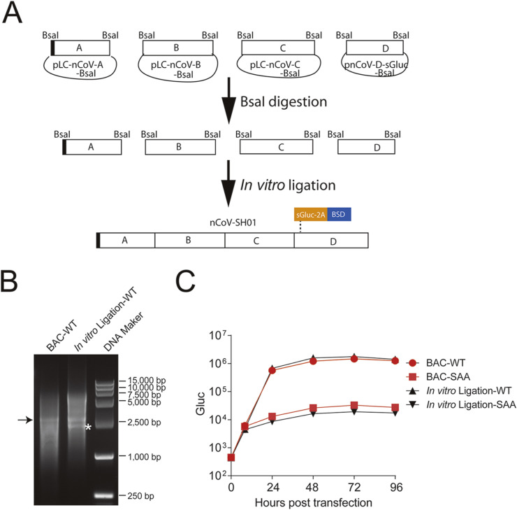
Vaccines and antiviral agents are in urgent need to stop the COVID-19 pandemic. To facilitate antiviral screening against SARS-CoV-2 without requirement for high biosafety level facility, we developed a bacterial artificial chromosome (BAC)-vectored replicon of SARS-CoV-2, nCoV-SH01 strain, in which secreted Gaussia luciferase (sGluc) was encoded in viral subgenomic mRNA as a reporter gene. The replicon was devoid of structural genes spike (S), membrane (M), and envelope (E). Upon transfection, the replicon RNA replicated in various cell lines, and was sensitive to interferon alpha (IFN-α), remdesivir, but was resistant to hepatitis C virus inhibitors daclatasvir and sofosbuvir. Replication of the replicon was also sensitive overexpression to zinc-finger antiviral protein (ZAP). We also constructed a four-plasmid in-vitro ligation system that is compatible with the BAC system, which makes it easy to introduce desired mutations into the assembly plasmids for in-vitro ligation. This replicon system would be helpful for performing antiviral screening and dissecting virus-host interactions.

Keywords: Antiviral agents; Replicon; SARS-CoV-2.


Conflict of interest statement

The authors declare no conflict of interest.

Figures

Fig. 1
Fig. 1
Schematic of construction of BAC-based replicon of SARS-CoV-2. Twenty fragments encompassing the whole viral genomes were amplified, cloned and sequenced. Four larger fragments A (1-8586 nt), B (8587-15102 nt), C (15,103-21562 nt) and D with deletion of structural protein genes and addition of reporter gene cassette were assembled and cloned. To facilitate cloning, a BamHI site (in bold) was introduced downstream the genome position of 21,562 (nt). In fragment A, T7 promoter (T7P) was added. In fragment D, an expression cassette containing secreted Gaussia luciferase (sGluc), foot-and-mouth disease virus (FMDV) 2 A peptide and blasticidin (BSD) was added. The 3′ viral genome was flanked with polyA30, hepatitis delta virus ribozymes (HDVr) and terminator sequence for T7 polymerase (T7T). Then the fragments were assembled and sequentially cloned into a modified BAC plasmid. Upon transfection into cells, the replicon RNA can be used as template for RNA replication or transcription to produce subgenomic RNA. The sGluc subgenomic RNA is translated to produce sGluc.
Fig. 2
Fig. 2
Replication of sgnCoV-sGluc in different cells. (A– D) Huh7, Huh7.5, Vero and BHK-21 cells were transfected with in-vitro transcribed replicon RNA (WT) or the nsp12 polymerase active-site mutant (SAA). An mRNA encoding the SARS-CoV-2 N protein was co-transfected or not. The luciferase activity in the supernatants was measured at the time points indicated. Medium was changed at all time point. A mock-transfection group in Fig 2A showed the background signals. Data are shown as mean ± SD (n = 3). (E–F) Replication of replicon RNA in Huh7 cells overexpressed with N protein. (E) Huh7 cells overexpressed with GFP protein or N protein were analyzed by Western blotting with the indicated antibodies. (F) Huh7-GFP and Huh7-N cells were transfected with replicon RNA (WT or SAA). The luciferase activity in the supernatants was measured at the time points indicated. Medium was changed at 8 h post transfection. Data are shown as mean ± SD (n = 3).
Fig. 3
Fig. 3
Sensitivity of the SARS-CoV-2 replicon to antiviral agents. (A) Huh7 cells were treated with remdesivir as the indicated concentrations. Four hours later, cells were co-transfected with replicon RNA (WT or SAA) and N mRNA. The luciferase activity in the supernatants was measured at the time points indicated. Medium was changed at 8 h post transfection. (B) The cell viabilities in (A) were measured at 72 h post transfection using the CCK8 kit. (C) Huh7 cells were co-transfected with replicon RNA (WT or SAA) and N mRNA. Eight hours later, medium was changed with remdesivir as the indicated concentration. The luciferase activity in the supernatants was measured at the time points indicated. Medium was changed at 8 h post transfection. (D) The cell viabilities in (C) were measured at 72 h post transfection using the CCK8 kit. (E) Huh7 cells were treated with remdesivir (100 nM), IFN-α (100 U/ml), daclatasvir (1 μM), sofosbuvir (10 μM), 2CMC (50 μM). Four hours later, cells were co-transfected with replicon RNA (WT or SAA) and N mRNA. The luciferase activity in the supernatants was measured at the time points indicated. Medium was changed at 8 h post transfection. Data are shown as mean ± SD (n = 3).
Fig. 4
Fig. 4
Sensitivity of SARS-CoV-2 replicon to overexpression with Zinc-finger antiviral protein (ZAP). (A) Huh7 cells overexpressed with ZAPL protein or GFP protein were analyzed by Western blotting with the indicated antibodies. (B) Huh7-GFP and Huh7-ZAPL cells were co-transfected with replicon RNA (WT or SAA) and N mRNA. The luciferase activity in the supernatants was measured at the time points indicated. Medium was changed at 8 h post transfection. Data are shown as mean ± SD (n = 3).
Fig. 5
Fig. 5
Assembly of SARS-CoV-2 replicon by in-vitro ligation. (A) Schematic of the in-vitro ligation system for SARS-CoV-2 replicon. (B) Gel analysis of RNA transcripts. About 2 μg of in-vitro transcribed RNAs were analyzed on a 0.7% native agarose gel. DNA maker are indicated. Because this is a native agarose gel, the DNA size is not directly corelated to the RNA size. Arrows indicates the replicon RNA transcript. Asterisk shows the shorter RNA transcript. (C) Huh7 cells were co-transfected with replicon RNA (WT or SAA) and N mRNA generated by BAC-based system or in-vitro ligation system. The luciferase activity in the supernatants was measured at the time points indicated. Medium was changed at 8 h post transfection. Data are shown as mean ± SD (n = 3).

Similar articles

Cited by

References

    1. Bonovas S., Piovani D. Compassionate use of remdesivir in covid-19. N. Engl. J. Med. 2020;382 - PubMed
    1. Chen L., Liu W., Zhang Q., Xu K., Ye G., Wu W., Sun Z., Liu F., Wu K., Zhong B., Mei Y., Zhang W., Chen Y., Li Y., Shi M., Lan K., Liu Y. RNA based mNGS approach identifies a novel human coronavirus from two individual pneumonia cases in 2019 Wuhan outbreak. Emerg. Microb. Infect. 2020;9:313–319. - PMC - PubMed
    1. Du X.T., Pan T.T., Xu J., Zhang Y., Song W.H., Yi Z.G., Yuan Z.H. Hepatitis C virus replicative double-stranded RNA is a potent interferon inducer that triggers interferon production through MDA5. J. Gen. Virol. 2016;97:2868–2882. - PubMed
    1. Gao M., Nettles R.E., Belema M., Snyder L.B., Nguyen V.N., Fridell R.A., Serrano-Wu M.H., Langley D.R., Sun J.H., O'Boyle D.R., 2nd, Lemm J.A., Wang C., Knipe J.O., Chien C., Colonno R.J., Grasela D.M., Meanwell N.A., Hamann L.G. Chemical genetics strategy identifies an HCV NS5A inhibitor with a potent clinical effect. Nature. 2010;465:96–100. - PMC - PubMed
    1. Gao Y., Yan L., Huang Y., Liu F., Zhao Y., Cao L., Wang T., Sun Q., Ming Z., Zhang L., Ge J., Zheng L., Zhang Y., Wang H., Zhu Y., Zhu C., Hu T., Hua T., Zhang B., Yang X., Li J., Yang H., Liu Z., Xu W., Guddat L.W., Wang Q., Lou Z., Rao Z. Structure of the RNA-dependent RNA polymerase from COVID-19 virus. Science. 2020;368:779–782. - PMC - PubMed

Publication types

MeSH terms