. 2021 Jan-Feb;14(1):88-96.
doi: 10.1016/j.brs.2020.11.010. Epub 2020 Nov 17.

Selective optogenetic stimulation of efferent fibers in the vagus nerve of a large mammal

Affiliations

Selective optogenetic stimulation of efferent fibers in the vagus nerve of a large mammal

Lindsea C Booth et al. Brain Stimul. 2021 Jan-Feb.

Abstract

Background: Electrical stimulation applied to individual organs, peripheral nerves, or specific brain regions has been used to treat a range of medical conditions. In cardiovascular disease, autonomic dysfunction contributes to the disease progression and electrical stimulation of the vagus nerve has been pursued as a treatment for the purpose of restoring the autonomic balance. However, this approach lacks selectivity in activating function- and organ-specific vagal fibers and, despite promising results of many preclinical studies, has so far failed to translate into a clinical treatment of cardiovascular disease.

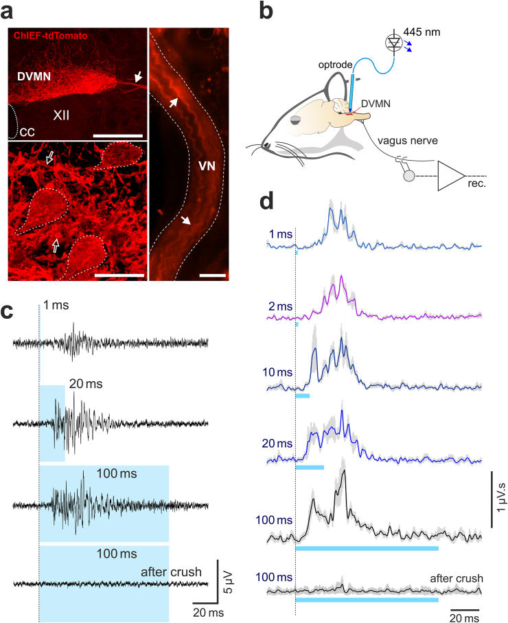
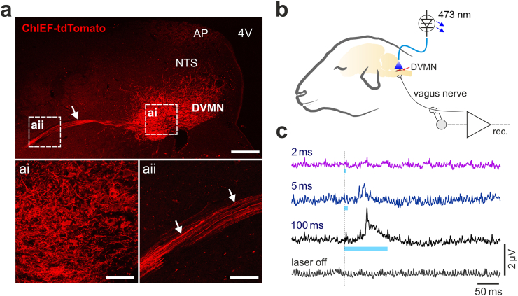
Objective: Here we report a successful application of optogenetics for selective stimulation of vagal efferent activity in a large animal model (sheep).

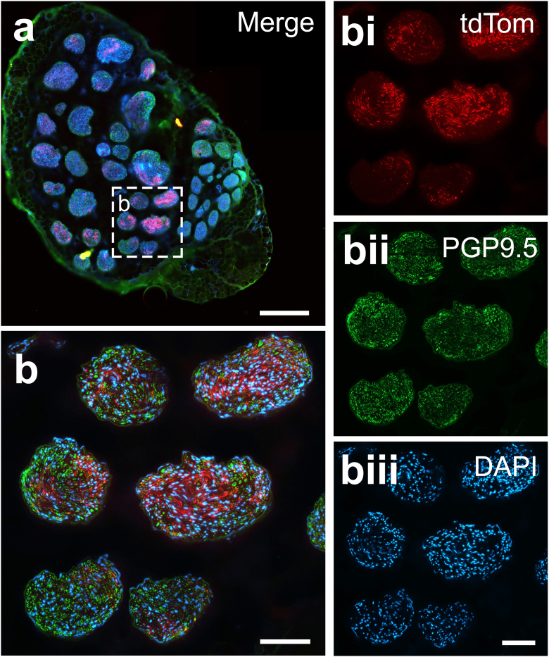
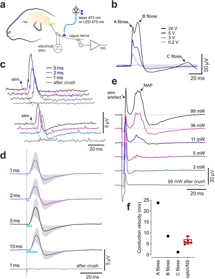
Methods and results: Twelve weeks after viral transduction of a subset of vagal motoneurons, strong axonal membrane expression of the excitatory light-sensitive ion channel ChIEF was achieved in the efferent projections innervating thoracic organs and reaching beyond the level of the diaphragm. Blue laser or LED light (>10 mW mm-2; 1 ms pulses) applied to the cervical vagus triggered precisely timed, strong bursts of efferent activity with evoked action potentials propagating at speeds of ∼6 m s-1.

Conclusions: These findings demonstrate that in species with a large, multi-fascicled vagus nerve, it is possible to stimulate a specific sub-population of efferent fibers using light at a site remote from the vector delivery, marking an important step towards eventual clinical use of optogenetic technology for autonomic neuromodulation.

Keywords: Autonomic nervous system; Brainstem; Neuromodulation; Optogenetic; Vagal preganglionic neurons; Vagus nerve stimulation.


Conflict of interest statement

Declaration of competing interest All authors declare no conflict of interest.

Figures

Fig. 1
Fig. 1
Optogenetic stimulation and properties of vagal preganglionic neurons of the dorsal motor nucleus of the vagus nerve (DVMN) in rats. (a) Photomicrographs of the coronal sections of the rat brainstem taken at low (top left) and high (bottom left) magnification illustrating representative examples of ChIEF-tdTomato expression in the DVMN (Bregma level: −13.8 mm). Neurons display specific membrane localization of the transgene expression. The image on the right illustrates DVMN neuronal projections expressing ChIEF-tdTomato visualized in a whole mount preparation of the right cervical vagus nerve. Solid arrows point at the projecting axons of the transduced DVMN neurons; open arrows point at severed neuronal processes within the nucleus; broken while lines outline transduced DVMN neurons. CC, central canal. VN, vagus nerve. XII, hypoglossal motor nucleus. Scale bars: 200 μm (top left), 20 μm (bottom left), 500 μm (right). (b) Schematic drawing of the experimental setup in an anaesthetized rat instrumented for stimulation of the DVMN neurons expressing ChIEF-tdTomato by application of blue laser light (via an implanted optrode) and recording of the efferent activity of the vagus nerve at the cervical level. (c) Stimulus-triggered averages and (d) rectified and smoothed averages (300 sweeps) of the evoked efferent vagus nerve mass action potentials induced by pulses of light of increasing duration delivered at 1 Hz to stimulate the DVMN vagal preganglionic neurons expressing ChIEF-tdTomato (n = 4).
Fig. 2
Fig. 2
Viral transduction and optogenetic stimulation of the DVMN vagal preganglionic neurons in sheep. (a) Photomicrographs of coronal sections of the sheep brainstem taken at low (top) and high (bottom) magnification illustrating representative examples of ChIEF-tdTomato expression in the DVMN. Arrows point at the projecting axons of the transduced DVMN neurons. 4V, fourth ventricle. AP, area postrema. NTS, nucleus of the solitary tract. Scale bars: 400 μm (top), 100 μm (ai, aii). (b) Schematic drawing of the experimental setup in an anaesthetized sheep instrumented for stimulation of the DVMN neurons expressing ChIEF-tdTomato by application of blue laser light and recording of the efferent activity of the vagus nerve at the cervical level. (c) Stimulus-triggered averages (300 sweeps) of the evoked efferent vagus nerve mass action potentials induced by pulses of light of increasing duration delivered at 1 Hz to stimulate the DVMN neurons expressing ChIEF-tdTomato in sheep.
Fig. 3
Fig. 3
Expression of ChIEF-tdTomato by vagal efferent fibers originating from the DVMN, visualized at the cervical level in sheep. (a) Representative cross-section of the whole cervical vagus nerve showing expression of ChIEF-tdTomato (red), Protein Gene Product 9.5 (PGP9.5) immunoreactivity (green) and 4′,6-diamidino-2-phenylindole (DAPI) staining (blue). Scale bar: 500 μm. (b) Photomicrograph of six fascicles taken at a higher magnification showing the expression of ChIEF-tdTomato (red), PGP9.5 immunoreactivity (green) and DAPI staining (blue) merged; bi, bii and biii show individual stains. Scale bars: 100 μm. (For interpretation of the references to colour in this figure legend, the reader is referred to the Web version of this article.)
Fig. 4
Fig. 4
Optogenetic stimulation and properties of vagal efferent fibers originating from the DVMN in sheep. (a) Schematic drawing of the experimental setup in an anaesthetized sheep instrumented for electrical or optical stimulation of the nerve and recording of vagal activity at the cervical level. (b) Averages of the evoked vagus nerve mass action potentials induced by whole-nerve electrical stimulation illustrating stimulus intensity-dependent recruitment of A, B and C fibres in sheep. (c) Representative examples of the recordings obtained in two individual animals and (d) group data (n = 4), illustrating stimulus-triggered averages of the evoked efferent vagus nerve mass action potentials, induced by pulses of laser light of increasing duration delivered to the nerve at the cervical level, proximal to the recording site. (e) Stimulus-triggered averages of the evoked efferent vagus nerve mass action potentials (MAP) induced by pulses of LED light delivered from a variable distance to the nerve in order to determine the light power threshold, required for activation of the transduced fibers. Light powers indicated are in mW per mm2. (f) Indicative values (black symbols) denoting the mean conduction velocities that define A, B and C vagal fibres and the group data illustrating the calculated velocities of action potential propagation, recorded in vagal efferent fibers originating from the DVMN in sheep (red symbols, n = 6).

References

    1. Famm K., Litt B., Tracey K.J., Boyden E.S., Slaoui M. Drug discovery: a jump-start for electroceuticals. Nature. 2013;496:159–161. - PMC - PubMed
    1. Bonaz B., Picq C., Sinniger V., Mayol J.F., Clarencon D. Vagus nerve stimulation: from epilepsy to the cholinergic anti-inflammatory pathway. Neuro Gastroenterol Motil. 2013;25:208–221. - PubMed
    1. Howland R.H. Vagus nerve stimulation. Curr Behav Neurosci Rep. 2014;1:64–73. - PMC - PubMed
    1. Yuan H., Silberstein S.D. Vagus nerve and vagus nerve stimulation, a comprehensive review: Part I. Headache. 2016;56:71–78. - PubMed
    1. Yuan H., Silberstein S.D. Vagus nerve and vagus nerve stimulation, a comprehensive review: Part II. Headache. 2016;56:259–266. - PubMed

Publication types

LinkOut - more resources