. 2021 Jan-Feb;14(1):69-73.
doi: 10.1016/j.brs.2020.11.009. Epub 2020 Nov 17.

White matter hyperintensities affect transcranial electrical stimulation in the aging brain

Affiliations

White matter hyperintensities affect transcranial electrical stimulation in the aging brain

Aprinda Indahlastari et al. Brain Stimul. 2021 Jan-Feb.

Abstract

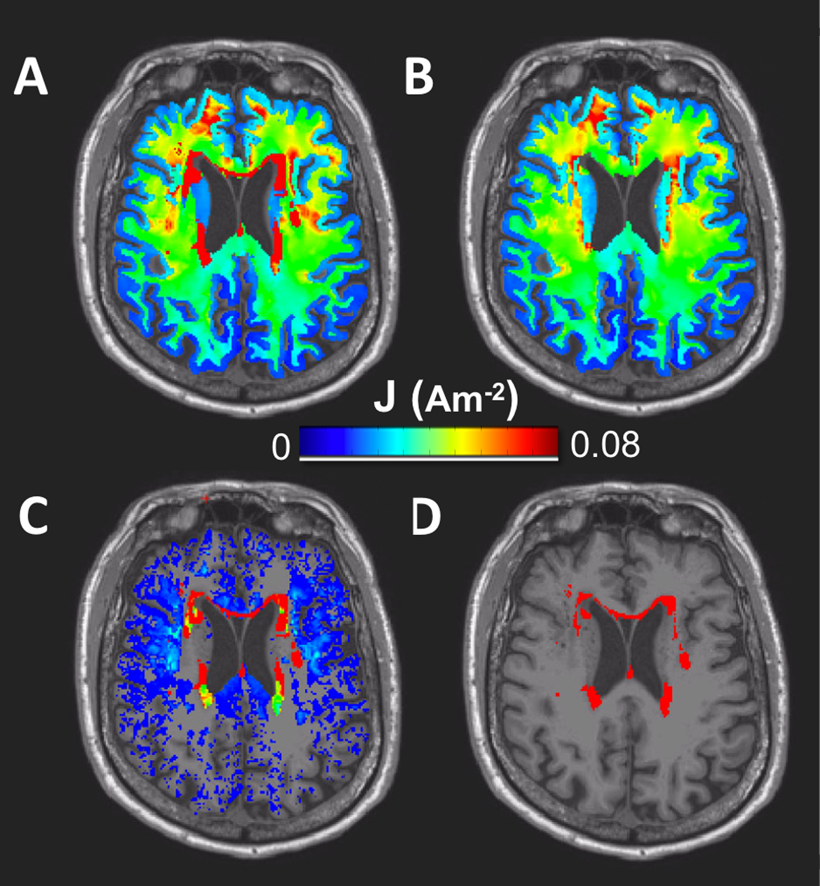
Background: White matter hyperintensities (WMH) are estimated to occur in greater than 63% of older adults over the age of 60 years. WMH identified in the T2-weighted FLAIR images can be combined with T1-weighted images to enhance individualized current flow models of older adults by accounting for the presence of WMH and its effects on delivered tES current in the aging brain.

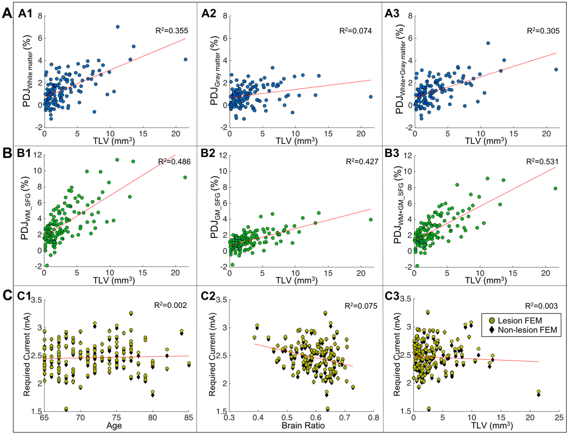
Methods: Individualized head models were derived from T1-weighted images of 130 healthy older adults (mean = 71 years). Lesions segmented from FLAIR acquisition were added to individualized models. Current densities were computed in the brain and compared between models with and without lesions.

Main results: Integrating WMH into the models resulted in an overall decrease (up to 7%) in median current densities in the brain outside lesion regions. Changes in current density and total lesion volume was positively correlated (R2 = 0.31, p < 0.0001).

Conclusions: Incorporating WMH into individualized models may increase the accuracy of predicted tES current flow in the aging brain.

Keywords: Aging; Finite element model; White matter hyperintensity; tES.


Conflict of interest statement

AUTHOR DECLARATION

The authors report no conflicts of interest.

Figures

Figure 1.
Figure 1.
Generated current density and lesion maps in the brain region (white and gray matter only). An axial view illustrates current density maps generated in A) lesion FEM B) non-lesion FEM, C) the subtraction of current density maps between lesion and non-lesion FEM, and D) location of the binarized lesion volume.
Figure 2.
Figure 2.
Scatter plots of current density distribution in the brain. A) Correlation plots between total lesion volume (TLV) and percentage differences in current density (PDJ) in white matter (A1), gray matter (A2) and white+gray matter (A3). B) Correlation plots between total lesion volume (TLV) and percentage differences in current density (PDJ) within the superior frontal gyrus (SFG) for white matter SFG (B1), gray matter SFG (B2) and white+gray matter SFG (B3). C) Required current dose to be adjusted in the aging brain relative to a 2mA stimulation in young adult brains for lesion FEM (yellow circles) and non-lesion FEM (black diamonds) for dose vs. age (C1), dose vs. brain ratio (C2), and dose vs. TLV (C3).

References

    1. Prins ND, Scheltens P. White matter hyperintensities, cognitive impairment and dementia: an update. Nat Rev Neurol 2015;11:157–65. doi:10.1038/nrneurol.2015.10. - DOI - PubMed
    1. Bakshi R, Ariyaratana S, Benedict RHB, Jacobs L. Fluid-attenuated inversion recovery magnetic resonance imaging detects cortical and juxtacortical multiple sclerosis lesions. Arch Neurol 2001. doi:10.1001/archneur.58.5.742. - DOI - PubMed
    1. O’Shea AM, Woods AJ. White Matter Hyper-intensities. In: Gu D, Dupre ME, editors. Encycl. Gerontol. Popul. Aging, Cham: Springer International Publishing; 2019, p. 1–5. doi:10.1007/978-3-319-69892-2_684-1. - DOI
    1. Yoshita M, Fletcher E, Harvey D, Ortega M, Martinez O, Mungas DM, et al. Extent and distribution of white matter hyperintensities in normal aging, MCI, and AD. Neurology 2006. doi:10.1212/01.wnl.0000249119.95747.1f. - DOI - PMC - PubMed
    1. Ye Q, Chen X, Qin R, Huang L, Yang D, Liu R, et al. Enhanced regional homogeneity and functional connectivity in subjects with white matter hyperintensities and cognitive impairment. Front Neurosci 2019. doi:10.3389/fnins.2019.00695. - DOI - PMC - PubMed

Publication types

LinkOut - more resources