Review
. 2021 Jan:267:120491.
doi: 10.1016/j.biomaterials.2020.120491. Epub 2020 Nov 5.

Trends of microneedle technology in the scientific literature, patents, clinical trials and internet activity

Affiliations
Review

Trends of microneedle technology in the scientific literature, patents, clinical trials and internet activity

Rohan S J Ingrole et al. Biomaterials. 2021 Jan.

Abstract

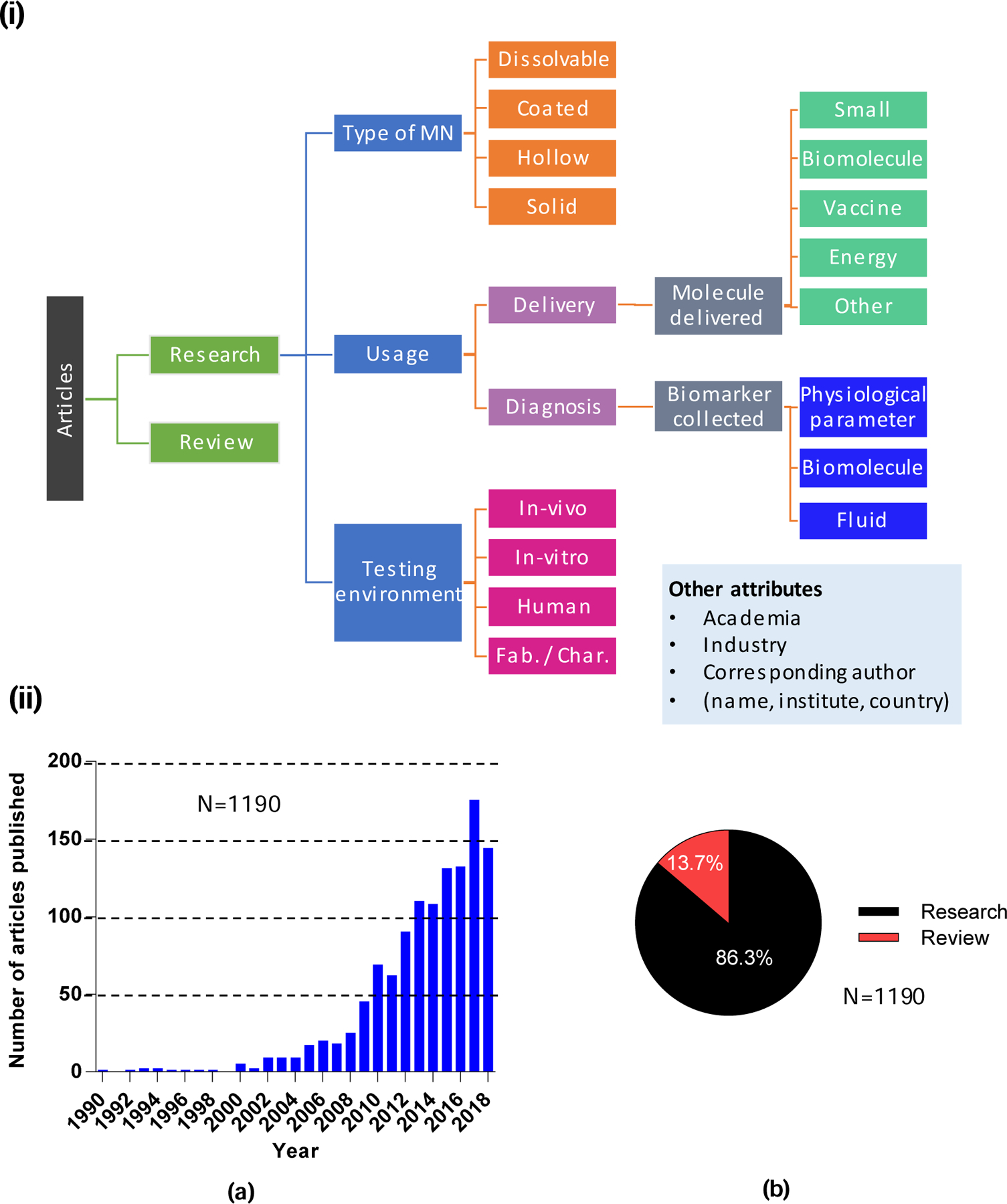
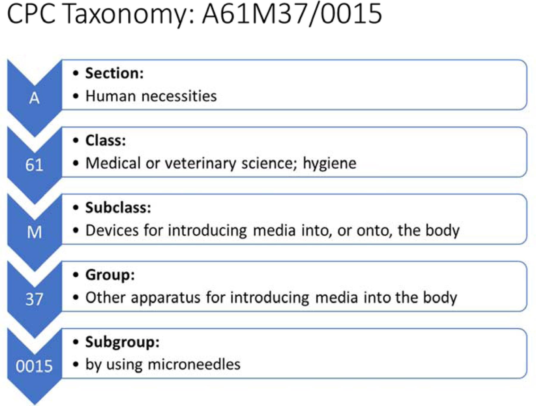
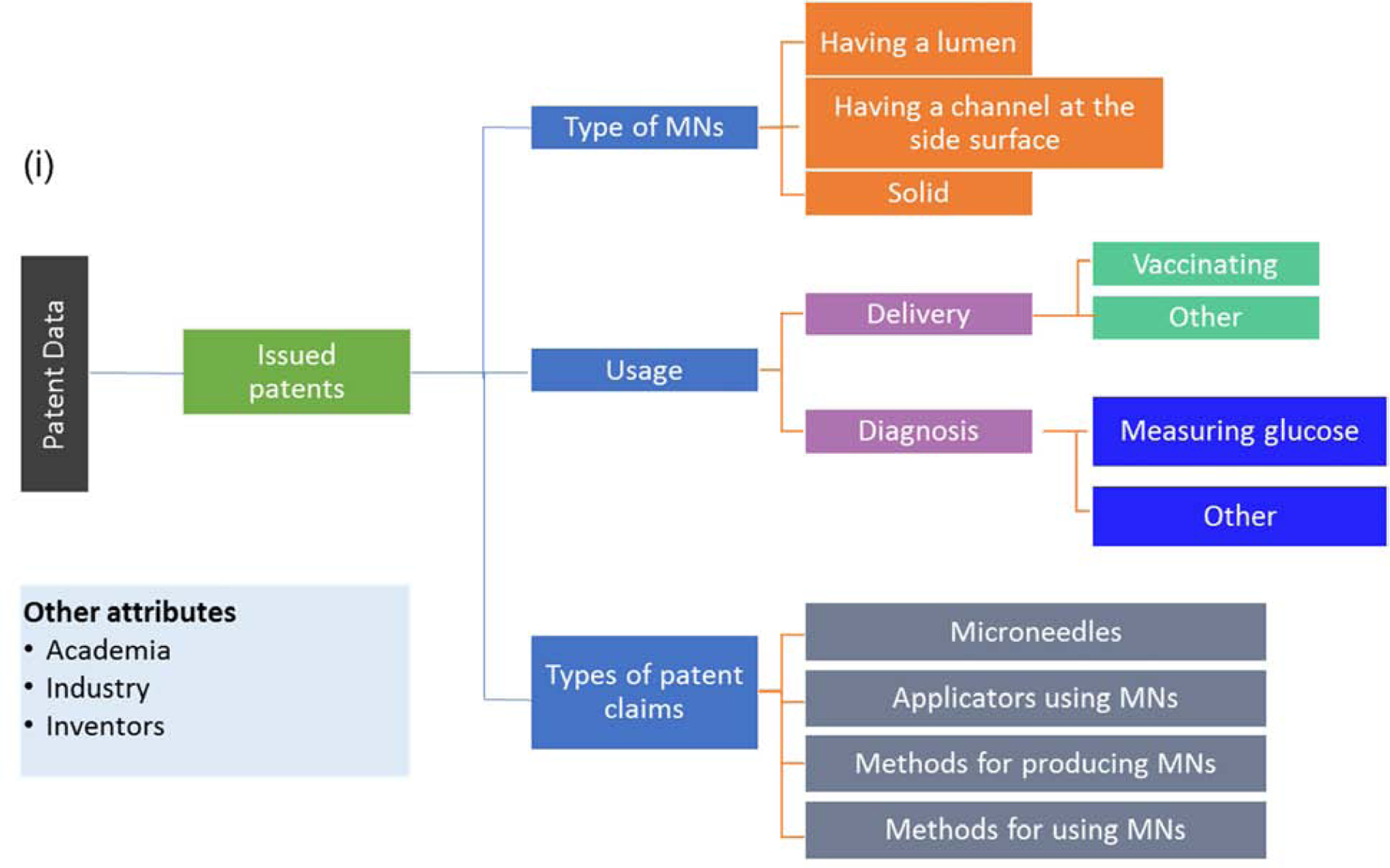
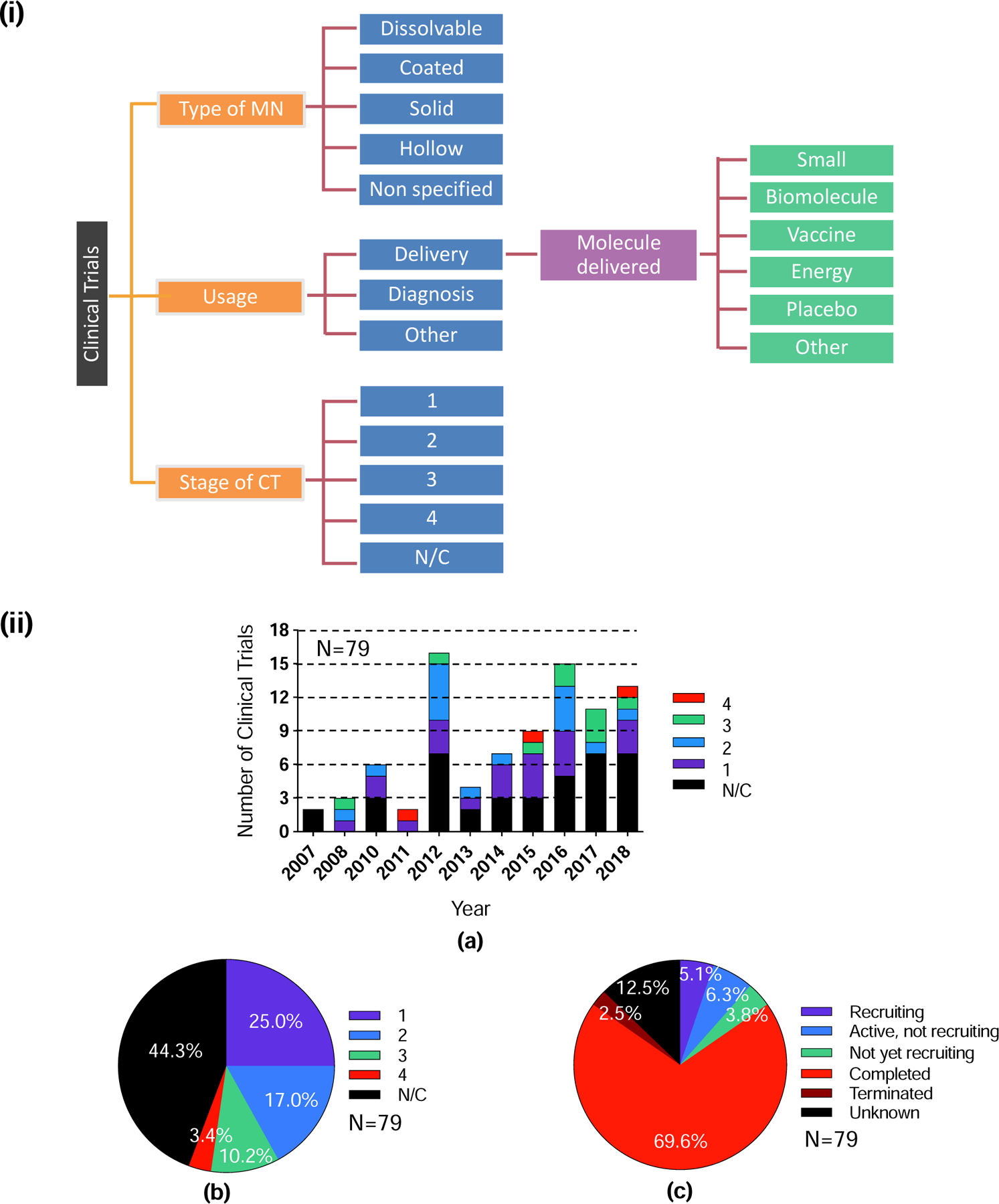
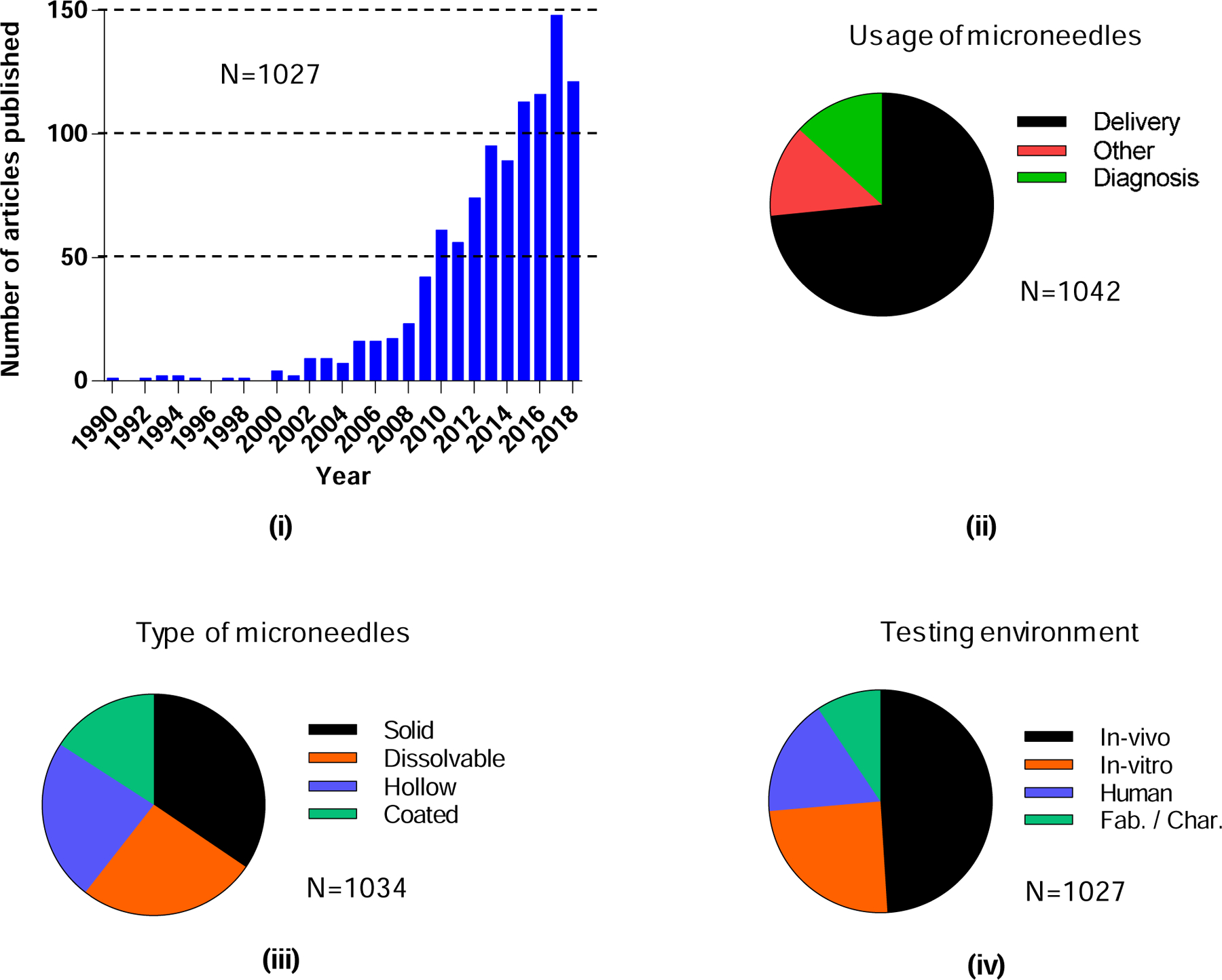
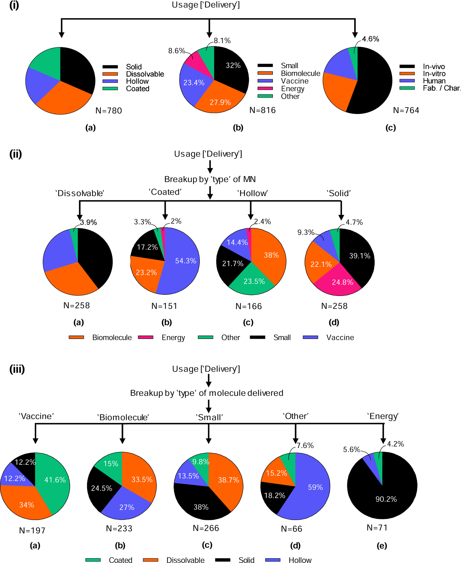
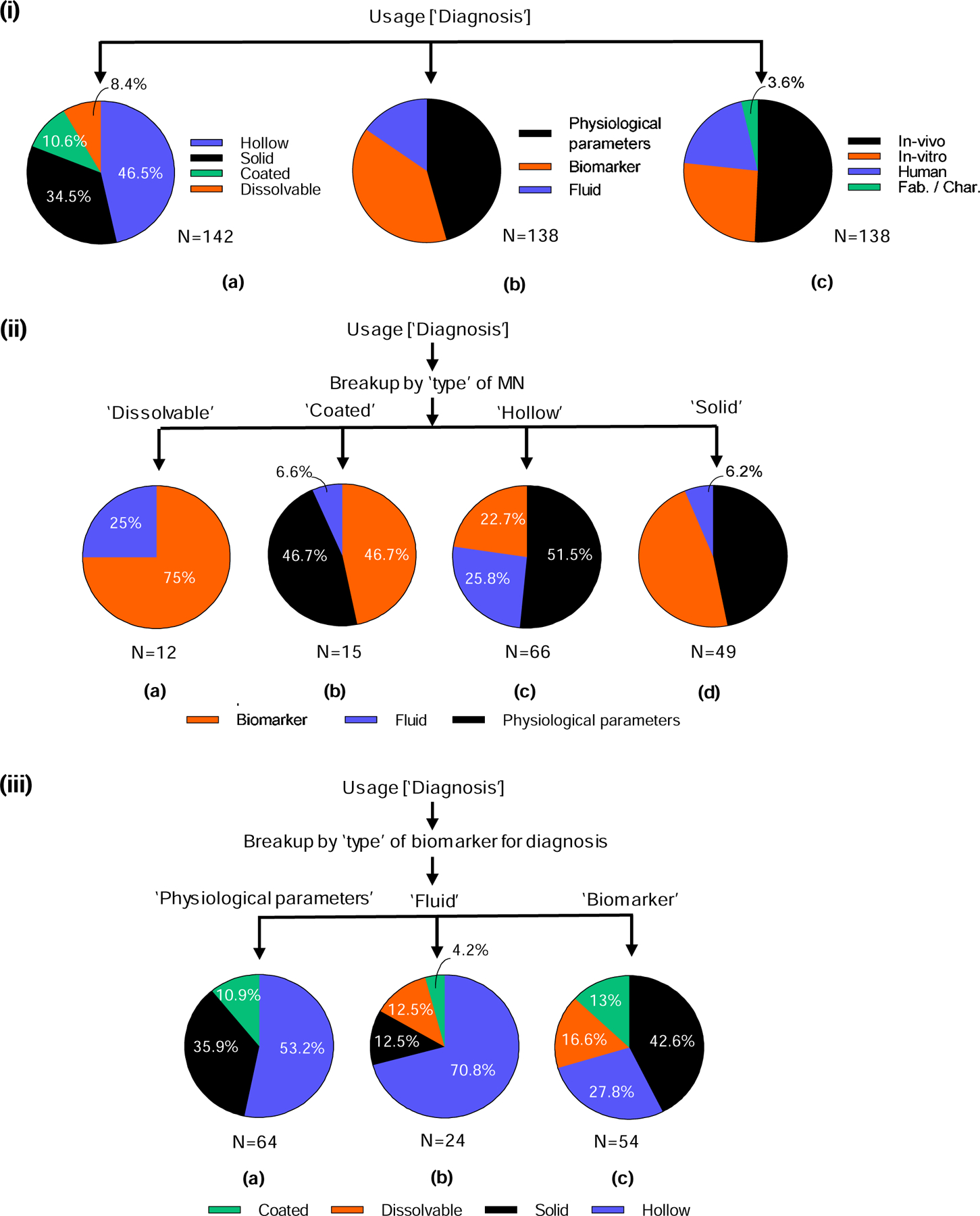
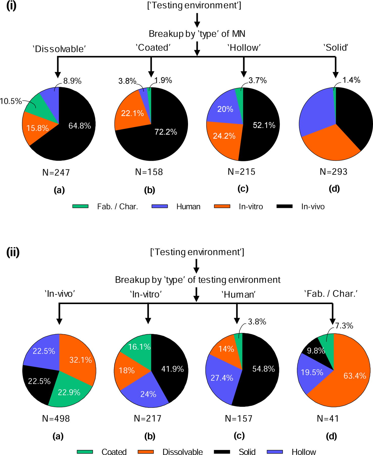
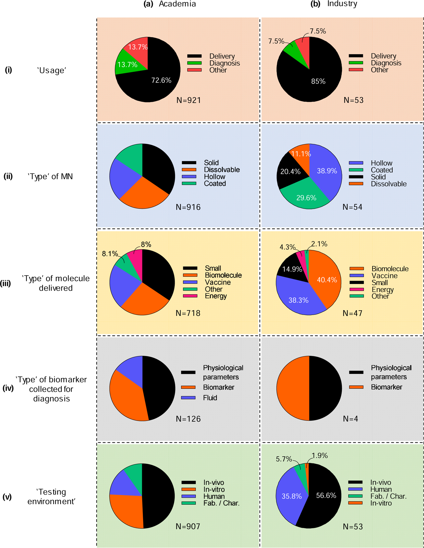
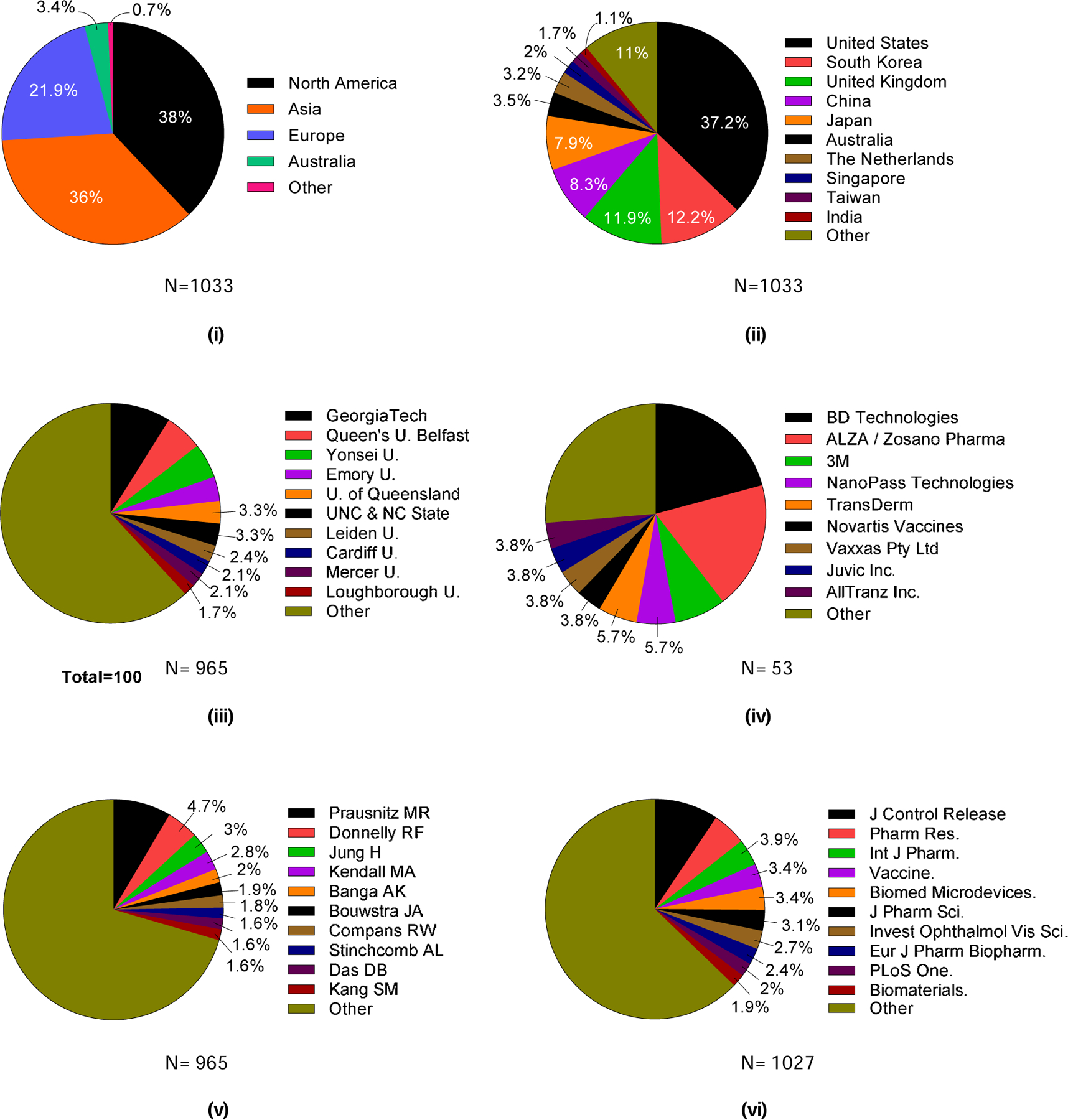
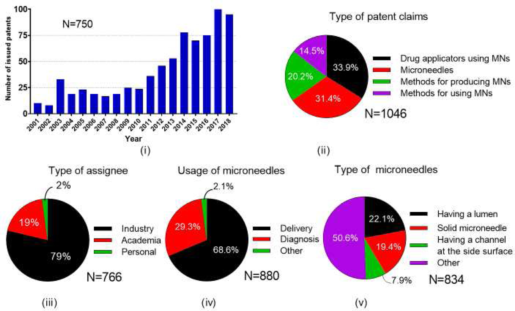
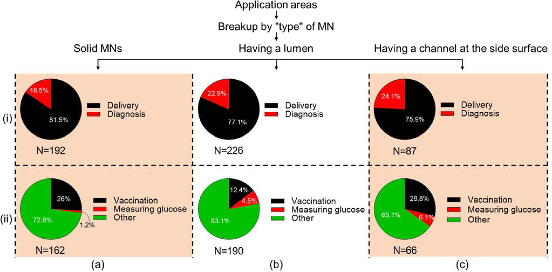
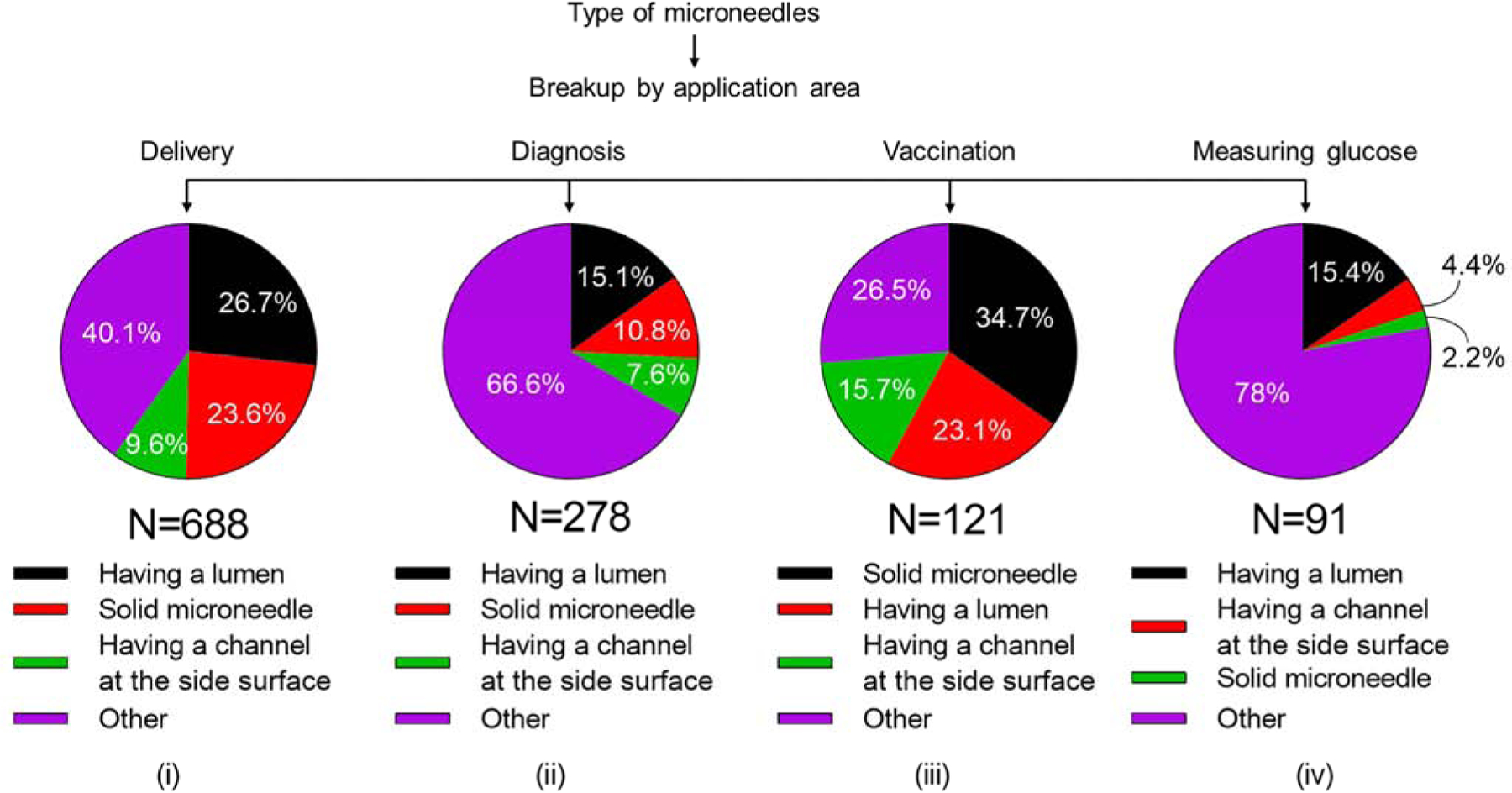
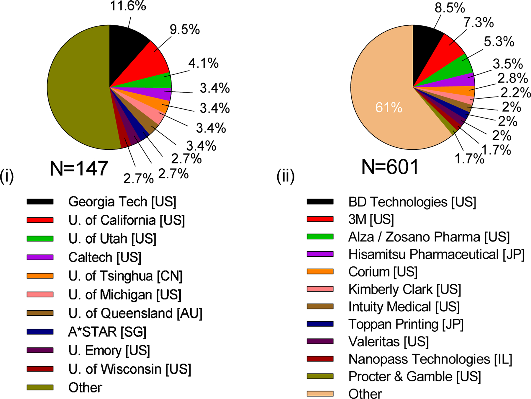
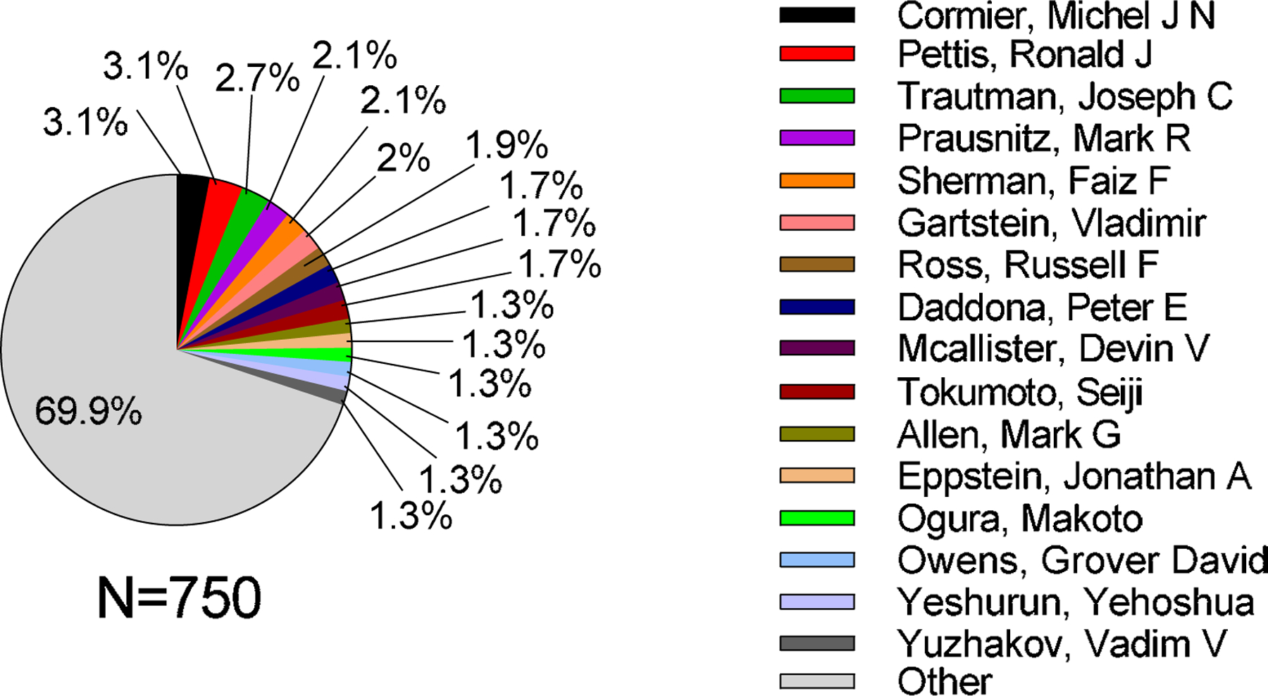
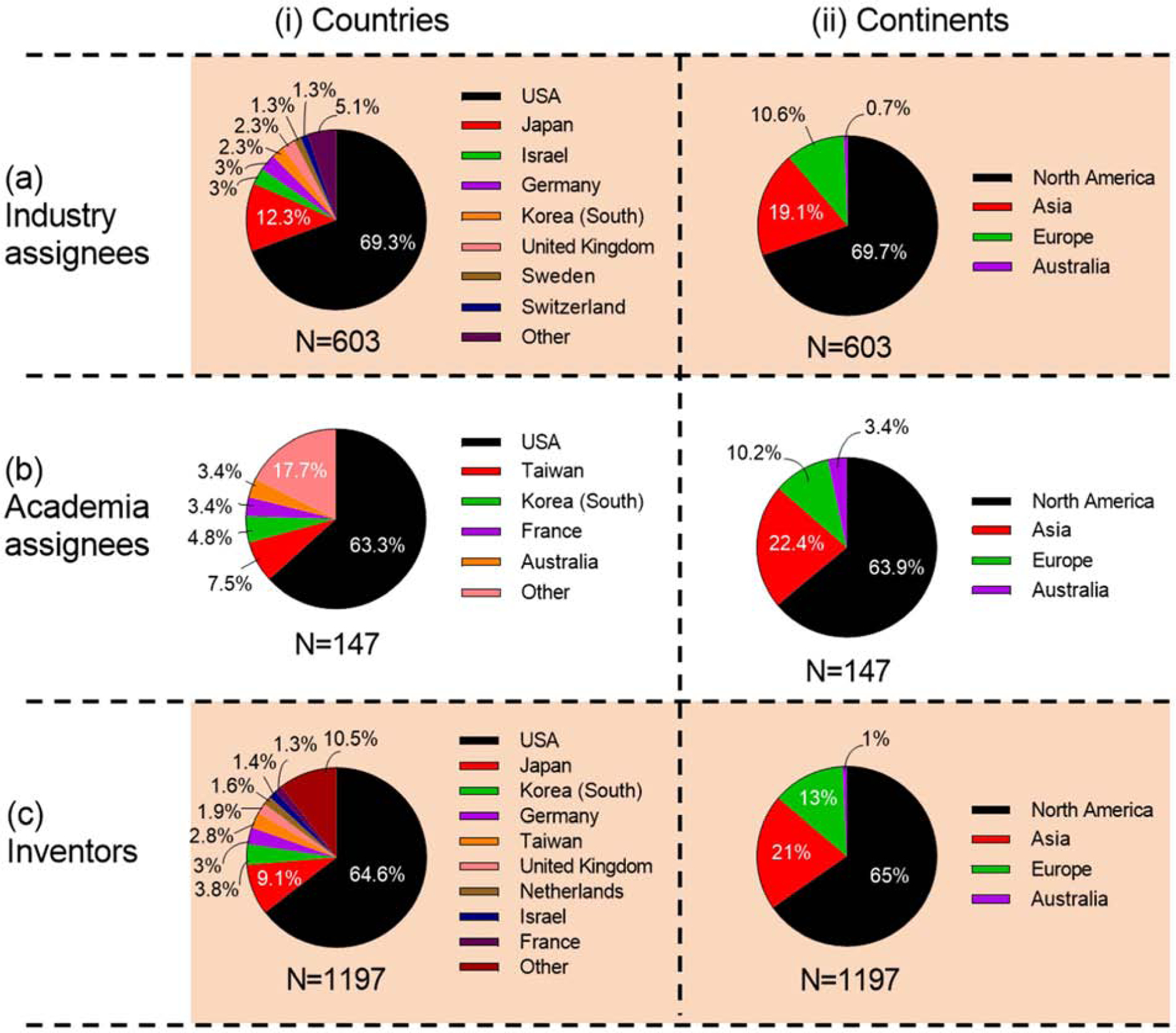
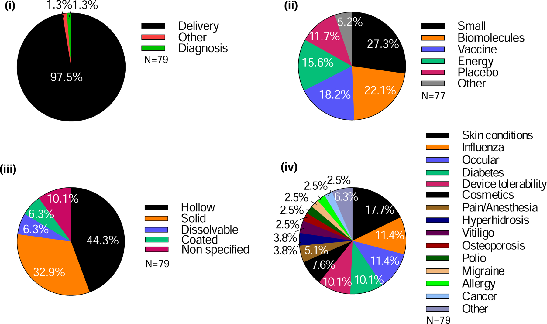
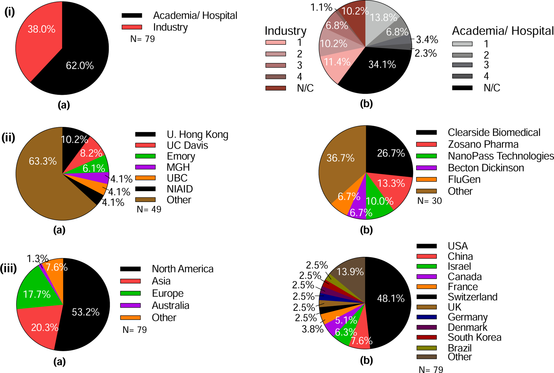
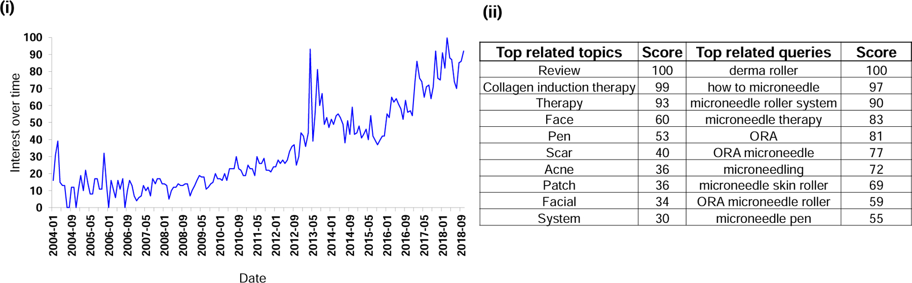
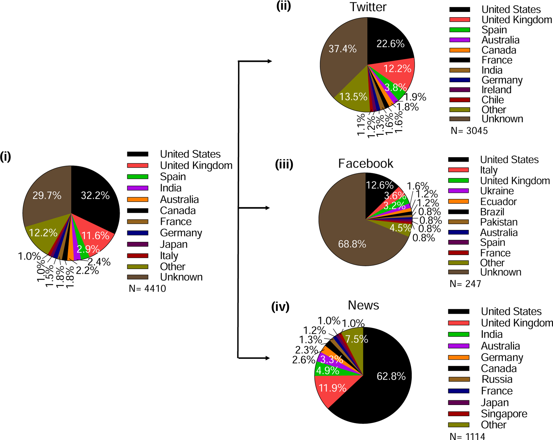
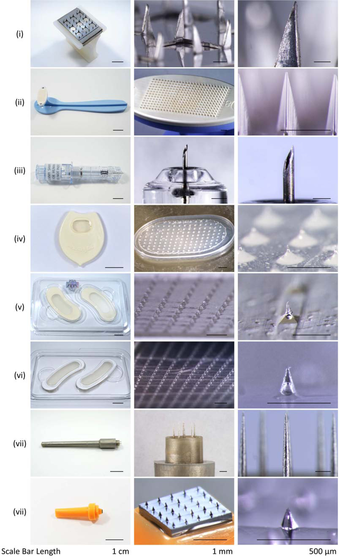
The powerful and intriguing idea that drives the emerging technology of microneedles-shrinking the standard needle to a micron scale-has fostered an entire field of microneedle study and subsequent exponential growth in research and product development. Originally enabled by microfabrication tools derived from the microelectronic industry, microneedles are now produced through a number of methods in a variety of forms including solid, coated, dissolvable, and hollow microneedles. They are used to deliver a broad spectrum of molecules, including small molecules, biomolecules, and vaccines, as well as various forms of energy into the skin, eye, and other tissues. Microneedles are also being exploited for use in diagnostics, as well as additional medical, cosmetic, and other applications. This review elucidates the relative roles of different aspects of microneedle technology development, as shown through scientific papers, patents, clinical studies, and internet/social media activity. Considering >1000 papers, 750 patents, and almost 80 clinical trials, we analyze different attributes of microneedles such as usage of microneedles, types of microneedles, testing environment, types of patent claims, and phases of clinical trials, as well as which institutions and people in academia and industry from different locations and in different journals are publishing, patenting, and otherwise studying the potential of microneedles. We conclude that there is robust and growing activity in the field of microneedles; the technology is rapidly developing and being used for novel applications to benefit human health and well-being.

Keywords: Clinical trial; Diagnostic device; Microneedle; Patent; Skin patch; Transdermal drug delivery.


Conflict of interest statement

7. Competing interest

M.R.P. serves as a consultant to companies, is a founding shareholder of companies, and is an inventor on patents licensed to companies developing microneedle-based products (Micron Biomedical, Clearside Biomedical). These potential conflicts of interest have been disclosed and are managed by Georgia Tech. H.S.G. is a co-inventor on a patent related to coated microneedles and has stock ownership in a startup company called Moonlight Therapeutics that is developing microneedles for food allergy immunotherapy. This conflict of interest has been disclosed and is being managed by Texas Tech University. No financial support was provided by Moonlight Therapeutics for this work.

⊠ The authors declare the following financial interests/personal relationships which may be considered as potential competing interests:

Figures

Figure 1.
Figure 1.
Overview of research papers included in the study. (i). General outline for the categorization of microneedles and their uses. (ii). Total number of research and review articles published on microneedles from 1990 until Oct. 1, 2018: a) number of yearly publications, and b) percentage of research versus review articles.
Figure 2.
Figure 2.
Taxonomy of a CPC symbol with definitions of categories and subcategories.
Figure 3.
Figure 3.
General outline of the categorization of microneedles and their uses.
Figure 4.
Figure 4.
Overview of clinical trials included in the study. (i) General outline of the categorization of microneedles and their uses in clinical trials. (ii) Total number of registered clinical trials involving the use of microneedles listed in ClinicalTrials.gov on Oct. 18, 2018 (N=79): a) classification of clinical trials based on number of clinical trials at various stages each year, b) percentage of clinical trials at each stage, c) percentage of clinical trials having different status. A full listing of clinical trials is shown in Table S2 in SI.
Figure 5.
Figure 5.
Total research papers published on microneedles. i) Number of yearly publications, and their classification based on ii) usage of microneedles, iii) type of microneedles, and iv) testing environment. “Fab./Char.” means fabrication or characterization of microneedles.
Figure 6.
Figure 6.
Analysis of microneedles used for delivery in the scientific literature. (i) Classification based on a) type of microneedle used for delivery, b) type of material or energy delivered, and c) testing environment. (ii) The different types of molecules delivered are identified for each type of microneedle: a) dissolvable, b) coated, c) hollow, and d) solid. (iii) The different types of microneedles are identified for each type of molecule delivered: a) vaccine, b) biomolecule, c) small, d) other and e) energy.
Figure 7.
Figure 7.
Analysis of microneedles used for diagnosis in the scientific literature. (i) Classification based on a) type of microneedle used for diagnosis, b) type of biomarker for diagnosis, and c) testing environment. (ii) The different types of biomarkers identified for each type of microneedle: a) dissolvable, b) coated, c) hollow and d) solid. (iii) The different types of microneedles identified for each type of biomarker: a) collection of physiological parameters, b) collection of fluid, and c) collection of biomarker.
Figure 8.
Figure 8.
Analysis of testing environment of microneedles in the scientific literature. (i) Different testing environments for each type of microneedle: a) dissolvable, b) coated, c) hollow, and d) solid. (ii) Different types of microneedles in each type of testing environment: a) in vivo, b) in vitro, c) in humans, and d) fabrication/characterization of microneedles.
Figure 9.
Figure 9.
Contributions on microneedles in the scientific literature in terms of (i) usage of microneedles, (ii) type of microneedle, (iii) type of molecule delivered, (iv) type of biomarker collected, and (v) testing environment, as contributed by (a) academia and (b) industry.
Figure 10.
Figure 10.
Contributions on microneedles in the scientific literature by location, institution, researcher, and journal. Contributions to the scientific literature on microneedles by location: (i) continents and (ii) top 10 countries. The 10 most prevalent contributors to the scientific literature on microneedles research by institution, researcher, and journal: (iii) universities among academic institutions, (iv) companies among industry institutions, (v) researchers, and (vi) journals. U = University. Georgia Tech = Georgia Institute of Technology. UNC = University of North Carolina. NC State = North Carolina State University.
Figure 11.
Figure 11.
Total number of issued US patents on microneedles. i) Number of yearly issued patents, and their classification based on ii) type of patent claims, iii) type of assignee, iv) usage of microneedles, and v) type of microneedles.
Figure 12.
Figure 12.
Analysis of application areas of different microneedle types in issued patents. (i) Delivery vs. diagnosis and (ii) vaccination vs. measuring glucose applications made with (a) solid microneedles, (b) microneedles having a lumen, (c) microneedles having a channel at the side surface.
Figure 13.
Figure 13.
Analysis of microneedle types used in (i) delivery, (ii) diagnosis, (iii) vaccination, and (iv) measuring glucose.
Figure 14.
Figure 14.
Contributions of microneedle patents invented in academia and industry. (i) Top universities in academia and (ii) top companies in industry patenting inventions on microneedles. U = University. Georgia Tech = Georgia Institute of Technology. Caltech = California Institute of Technology. A*STAR = The Agency for Science, Technology and Research. BD = Becton Dickinson and Company.
Figure 15.
Figure 15.
Analysis of microneedle patents by the top inventors. “Other” represents the sum of patents that were not issued to the listed inventors.
Figure 16.
Figure 16.
Contributions of microneedle patents by countries and continents of assignees. (i) Countries and (ii) continents of assignees from (a) industry, (b) academia, and (c) overall.
Figure 17.
Figure 17.
Analysis of attributes of clinical trials involving the use of microneedles. (i) Purpose of study, (ii) type of molecule delivered, (iii) type of microneedle, and (iv) indications to be treated.
Figure 18.
Figure 18.
Analysis of clinical trials involving the use of microneedles in terms of sponsor type, organization, and location. (i) Clinical trials sponsored by academia/hospital versus industry. (a) Percentage of clinical trials carried out by academia/hospital or industry. b) Detailed analysis of stages of clinical trials sponsored by academia/hospital or industry. (ii) Institutions sponsoring microneedle clinical trials from (a) academia/hospital or (b) industry that carried out more than one clinical trial. (iii) Location of microneedle clinical trials sponsors, in terms of (a) Continents and (b) Countries. UC Davis = University of California, Davis, MGH = Massachusetts General Hospital, UBC = University of British Columbia, NIAID = National Institute of Allergy and Infectious Diseases. Note, the percentages shown on the charts relate to the total number of studies (n=79) in (i) and (iii) and the number of studies sponsored by academia (n=49) (a) and industry (n=30) (b) in (ii).
Figure 19.
Figure 19.
Interest in microneedles as determined by searches on Google. (i) Interest in microneedles via searches over time. The value for search interest over time is relative to the highest point on the chart. (ii) Top topics and queries related to the microneedle search, i.e., users searching for microneedles also searched for these topics or queries.
Figure 20.
Figure 20.
Demographics of metrics showing online mentions of microneedle or microprojection by various media. (i) Analysis of all mentions. Analysis of mentions posted on (ii) Twitter, (iii) Facebook, and (iv) in the news media.
Figure 21.
Figure 21.
Representative examples of microneedle products approved for medical use or sold as cosmetics. (i) Sterile Multipuncture Device, Organon Teknika Corporation (Durham NC, USA). (ii) Microchannel Skin System, 3M (St. Paul, MN, USA). (iii) Soluvia microinjection system, BD (Franklin Lakes, NJ, USA). (iv) MicroHyala, CosMED Pharmaceutical (Kyoto, Japan). (v) Wellage Hyaluronic Acid Micro Needle Patch, Hugel (Chuncheon, South Korea). (vi) Reviewcell Snow White Hyaluronic Sheet, Soya Greentech (Seoul, South Korea). (vii) Dermastamp, Dermaroller (Wolfenbüttel, Germany). (viii) Liteclear Acne Treatment System, Nanomed Skincare (Cupertino, CA, USA).

References

    1. Ma G, Wu C, Microneedle, bio-microneedle and bio-inspired microneedle: A review, Journal of Controlled Release 251 (2017) 11–23. - PubMed
    1. Prausnitz MR, Engineering Microneedle Patches for Vaccination and Drug Delivery to Skin, Annual review of chemical and biomolecular engineering 8(1) (2017) 177–200. - PubMed
    1. van der Maaden K, Luttge R, Vos PJ, Bouwstra J, Kersten G, Ploemen I, Microneedle-based drug and vaccine delivery via nanoporous microneedle arrays, Drug delivery and translational research 5(4) (2015) 397–406. - PMC - PubMed
    1. Pastore MN, Kalia YN, Horstmann M, Roberts MS, Transdermal patches: history, development and pharmacology, British journal of pharmacology 172(9) (2015) 2179–209. - PMC - PubMed
    1. Prausnitz MR, Langer R, Transdermal drug delivery, Nature biotechnology 26(11) (2008) 1261–8. - PMC - PubMed

Publication types