Review
. 2021 Mar;99(3):750-777.
doi: 10.1002/jnr.24752. Epub 2020 Nov 20.

Severe acute respiratory syndrome coronavirus 2 infection reaches the human nervous system: How?

Affiliations
Review

Severe acute respiratory syndrome coronavirus 2 infection reaches the human nervous system: How?

Vladimir N Uversky et al. J Neurosci Res. 2021 Mar.

Abstract

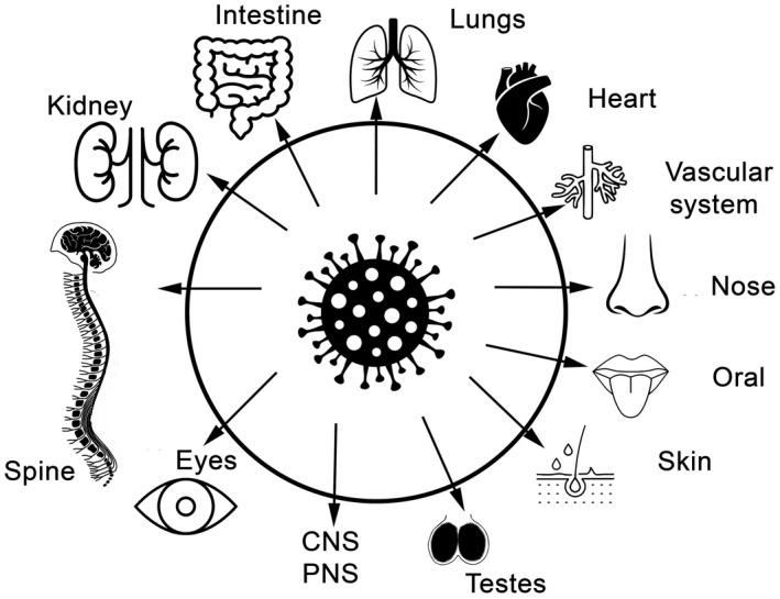
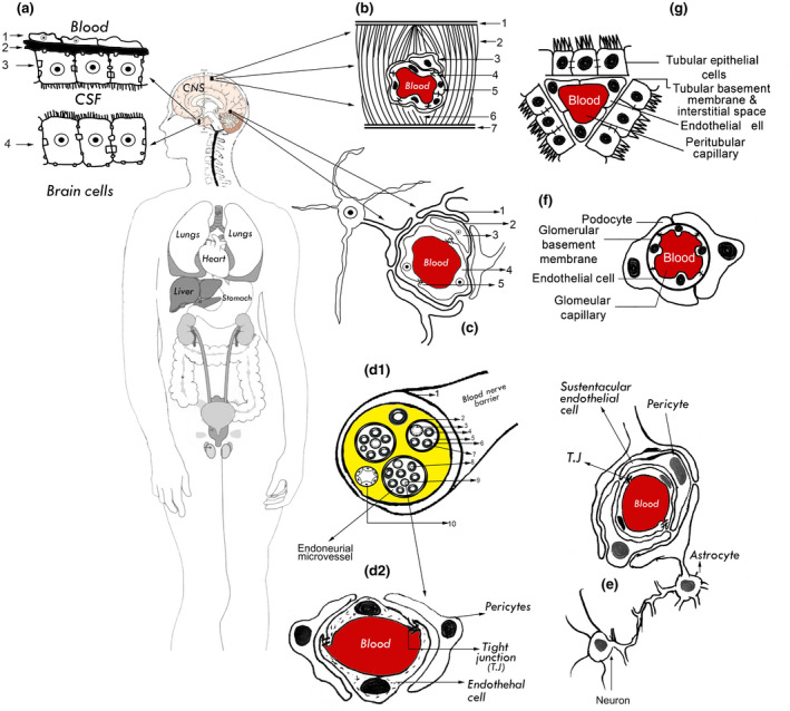
Without protective and/or therapeutic agents the severe acute respiratory syndrome coronavirus 2 (SARS-CoV-2) infection known as coronavirus disease 2019 is quickly spreading worldwide. It has surprising transmissibility potential, since it could infect all ages, gender, and human sectors. It attacks respiratory, gastrointestinal, urinary, hepatic, and endovascular systems and can reach the peripheral nervous system (PNS) and central nervous system (CNS) through known and unknown mechanisms. The reports on the neurological manifestations and complications of the SARS-CoV-2 infection are increasing exponentially. Herein, we enumerate seven candidate routes, which the mature or immature SARS-CoV-2 components could use to reach the CNS and PNS, utilizing the within-body cross talk between organs. The majority of SARS-CoV-2-infected patients suffer from some neurological manifestations (e.g., confusion, anosmia, and ageusia). It seems that although the mature virus did not reach the CNS or PNS of the majority of patients, its unassembled components and/or the accompanying immune-mediated responses may be responsible for the observed neurological symptoms. The viral particles and/or its components have been specifically documented in endothelial cells of lung, kidney, skin, and CNS. This means that the blood-endothelial barrier may be considered as the main route for SARS-CoV-2 entry into the nervous system, with the barrier disruption being more logical than barrier permeability, as evidenced by postmortem analyses.

Keywords: COVID-19; SARS-CoV-2; blood-brain barrier; blood-nerve barrier; blood-nervous system barrier; bloodcerebrospinal-fluid-barrier; double membrane vesicles cargo route; lymphatic brain drainage route; macrophage/monocytes cargo route; neurotropic virus; nicotinic acetylcholine receptor; olfactory route; peripheral nerve or neuronal retrograde route.


Conflict of interest statement

The authors declare that there are no conflict of interest.

Figures

FIGURE 1
FIGURE 1
Respiratory and extra respiratory organ/system COVID‐19 prevalence
FIGURE 2
FIGURE 2
Diagram for human blood–nervous system barriers. (a) Blood–cerebrospinal fluid (BCSFB)/CP barrier (1. Fenestrated endothelium, 2. Interstitial matrix, 3 and 4. Choroid plexus epithelium, 5. Brain cells). (b) Blood–cerebrospinal fluid (BCSFB)/meningeal barrier (1. arachnoid, 2. Trabeculae cross‐section, 3. Pericyte, 4. Epithelial cell tight junction, 5. Epithelial cell, 6. CSF, 7. Pia Mater). (c) Blood‐brain barrier (1. Astrocyte, 2. Basement membrane, 3. Pericyte, 4. Endothelial cell [non‐fenestrated], 5. Tight junction). (d1) Blood–nerve barrier BNB (cross section) (1. Epineurium, 2. Perineurium, 3. Endoneurial vessel, 4. Basal lamina. 5. Endoneurium, 6. Myelin, 7. Nucleus of Schwann cell, 8. Axon, 9. Endoneurial endothelial cells of microvessel, 10. Epineurium blood vessel). (d2) Blood–nerve barrier. (e) Blood olfactory nerve barrier. (f) Inside the glomeruli barriers both of glomerular endothelial and epithelial cells (known as podocytes) cross talk occurs, where they share the glomerular basement membrane. (g) inside the glomerular both of peritubular capillary endothelial and tubular epithelial cells cross talk also occurs through the barrier where they are separated by a tubular basement membrane and interstitial space. Both of kidney epithelial (podocytes and tubular) are breached with SARS‐CoV‐2 in COVID‐19 patients (reviewed in (Elrashdy et al., 2020)). From her the viral and/or its components can spread from renal anastomosis into CNS via nerve supply. Although the cross talk between human organs in health and diseases is a very complicated processes go through huge number of mechanisms, but it well documented in a dramatically pattern (Armutcu, ; Lu et al., 2015) [Color figure can be viewed at wileyonlinelibrary.com]
FIGURE 3
FIGURE 3
Classification of COVID‐19 disease stages. The figure illustrates three escalating phases of COVID‐19 disease progression, with hypothesis of blood–endothelial/epithelial barriers integrity/permeability scale associated with age and comorbidities diseases over the three stages. The blood–endothelial barriers are representative for all body barriers and specifically for blood–nervous system barrier (BNSB). Progressive increase in inflammatory cytokine and chemokines eventually leads to cytokine storm in a profile similar to in sepsis cases, which eventually leads to endothelial barrier dysfunction. Many other biomarkers molecules (in addition to the cytokine storm elements) have a direct effect on the BNSB as discussed in text. The times on the x axis are approximate. The figure designed based on and adapted from (Akhmerov & Marban, ; Delaney & Campbell, ; Doran et al., ; Elrashdy et al., ; Li, Liu, et al., ; Siddiqi & Mehra, 2020). + to >4+ indicative for barrier integrity/permeability like scale, IIA (stage II without hypoxia) IIB (Stage II with hypoxia). Tumor necrosis factor (TNF‐α), interleukin 1β (IL‐1β), IL‐6, GCSF: granulocyte‐colony stimulating factor, interferon gamma‐induced protein‐10, monocyte chemoattractant protein‐1, and MCA‐protein 1: macrophage inflammatory proteins 1‐α [Color figure can be viewed at wileyonlinelibrary.com]

Similar articles

Cited by

References

    1. Abassi, Z. , Knaney, Y. , Karram, T. , & Heyman, S. N. (2020). The lung macrophage in SARS‐CoV‐2 infection: A friend or a foe? Frontiers in Immunology, 11, 1312. 10.3389/fimmu.2020.01312 - DOI - PMC - PubMed
    1. Abbott, N. J. (2002). Astrocyte‐endothelial interactions and blood‐brain barrier permeability. Journal of Anatomy, 200(6), 629–638. - PMC - PubMed
    1. Abdul Razzak, R. , Florence, G. J. , & Gunn‐Moore, F. J. (2019). Approaches to cns drug delivery with a focus on transporter‐mediated transcytosis. International Journal of Molecular Sciences, 20(12), 3108. 10.3390/ijms20123108 - DOI - PMC - PubMed
    1. Ackermann, M. , Verleden, S. E. , Kuehnel, M. , Haverich, A. , Welte, T. , Laenger, F. , Vanstapel, A. , Werlein, C. , Stark, H. , Tzankov, A. , & Li, W. W. (2020). Pulmonary vascular endothelialitis, thrombosis, and angiogenesis in Covid‐19. New England Journal of Medicine, 383(2), 120–128. - PMC - PubMed
    1. Agosti, E. , Giorgianni, A. , D'Amore, F. , Vinacci, G. , Balbi, S. , & Locatelli, D. (2020). Is Guillain‐Barrè syndrome triggered by SARS‐CoV‐2? Case report and literature review. Neurological Sciences. 10.1007/s10072-020-04553-9 - DOI - PMC - PubMed

MeSH terms

Substances