Review
. 2020 Nov 18;6(4):293.
doi: 10.3390/jof6040293.

Molecular Tools for Detection and Identification of Paracoccidioides Species: Current Status and Future Perspectives

Affiliations
Review

Molecular Tools for Detection and Identification of Paracoccidioides Species: Current Status and Future Perspectives

Breno Gonçalves Pinheiro et al. J Fungi (Basel). .

Abstract

Paracoccidioidomycosis (PCM) is a mycotic disease caused by the Paracoccidioides species, a group of thermally dimorphic fungi that grow in mycelial form at 25 °C and as budding yeasts when cultured at 37 °C or when parasitizing the host tissues. PCM occurs in a large area of Latin America, and the most critical regions of endemicity are in Brazil, Colombia, and Venezuela. The clinical diagnosis of PCM needs to be confirmed through laboratory tests. Although classical laboratory techniques provide valuable information due to the presence of pathognomonic forms of Paracoccidioides spp., nucleic acid-based diagnostics gradually are replacing or complementing culture-based, biochemical, and immunological assays in routine microbiology laboratory practice. Recently, taxonomic changes driven by whole-genomic sequencing of Paracoccidioides have highlighted the need to recognize species boundaries, which could better ascertain Paracoccidioides taxonomy. In this scenario, classical laboratory techniques do not have significant discriminatory power over cryptic agents. On the other hand, several PCR-based methods can detect polymorphisms in Paracoccidioides DNA and thus support species identification. This review is focused on the recent achievements in molecular diagnostics of paracoccidioidomycosis, including the main advantages and pitfalls related to each technique. We discuss these breakthroughs in light of taxonomic changes in the Paracoccidioides genus.

Keywords: Paracoccidioides brasiliensis; Paracoccidioides lutzii; diagnosis; endemic mycosis; epidemiology; molecular diagnostics; paracoccidioidomycosis; systemic mycosis.


Conflict of interest statement

The authors declare no conflict of interest. The funders had no role in the design of the study; in the collection, analyses, or interpretation of data; in the writing of the manuscript, or in the decision to publish the results.

Figures

Figure 1
Figure 1
Phylogenetic tree inferred using the maximum likelihood method and Kimura 2-parameter model of the partial sequences of the immunodominant antigen GP43, ADP ribosylation factors (ARF), and tubulin alpha-1 chain (TUB1) of Paracoccidioides isolates. The P. brasiliensis complex is composed of the cryptic species S1, PS2, PS3, and PS4. Paracoccidioides lutzii appear as a divergent genetic group, apart from the P. brasiliensis complex. Numbers close to the branches represent bootstraps values (maximum likelihood/neighbor joining, respectively). Bootstraps higher than 80 based on 1000 replications are represented in bold branches. Sequences were described previously by Matute et al. [14], Teixeira et al. [15], and Hahn et al. [20].
Figure 2
Figure 2
Distribution patterns of Paracoccidioides species in South America, based on epidemiological reports in the literature [19,23,24,27,28]. (a) P. brasiliensis sensu stricto (S1); (b) P. americana (PS2); (c) P. restrepiensis (PS3), and P. venezuelensis (PS4); (d) P. lutzii.
Figure 3
Figure 3
Flowchart for laboratory diagnosis of paracoccidioidomycosis (PCM). BAL: Bronchoalveolar lavage; H&E: Hematoxylin and eosin staining; PAS: Periodic acid–Schiff; GMS: Gomori methenamine silver; ELISA: enzyme-linked immunosorbent assay; DID: double immunodiffusion; CIE: counterimmunoelectrophoresis reaction; WB: Western blot.
Figure 4
Figure 4
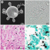
Morphological aspects of Paracoccidioides yeast. (a) Paracoccidioides lutzii showing multiple buds with a “steering wheel” shape in vitro (scanning electron microscopy); (b) The positive direct mycological examination of pus showing large yeasts (5–15 µm) that have a thick, birefringent cell wall with single or multiple buds; (c) Paracoccidioides spp. in tissue (arrows) stained by hematoxylin and eosin (HE); (d) Paracoccidioides spp. in tissue stained by Grocott’s methenamine silver (GMS) showing a “Mickey Mouse” shape (arrow).
Figure 5
Figure 5
Major developments in the identification and molecular/proteomic characterization of Paracoccidioides species. PCM: paracoccidioidomycosis; Pb: Paracoccidioides brasiliensis; qPCR: quantitative real-time polymerase chain reaction; LAMP: loop-mediated isothermal amplification; FFPE: formalin-fixed paraffin-embedding; MLSA: multilocus sequence analysis; SSR: single sequence repeats; Pb: Paracoccidioides brasiliensis; SnaPshot: single-nucleotide polymorphism (SNP) genotyping; MALDI-ToF: matrix-assisted laser desorption/ionization time-of-flight mass spectrometry; PCR-RFLP: polymerase chain reaction-restriction fragment length polymorphism; TUB1: tubulin alpha-1 chain; FISH: fluorescence in situ hybridization; FT-IR: Fourier-transform infrared spectroscopy.
Figure 6
Figure 6
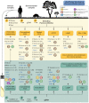
Schematic representation of Paracoccidioides spp. molecular detection/identification pipeline, directly from clinical and/or environmental samples (yellow panel) or from gDNA extracted from cultured isolates (green panel). Breakthroughs are listed, as well as their targeted genes and species discrimination power. PCR, polymerase chain reaction; RFLP, restriction fragment length polymorphism; qPCR, quantitative real-time polymerase chain reaction; FISH, fluorescent in situ hybridization; LAMP, loop-mediated isothermal amplification.

Similar articles

Cited by

References

    1. Shikanai-Yasuda M.A., Mendes R.P., Colombo A.L., Queiroz-Telles F., Kono A.S.G., Paniago A.M.M., Nathan A., Valle A., Bagagli E., Benard G., et al. Brazilian guidelines for the clinical management of paracoccidioidomycosis. Rev. Soc. Bras. Med. Trop. 2017;50:715–740. doi: 10.1590/0037-8682-0230-2017. - DOI - PubMed
    1. Do Amaral C.C., Fernandes G.F., Rodrigues A.M., Burger E., de Camargo Z.P. Proteomic analysis of Paracoccidioides brasiliensis complex isolates: Correlation of the levels of differentially expressed proteins with in vivo virulence. PLoS ONE. 2019;14:e0218013. doi: 10.1371/journal.pone.0218013. - DOI - PMC - PubMed
    1. Ambrósio A., Camelo C., Barbosa C., Tomazatti F., Brazões F., Veloso J., Rodrigues G., Rodrigues L., Oliveira P., Aguiar R., et al. Paracoccidioidomycosis disease (Lutz-Splendore-Almeida disease): Additional workup, differential diagnosis, cure control. RMMG-Rev. Méd Minas Gerais. 2014;24:81–92. doi: 10.5935/2238-3182.20140021. - DOI
    1. Restrepo A., Gómez B.L., Tobón A. Paracoccidioidomycosis: Latin America’s own fungal disorder. Curr. Fungal Infect. Rep. 2012;6:303–311. doi: 10.1007/s12281-012-0114-x. - DOI
    1. Griffiths J., Lopes Colombo A., Denning D.W. The case for paracoccidioidomycosis to be accepted as a neglected tropical (fungal) disease. PLoS Negl. Trop. Dis. 2019;13:e0007195. doi: 10.1371/journal.pntd.0007195. - DOI - PMC - PubMed

LinkOut - more resources