. 2020 Nov 18;21(22):8687.
doi: 10.3390/ijms21228687.

Substrate Specificity and Structural Modeling of Human Carboxypeptidase Z: A Unique Protease with a Frizzled-Like Domain

Affiliations

Substrate Specificity and Structural Modeling of Human Carboxypeptidase Z: A Unique Protease with a Frizzled-Like Domain

Javier Garcia-Pardo et al. Int J Mol Sci. .

Abstract

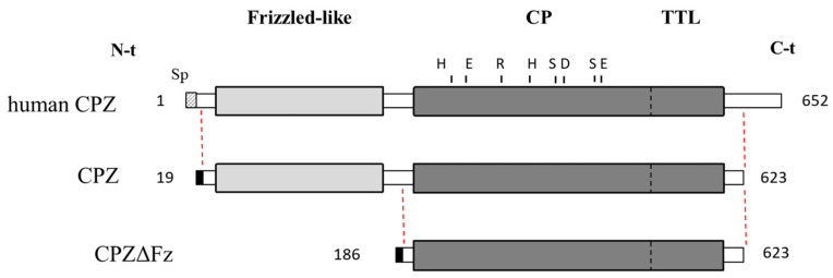
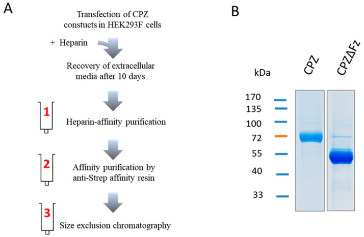
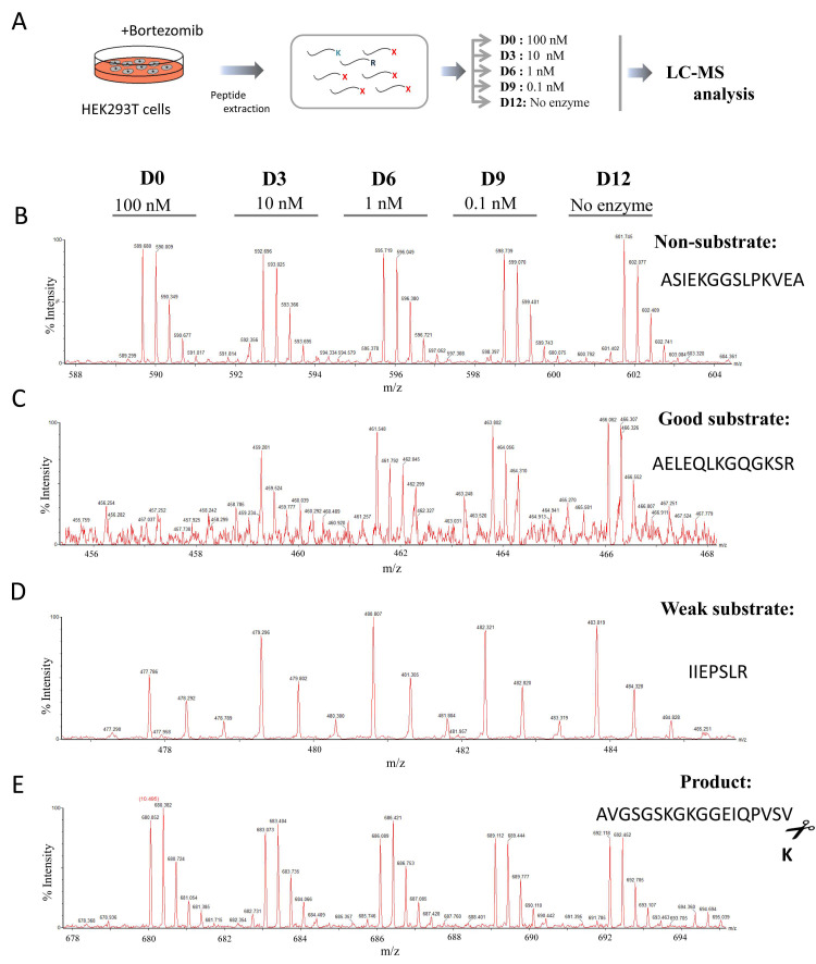
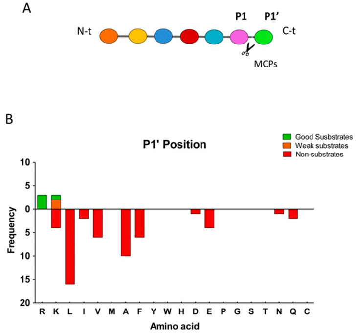
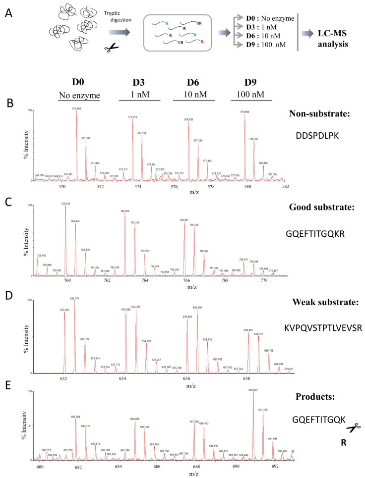
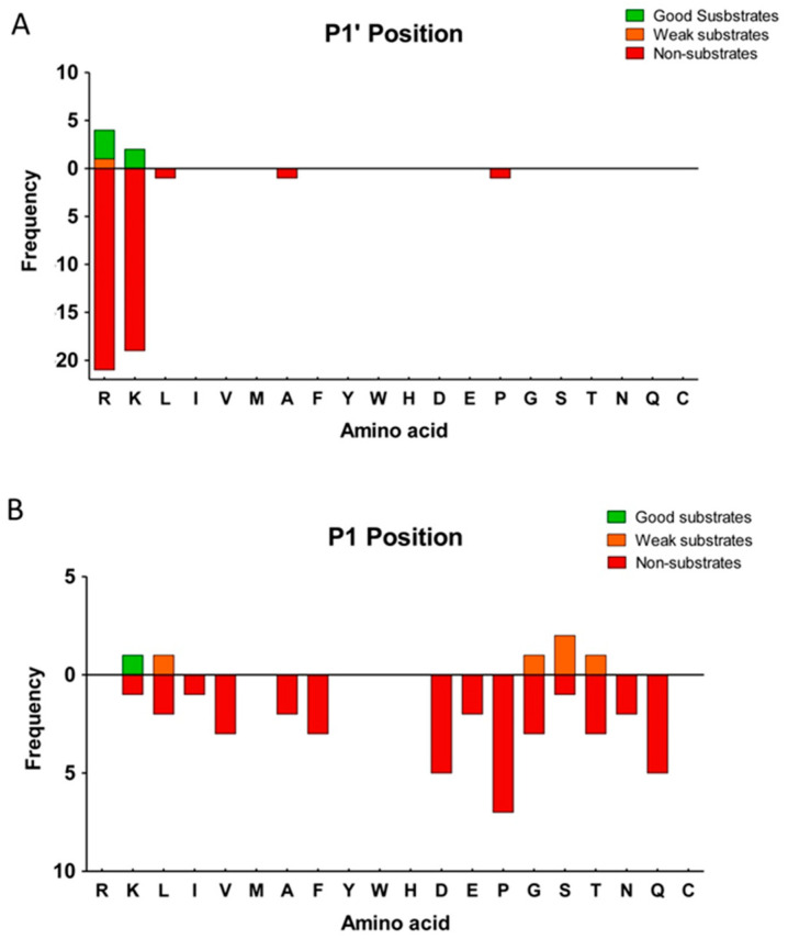
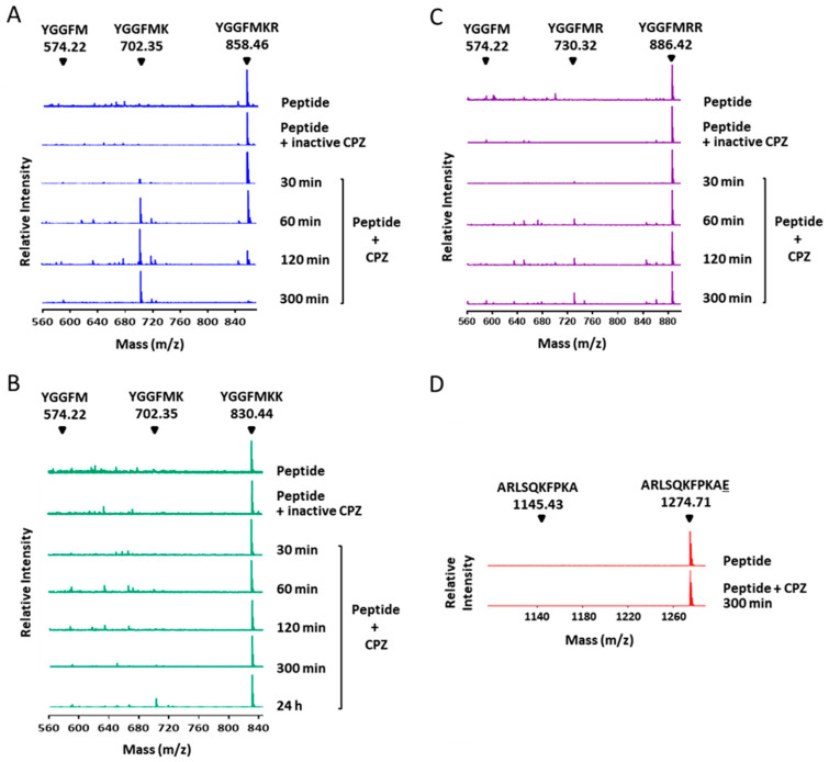
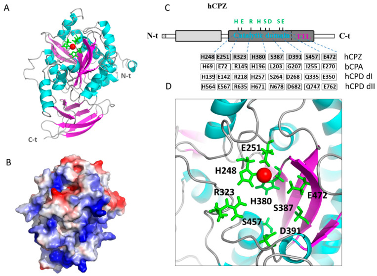
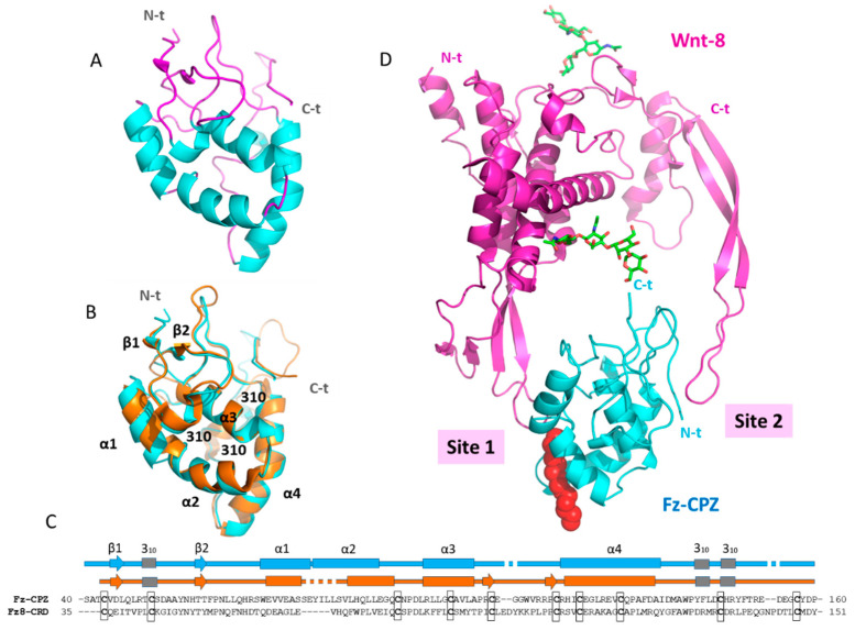
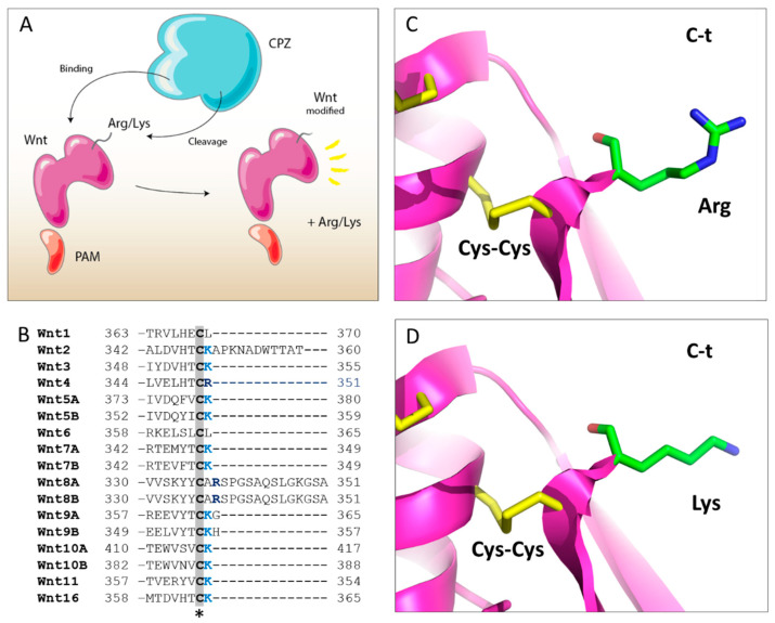
Metallocarboxypeptidase Z (CPZ) is a secreted enzyme that is distinguished from all other members of the M14 metallocarboxypeptidase family by the presence of an N-terminal cysteine-rich Frizzled-like (Fz) domain that binds Wnt proteins. Here, we present a comprehensive analysis of the enzymatic properties and substrate specificity of human CPZ. To investigate the enzymatic properties, we employed dansylated peptide substrates. For substrate specificity profiling, we generated two different large peptide libraries and employed isotopic labeling and quantitative mass spectrometry to study the substrate preference of this enzyme. Our findings revealed that CPZ has a strict requirement for substrates with C-terminal Arg or Lys at the P1' position. For the P1 position, CPZ was found to display specificity towards substrates with basic, small hydrophobic, or polar uncharged side chains. Deletion of the Fz domain did not affect CPZ activity as a carboxypeptidase. Finally, we modeled the structure of the Fz and catalytic domains of CPZ. Taken together, these studies provide the molecular elucidation of substrate recognition and specificity of the CPZ catalytic domain, as well as important insights into how the Fz domain binds Wnt proteins to modulate their functions.

Keywords: Wnt signaling; carboxypeptidase Z; cysteine rich domain; frizzled; growth factor; metallocarboxypeptidase; substrate specificity.


Conflict of interest statement

The authors declare no conflict of interest. The funders had no role in the design of the study; in the collection, analyses, or interpretation of data; in the writing of the manuscript, or in the decision to publish the results.

Figures

Figure 1
Figure 1
Schematic representation of human metallocarboxypeptidase Z (CPZ) and its recombinant forms. Full-length human CPZ (UniProtKB accession number Q66K79) and the recombinant CPZ forms used in this work are represented. Key residues essential for the catalytic mechanism are indicated: His69, Glu72, Arg145, His198 and Glu270 (numbering based on the mature form of bovine CPA, by convention in the field). Positions of residues Ser387, D391 and Ser457 of CPZ (equivalent to Leu203, Gly207 and Ile255 in bovine CPA, respectively) are also indicated. The recombinant form CPZ lacks 29 residues in its C-terminal region. The recombinant form CPZΔFz, in addition to the C-terminal truncation, lacks 167 residues in its N-terminal tail, corresponding to the Frizzled-like (Fz) domain.
Figure 2
Figure 2
Recombinant expression and purification of human CPZ. (A) Schematic diagram of the experimental strategy used for the recombinant expression and purification of CPZ and CPZΔFz. Protein expression was performed by high-level transient transfection in suspension-growing HEK (Human Embryonic Kidney) 293F cells, followed by the addition of heparin at 48 h post-transfection. For protein purification, the extracellular medium was collected after 10-days incubation, and recombinant proteins were purified in three purification steps by heparin-affinity chromatography, affinity chromatography using anti-strep tag affinity resin, and by size-exclusion chromatography; (B) SDS–PAGE showing the purity and size of the recombinant proteins.
Figure 3
Figure 3
Quantitative peptidomics analysis of CPZ substrate specificity using peptides extracted from HEK293T cells treated with bortezomib. (A) Quantitative peptidomics analysis scheme and (B–E) representative results. After incubation of the peptides with CPZ at different concentrations, the individual samples were labeled with one of five stable isotopic TMAB tags (D0 for 100 nM CPZ; D3 for 10 nM CPZ; D6 for 1 nM CPZ; D9 for 0.1 nM CPZ; D12 for samples without enzyme). All five reactions were pooled and analyzed by LC-MS. Representative examples are shown for (B) non-substrates, (C) good substrates, (D) weak substrates and (E), products of CPZ.
Figure 4
Figure 4
Analysis of the substrate preferences of CPZ from the peptidomics experiment using the HEK293T-derived peptide library. (A) Schematic representation of relevant residues involved in a typical MCPs cleavage, according to the model proposed by Schechter and Berger [32]; (B) Substrate preference of CPZ at the C-terminal (P1′) position. The frequency for each amino acid found in P1′ is indicated for good substrates, weak substrates, and non-substrates.
Figure 5
Figure 5
Quantitative peptidomics analysis scheme for the characterization of the substrate specificity of CPZ using a tryptic peptide library. (A) Schematic representation of the quantitative peptidomics analysis and (BE) representative results. Tryptic peptides were prepared from the digestion of five selected proteins with trypsin. The resultant peptide library was aliquoted and incubated with no enzyme or different CPZ concentrations (i.e., 1, 10, and 100 nM) for 16 h at 37 °C. After incubation samples were labeled with one of four stable isotopic TMAB tags as follows: D0 for the sample without enzyme; D3 for 1 nM CPZ; D6 for 10 nM CPZ; and D9 for 100 nM CPZ. All the individual reactions were pooled and analyzed by LC-MS. Representative examples are shown for (B) non-substrates, (C) good substrates, (D) weak substrates, and (E) products of CPZ.
Figure 6
Figure 6
Analysis of the substrate preferences of CPZ determined using the tryptic peptide library. Substrate preferences of CPZ at (A) C-terminal (P1′) and (B) penultimate (P1) positions. The frequency for each amino acid present in P1 or P1′ is indicated for good substrates, weak substrates, and non-substrates. For P1 analysis, only substrates with permissive P1′ amino acids according to the results from (A) were considered.
Figure 7
Figure 7
Substrate specificity of CPZ against Met-enkephalin-derived peptides. MALDI-TOF MS spectra of Met-enkephalin-derived peptides (A) YGGFMKR, (B) YGGFMKK, and (C) YGGFMRR treated with 100 nM CPZ at 37 °C with different incubation times. The peaks with a mass of 858.46 Da, 830.44 Da and 886.42 Da correspond to the peptides YGGFMKR (theoretical monoisotopic MH+ mass = 858.41 Da), YGGFMKK (theoretical monoisotopic MH+ mass = 830.41 Da) and YGGFMRR (theoretical monoisotopic MH+ mass = 886.42 Da) respectively. The peak with a mass of 702.35 Da generated in the presence of CPZ corresponds to the peptide YGGFMK (theoretical monoisotopic MH+ mass = 702.31 Da) produced by the cleavage of the C-terminal Arg or Lys amino acids from the peptides YGGFMKR and YGGFMKK, respectively. The peak with a mass of 730.32 Da generated in the presence of CPZ corresponds to the peptide YGGFMR (theoretical monoisotopic MH+ mass = 730.32 Da) produced by the cleavage of the C-terminal Arg from the peptide YGGFMRR. The position for the peptide YGGFM (with mass of 574.22 Da) is indicated. CPZ was inactivated by incubating the enzyme for 15 min at 90 °C to be used as a control reaction (peptide + inactive CPZ). Control samples were incubated for 24 h at 37 °C; (D) Spectra of the synthetic peptide ARLSQKFPKAE after 300 min of incubation in the absence (peptide) or in the presence of 100 nM CPZ (peptide + CPZ). Numbers above the major peaks indicate the monoisotopic masses of the MH+ ion (m/z) for the full-length peptide (mass = 1274.71 Da) or the peptide without its C-terminal residue, ARLSQKFPKA (mass = 1145.43 Da).
Figure 8
Figure 8
Structural modeling of the catalytic and transthyretin-like (TTL) domains of human CPZ. (A) Ribbon representation of human CPZ structure showing the central catalytic moiety at the top and the C-terminal TTL domain at the bottom. The side chains of the three residues involved in the Zn2+ binding (i.e., His248 Glu251, His380) are indicated in green; (B) Electrostatic surface potential distribution of the catalytic domain of human CPZ (in the same orientation as panel A). Blue indicates positive and red indicates negative charge potential; (C) Linear representation of the full-length human CPZ, showing the location of relevant amino acids involved in the catalytic mechanism and substrate binding (Arg323, Ser387, Asp391, Ser457, and Glu472), as well as in zinc binding (His248 Glu251, His380). Residues found in equivalent positions in bovine CPA (bCPA, as reference), in domain I of human CPD (hCPDdI) and domain II of human CPD (hCPDdII) are indicated; (D) Magnification of the active site of human CPZ, showing the location of these residues important for the catalytic mechanism and substrate specificity determination. The Zn2+ metal ion is shown in all representations as a red sphere.
Figure 9
Figure 9
Structural modeling of the Fz domain of human CPZ and its interaction with Wnt-8. (A) Ribbon representation of the highly conserved N-terminal Fz domain of human CPZ; (B) Structural comparison of the Fz domain of human CPZ (Fz-CPZ, represented in blue) and the cysteine-rich domain of mouse Fz8 (Fz8-CRD, represented in orange); (C) Structure-based sequence alignment of Fz-CPZ and Fz8-CRD (represented in blue and orange respectively); (D) Ribbon representation of Wnt-8 in a proposed complex with the Fz domain of CPZ. The extended palmitoleic acid (PAM) group is represented with red spheres, and the most important sites of interaction between Fz-CPZ and Fz8-CRD are indicated as Site 1 and Site 2. For all proteins, the N-terminus and C-terminus are indicated as N-t and C-t, respectively.
Figure 10
Figure 10
Comparison of the C-terminal region of human Wnt proteins. (A) Proposed model for the proteolytic cleavage of Wnt proteins by CPZ. (B) Sequence alignment of the C-terminal region of all human Wnt proteins. The star indicates the location of the last conserved cysteine residue, involved in a disulfide bridge formation that links the last two β-sheet regions. The C-terminal Lys and Arg residues located immediately after the last Cys residue in the majority of human Wnt proteins are shown in bright blue or dark blue, respectively; (C,D) Structural modeling of the C-terminal tail of XWnt-8, showing the location of the C-terminal residues identified in the majority of human Wnt proteins. Models were generated by mutating the last Ala residue found in XWnt-8 for Arg or Lys amino acids (C and D, respectively). The last Cys-Cys bond formed in XWnt-8 between Cys295 and Cys337 is indicated.

Similar articles

Cited by

References

    1. Arolas J.L., Vendrell J., Aviles F.X., Fricker L.D. Metallocarboxypeptidases: Emerging drug targets in biomedicine. Curr. Pharm. Des. 2007;13:349–366. doi: 10.2174/138161207780162980. - DOI - PubMed
    1. Reznik S.E., Fricker L.D. Carboxypeptidases from a to Z: Implications in embryonic development and wnt binding. Cell Mol. Life Sci. 2001;58:1790–1804. doi: 10.1007/PL00000819. - DOI - PMC - PubMed
    1. Rawlings N.D. Twenty-five years of nomenclature and classification of proteolytic enzymes. Biochim. Biophys. Acta Proteins Proteom. 2020;1868:140345. doi: 10.1016/j.bbapap.2019.140345. - DOI - PubMed
    1. Reverter D., Maskos K., Tan F., Skidgel R.A., Bode W. Crystal structure of human carboxypeptidase M, a membrane-bound enzyme that regulates peptide hormone activity. J. Mol. Biol. 2004;338:257–269. doi: 10.1016/j.jmb.2004.02.058. - DOI - PubMed
    1. Garcia-Pardo J., Grana-Montes R., Fernandez-Mendez M., Ruyra A., Roher N., Aviles F.X., Lorenzo J., Ventura S. Amyloid formation by human carboxypeptidase D transthyretin-like domain under physiological conditions. J. Biol. Chem. 2014;289:33783–33796. doi: 10.1074/jbc.M114.594804. - DOI - PMC - PubMed

LinkOut - more resources